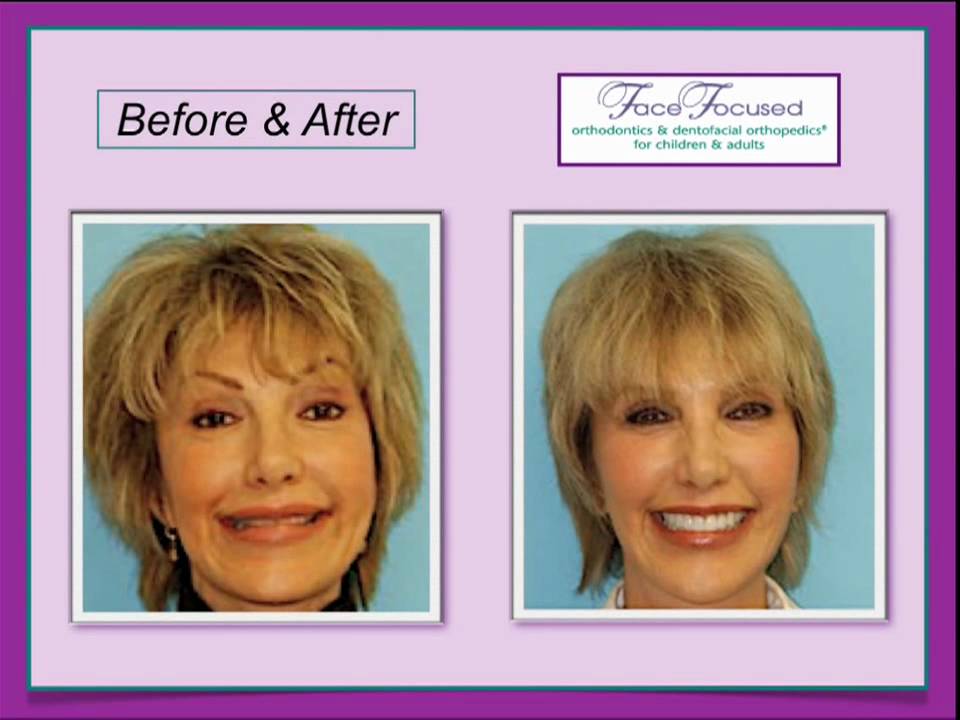
In this 2010 case‑study video, Dr. Bill Hang presents the comprehensive orthodontic treatment of a 55‑year‑old woman who sought a broader, more attractive smile before receiving veneers. The patient also suffered from jaw and neck pain caused by nighttime grinding and a chronic head‑tilt posture used to keep her airway open. Dr. Hang explains the pre‑treatment problems—narrow upper and lower arches, backward‑tipped front teeth, limited smile display, and airway‑related head posture—then outlines a four‑fold orthodontic plan: (1) dramatically broaden both arches, (2) open the bite to create vertical space, (3) angle the upper front teeth forward to improve visibility and brightness, and (4) idealize gum‑tissue height while creating precise inter‑tooth spacing for optimal veneer dimensions. Before‑and‑after images illustrate how arch widening eliminated the head‑tilt, reduced muscular pain, increased tongue space, and allowed the restorative dentist to place veneers of ideal size, shape, and color. The final result is a full‑width, youthful smile with all teeth visible, balanced gum lines, and resolved functional complaints. Dr. Hang emphasizes that meticulous orthodontic preparation is essential for successful restorative dentistry.
Chapters
This case study describes a 55-year-old female who came to us seeking to have her smile broadened before having veneers placed on her teeth. Her primary purpose for seeking treatment was aesthetics. However, she also suffered from jaw pain as a result of grinding her teeth at night and neck pain due to tilting her head back to keep her airway open. We believed that our treatment would alleviate those symptoms as well. This picture shows the patient in profile before her orthotic treatment began. The purple arrow and red line illustrate how she tilts her head back as she looks forward. As you can see from this diagram, tilting the head back opens the airway and makes it easier for patients like this to breathe. Over time, As a patient continues to keep his or her head tilted back, the neck and shoulder muscles can become chronically sore. However, by moving the forehead into a vertical position, the tongue and soft palate fall back into the airway, creating a blockage. That blockage makes breathing difficult. This picture shows her in full-face smile. She does not have a particularly attractive smile. There are 2 primary reasons for this. First, her teeth are located too far back in her face, which makes them barely visible when she smiles. Second, her teeth are tipped back, which causes her smile to appear very dark. This picture is a view of her upper teeth from below before she received orthodontic treatment. You can see that her teeth are out of alignment and her upper front teeth are tipped back from an ideal position. She also has a narrow upper arch, which is creating several problems for her that I will explain later on in the program. This picture is a view of her lower teeth from above. Again, the teeth are not ideally aligned and the arch is narrow. From this angle, you can see from the right side how her teeth fit together before she was treated. If you look closely at the teeth that I've numbered 1 through 4, you'll notice that they are tipped back. She would have a far more attractive smile if these teeth were angled forward slightly. As you look at her teeth from the front, you'll notice that her side teeth were less visible because her arch was narrow. This is a typical pattern that we see in many patients. If this patient were to have restorative dentistry done on arches that are narrow like this, she would not have attained the broad, full smile that she wanted. From the left, you can see that her upper teeth overlapped her lower teeth excessively, which took away from the attractiveness of her smile. The orthodontic treatment plan for this patient was to align her upper and lower arches and broaden the arches dramatically. We also wanted to make her upper front teeth appear more visually pleasing by angling them properly and bringing them forward in her face. The red lines in these before and after pictures point out a major benefit of this type of treatment. Her head is no longer tipped back because she no longer needs to keep her chin forward to keep her airway open. Therefore, her jaw and neck muscles are no longer tender. The after picture on the right side of your screen shows why proper orthodontic care is such a critical part of restorative dentistry. The orthodontics broadened her smile and brought her teeth forward, giving her a smile complete to the corners of the mouth. In cases like this, we believe that tiny details are of the utmost importance. Our goal is to lay a perfect foundation for the restorative dentist who will create veneers to achieve the best possible result. In this case, we focused on 4 critical facets of her treatment to ensure that her veneers would give her the result she was seeking. First, you'll notice a small space, pointed out by the purple arrow, behind her lateral incisor and in front of her eye tooth. We created that space intentionally so that veneers of ideal size and shape could be constructed for those teeth. Second, as this picture set shows, we opened her bite significantly, giving the restorative dentist the foundation he needed to ideally place her veneers. Third, we angled her teeth properly so that when the veneers are placed, her smile will be brighter. Finally, we idealized her gum tissue height. Gum tissue height describes the point at which the top of the teeth meet the bottom of the gum tissue. Gum tissue heights should be symmetrical and in a proportion that pleases the eye. The change in gum tissue height is highlighted by the black shapes on your screen. In summary, because her jaws have been widened, her smile is now broad and complete to the corners of the mouth. Instead of having only the front 6 teeth visible when she smiles, all of the teeth are now visible. As you can also see, the gum tissue height is idealized and the restorative dentist created veneers of ideal size and color. This before and after set of pictures shows the entire lower arch. The red line illustrates better tooth alignment and the black line shows a broadened lower arch. It's my feeling that the broadening of the arches, which increases the tongue space, can often lead to a reduction or elimination of the patient's grinding habit at night. These 2 pictures show the before and after of the patient's upper teeth from below. The red line shows the improved alignment that we achieved during her treatment, and the black line shows the broadening of her arch. This side-by-side before and after picture set shows the final result that was achieved. The dramatic improvement of her smile would not have been possible without the orthodontic preparation of her case through broadening her smile, improving the angulation of her upper front teeth, and creating the proper space for ideal veneer placement. She now has a broader, brighter, more youthful look to her smile. This patient was very happy with her aesthetic results. She was also pleasantly surprised to have her neck and jaw pain alleviated as a result of the treatment. I'm Dr. Bill Hang. Thanks for taking the time to watch and listen to this case study.