How to Recognize Right Bundle Branch Block
A clinician-led tutorial explaining how to recognize right bundle branch block (RBBB) on ECG. Covers normal ventricular conduction, how RBBB alters activation (delayed right ventricular depolarization), the characteristic ECG patterns (RSR' in V1; wide terminal S in V6/Lead I; widened QRS typically ~0.14–0.16s), and clinical context (mostly benign causes, possible post‑surgical or septal MI implications).
Chapters
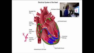
Today's talk is about how to recognize right bundle branch block. Before we talk about right bundle branch block, let's review normal conduction so we can see the difference between normal and abnormal. We're going to talk about conduction through the ventricles only. So I'll start here in the AV node. And in normal conduction, the impulse comes out of the AV node down the bundle of his or the common bundle and enters both of the bundle branches simultaneously. I can't draw them simultaneously, so I'll just show you both. And this would happen very fast because the whole idea of having bundle branches is to have a fast racetrack system that carries the impulse really quickly, with the overall goal being depolarize all the cells in both ventricles in a simultaneous manner. Get the ventricles to conduct fast, contract fast, make a narrow qrs. Narrow QRS indicates we have much better cardiac output than wide qrs. So that's our goal. When one of the bundle branches is blocked, there's going to be a disruption in that fast snappy conduction and the fast snappy contraction, and we're going to get a wider QRS and sometimes less cardiac output. Luckily, in right bundle branch block, that isn't nearly as much of a problem as it is in left bundle branch block. So to illustrate it going to put a block, a blockage across the right bundle. Now, the cause of that, there are many causes. Most of them in right bundle branch block are benign. Many people have right bundle branch block from birth. It's not a problem. Most right bundle branch blocks cause very little if any clinical problems. So not to worry about most of them. This one may be caused by an mi, which that would be about the only really serious thing I can think of that we would need to worry about. So if we have a patient with a septal MI and the tissue in that area is dying, the right bundle branch could be killed. And the danger there is that the left bundle branch is right next door to it and it could die as well. And if it did, then the patient would experience a complete heart block, which could be life threatening. But we're going to confine this discussion to right bundle branch block and how to recognize it. And so to illustrate it, I've got the block in place and now I'm going to show normal conduction proceeding down through the left ventricle. Now, this is great. This is what we want. We want the left ventricle to depolarize normally. And it does. And there's no issues with timing no issues with contractions. So the heart's pretty happy at this point. The body's pretty happy because your left ventricle is the part of the heart that supplies your body with cardiac output. But the all or none principle comes in here and. And the right ventricle is sitting over there saying, well, you know, I would like to contract two. And I haven't received the message. The all or none principle promises that the impulse will travel cell by cell if it can't travel on the bundle branches. So from the left ventricle, that electrical impulse is going to spread across the right ventricle, and it doesn't take long, but it will depolarize the right ventricle. In right bundle branch block, we will get a slightly wide qrs, but it's only the second part of the QRS that's widened. If we take a view of the heart from V1 position, remember you're on the right side of the heart or the right side of the chest. V1 will see that left ventricle depolarizing completely normally. And so the septum depolarizes. That gives us an upright little R wave. And then the left ventricle depolarizes and gives us an S wave. And that's because the left ventricle depolarizes away from V1. So we get a negative wave. But then the right ventricle depolarizes late. Normally it would happen at the same time as the left ventricle, but now it's late, and so we get an extra wave. And that extra Wave comes toward V1 through the right ventricle and produces a tall R wave. It's usually pretty big. It seems out of context when the right ventricle is such a small area. But the reason that extra wave is big is because nothing else is happening to affect that lead. Nothing else is giving that lead any information. The only thing happening is this wave traveling through the right ventricle. So it's pretty pronounced, and that is called R prime. So. So we have a little R wave septum. Little or big S wave, doesn't matter. And the S wave is the left ventricle. And then we have an R prime because all upright waves are called R. So this one has to be called R prime. And that represents the right ventricle depolarizing late. The overall effect on the QRS is that it is wide, but only because we're combining the measurements of the S and the R prime. The important part of the depolarization, the left Ventricle isn't wide, it's normal. And the good news here, there's a lot of good news with right bundle branch block. But the good news here is that the ST segment, this part belongs to the left ventricle. And so the ST segment will be normal, just like the left ventricular part of this QRS is normal. And we can use this ECG to look for mi, which is a problem. With left bundle branch block, we can't use the ECG to look for mi, but with right bundle branch block, that rule doesn't exist. To just complete the picture, we'll go over here to V6.
And by the way, V6 and lead one are really similar. Lead one is on the left arm, and V6 is just below the left arm and the subaxillary or the mid axillary line. And so they really look a lot alike and you can use them interchangeably. But those two leads in right bundle branch block should be kind of the opposite of what V1 shows. So what you're going to see is an initial perfect left ventricular R wave, and that's what V6 would look like all by itself within a normal ECG lead. I will look pretty much the same way, just a nice little narrow R wave. That's what they look like normally. But again, we have the secondary wave going through the right ventricle late should have gone at the same time as the left, but it didn't. And we get a wide little S wave at the end. And so that would happen in both leads 1 and V6, that wide little S wave now. Now with the addition of the wide little S wave, we now have a wide qrs. The main part of the qrs, which is the left ventricle, isn't wide. It's functioning normally, it's contracting normally. And the ST segment belongs to that, so it's normal. The only thing abnormal here is this wide little S wave, which widens the qrs usually only to about maybe 0.14. Between 0.16, not a huge amount of widening. And an interesting thing that happens with right bundle branch block is that the T wave will go opposite that extra wave. So in V6, the T wave would be upright. And over here in V1, I'll draw it again, the T wave would be negative, and that's normal.
So let's take a look at an ECG with right bundle branch block. This ECG was taken from a healthy young firefighter when he was a baby. He had a heart defect that he was born with that was successfully repaired with surgery, and he was either born with a right bundle branch block or it was accidentally cut during the surgery. It doesn't matter. In his case, it's a benign condition and his heart got a very good repair from the surgery, and he's healthy. So just want to emphasize that right bundle branch block is not always a sign of something being wrong. It's just interesting. You get to see the left ventricle and the right ventricle separately. So it's an interesting thing. The right bundle branch block is actually present in every lead. This isn't like an MI that happens to just part of the heart. It's an overall pattern. But traditionally we know that V1 and V6 show the pattern so clearly, and it's so easy to read in those leads that we tend to look in those leads. Lead I also is a good lead for bundle branch block because it imitates V6. So if you haven't done a 12 lead yet, you may see the bundle branch block criteria of right bundle branch block. The first criteria for any bundle branch block is that the QRS has to be wide. In this case, the width comes from the addition of the right ventricular wave, the R prime. Without that, we would have had a normal narrow qrs. But we do meet the criteria for wide QRS in this case. And so we move on to the next criteria. The next criteria is we must have a supraventricular rhythm. That means it's not a ventricular rhythm. It comes from above the ventricles. Very easy to see. In this case, because this young man is in normal sinus rhythm, we have P waves. There will be times when people don't have P waves and still have supraventricular rhythms. It can be a little more difficult to diagnose left bundle branch block in cases like junctional rhythm, atrial fibrillation, or in tachycardias, where the P waves have disappeared into the T waves. So those can complicate your diagnosis a little bit. This one's easy. We have P waves, we have sinus rhythm. So the first two criteria have been met. Wide qrs, supraventricular rhythm. Now, to determine that this bundle branch block is a right bundle branch block, Simply look at V1 and you're looking for the RSR prime pattern. This young man has a very small r wave in v1. That's normal, or it can be lead placement. But if I look in V2, I can see it progressing. So I know the R wave exists. And then we have an S wave, which is normal. That's the left ventricle depolarizing. And here's the big clincher. This proves now that we have a right bundle branch block. We have an r prime in v1. To confirm it, we always want to check lead v6 and. Or lead one. And v6 has that wide little S wave, fat little S. And lead one also has it. So we don't need to go any further. We found all the things we need to find to prove this is right bundle branch block. Remember that in right bundle branch block, the T wave will point opposite the extra or right ventricular wave. So in this case, in v1, it's negative. And over here, in lead one, the extra wave goes down, so the T wave goes up, and that's normal. In fact, if you see the opposite of that, that can be abnormal. But we're talking about normal right now. And also remember that the ST segments belong to the left ventricle, not the right, and so you can use them to screen for stemi. And in this case, obviously this young man's healthy. He doesn't have any symptoms and he doesn't have any stemi. So that's it for today's ECG training, and I hope you enjoyed it. I hope it helps.
More from Peter




Melissa Olen MSN, FPN-C, CCRN: Sudden Cardiac Arrest Training in Schools (Project ADAM) 4/25/25



Pediatric Hospital Readiness: How Teamwork & CPR Metrics Drive Better Outcomes


Chelsea Kadish, MD & Kyle Goodknight, EMT-P, RT: Pediatric CPAP in EMS (3/14/25)