How to Recognize Wide Complex Tachycardia
Peter Antevy teaches how to tell ventricular tachycardia from supraventricular tachycardia with a wide complex using ECG features. The session covers specific criteria for recognizing each condition and how to differentiate them in practice.
Chapters
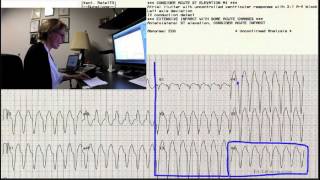
In today's ECG training, we're going to discuss wide complex tachycardia and how to tell the difference between a supraventricular tachycardia with a wide complex like left bundle branch block and V tach. First thing that's most important to know, the most important rule to follow is, in the field especially, all wide complex tachycardias are V tach until proven otherwise. So the most important rule is all wide complex tachycardias are V tach until proven otherwise. Now, if you follow that rule, you'll treat 99% of your patients correctly and safely and will have no problems because your wide complex tachycardia protocol is your V tach protocol. And it really works very well. For V tach and for SVT with wide qrs, the treatment is pretty much the same and is dependent on whether the patient is stable or unstable. That being said, some of your wide complex tachycardias are going to turn out to be SVT with a wide qrs, usually due to left bundle branch block. In the first example I'm showing you here, we're not even sure if that's what this one is. It has a lot of the features of V tach. And so in the field, I would definitely treat this as V tachycardia and I would treat it depending on whether the patient's stable or unstable. If they're stable, they're going to get amiodarone. If they're unstable, they're going to get immediate cardioversion. If it turns out they were svt, those two treatments are still appropriate. But I want to show you what happened with this patient. We saw the wide complexes. We don't see P waves associated. So that's a big red flag that we're probably looking at a V tach. If you look closely though, over here at V5, some people would suggest that there might be P wave activity here. Is that P wave or is it part of the qrs? If you line it up with what's above, appears that it's more part of the qrs. Another possibility is that the P waves have a first degree heart block and they're in the T waves. This is a fast rate and the P waves could easily have gone back into the T waves. So the first choice is to treat this as a V tach. The interesting thing about this patient is that after the rate was slowed down, either with medication or cardioversion, I don't remember, but the rate was slowed down and the patient developed a sinus Rhythm which you can see here superimposed on this picture. This is a later rhythm strip and we clearly have a sinus rhythm with a probably a first degree block. But we see P waves very clearly. And what's interesting about this is the QRS morphology hasn't changed from the tachycardia. So lead two looks like this. And then if you look down here at the lead two when the patient was going fast, it looks pretty much the same. V1, here's the morphology. It's probably a left bundle branch block. And here's V1 when the patient was going fast. It really, really looks pretty much the same. So the conclusion could be then that they were always in a left bundle branch block because the QRSS didn't change and this never was a V tach. But that's alright, they were treated appropriately and things turned out okay. There is a little bit of an issue with wide complex tachycardia. When you have a sinus tachycardia with left bundle branch block or something else causing wide complex, there's a chance of treating with cardioversion or medicines that aren't needed because it's a sinus tach. That's another topic. We'll talk about it some other day. It's more rare, but it does happen that people mistake sinus tach for V tach. But now we're talking about mistaking SVT for V tach. I just wanted to assure you that if you do get them wrong, as long as you follow the rule to treat as V tach unless you know for sure, otherwise your patient will be safe and you'll give good treatment. But it is interesting to see how we know the difference. So I'm going to show you another strip. Now. This ECG is interesting because I left the interpretation at the top because it's all wrong. Everything's wrong about it. And I just want to remind you that the interpretation isn't always right and you should always look at the ECG yourself.
This is a V tach and it was confirmed and treated as V tach and treated successfully. There are some features of this ECG that tell us it's V tach and that aren't 100% accurate, but they do increase the chances of it being V tach very greatly. And the more of these features you see, the more likely it is you're dealing with a V tach. There are many of those ECG features that we could talk about, but I'm not going to go into all of them that I know, but I am going to show you some that I think will help you. The ones that are practical and easy to remember. And when you are looking at a wide complex tachycardia, they can really help you quickly decide that you are dealing with a V tach. First thing I wanted to point out is that multiple leads are very helpful. Some people feel that if the patient's in a tachycardia, especially if they are a little unstable, they don't have time to do a 12 lead. But your rhythm strip has two leads and that's good to look at. But the more leads the better. And as evidence of that, I want to show you lead I in this patient. So over here you may not. If you were just monitoring in lead I, if that's all you saw, you may not recognize that this is a V tach. It doesn't look typical, but look at the rest of the ecg. The rest of the ECG looks very typical. I've even had people say, oh, I think those are p waves in v1. So this can't be a V tach. But let me show you why it is a V tach.
One of the clues is that.
V tach is very, very likely when you have a very wide monomorphic or 1 phase QRS. In this case, the width of the QRS that they give it as 138 milliseconds. I don't believe that's accurate. When I look at this qrs, I believe it starts right about there. If you can see the little blue dot that I'm making, and it ends right about there. And I think that's at least four little blocks. And so four little blocks would be 0.16. Either way, this is wide, but wide, wide or extremely wide. QRS definitely favors the diagnosis of, of V tach.
The next thing that doesn't always show, but when it does, it's a really good indicator of V tach and can make you very confident is when you look at the precordial leads, which are the V leads, the chest leads that you put on the patient's anterior chest V1 through V6, when all of those leads point in the same direction. The word for that is for the words are precordial concordance. They might all point upward, or they might all point downward, but either way, precordial concordance is a strong indicator of V tach. And in this case they're all pointing downward. This is a V tach. Another sign of V tach is simply looking at V6. And if V6 is negative, strongly suggestive of V tach. When we have a wide complex tachycardia that is not V tach, it's usually one of the bundle branches, left bundle branch block or right bundle branch block. Neither of those have a negative V6. Both left bundle branch block and right bundle branch block have a positive or upright V6. The normal ECG has a positive or upright V6. There are very few conditions that cause a negative V6 other than V tach. So that's a real quick and easy one that you can remember for the field. In a wide complex tachycardia, a negative V6 strongly suggests V tach.
Another sign of V tach is an extreme axis. Now, some people say, oh, I don't need to know about axis, or axis doesn't mean anything in the field, but it is really helpful to understand axis. And all that means all axis is, is a determination of the direction the electricity is flowing through the heart. So for most of us in the normal heart, the electricity or the wave of depolarization travels down and to the left toward our left leg. In a normal ecg, this electricity traveling downward toward the left leg produces a strongly positive Lead ii. So Lead II would have a QRS that's upright and fairly tall and nice and.
Positive looking. In this case, you see, lead 2 is clearly negative. Not only lead 2, but lead III and AVF are also negative. 2, 3, and AVF all come from the left leg electrode. They're all looking up from the bottom of the patient or from the floor up toward the heart. A normal heart will depolarize toward the floor. This heart is depolarizing completely backward, away from the floor, away from the foot. And we can confirm that by looking at the two shoulder leads. AVR from the right arm and AVL from the left arm. These both come from the shoulders and look down at the heart. In this case, AVR is strongly positive. That's completely abnormal. That would never happen in a normal rhythm. And AVL is positive. So as long as we've checked our electrodes and we know we put the right arm on the right arm and the left arm electrode on the left arm, then the only conclusion we can draw here is that this impulse started near the bottom of the heart and is traveling backward. It's traveling toward AVR, toward AVL, and away from the foot leads 2, 3, and AVF. So that backward direction or backward axis is very strongly suggestive of V tach.
Another thing that can cause backward or very extreme axis usually Extreme left would be a pacemaker that's pacing the right ventricle. Usually in a pacemaker, AVR won't necessarily be this strongly positive, but AVL will, because we're pacing from the right ventricle toward the left shoulder. But in this case, this isn't a pacemaker. This is a tachycardia. And. And AVR is strongly positive. So the only conclusion is V tach. Another sign of ventricular tachycardia, which doesn't show in this ecg, is AV dissociation. If you happen to see P waves, but they're not associated with the qrss, they might pop up at random places, you might see them in T waves, but you can march out a few enough to convince yourself that those are regular P waves, then that would be strongly suggestive of V tach. In other words, the sinus node is still firing, the atria is still responding. But the ventricular tachycardia, the focus in the ventricles, has taken over control of the heart, and it's completely ignoring what's going on in the atria. So the P waves and the QRS would have nothing to do with each other, and you would only see P waves now and then because of this fast rhythm overriding them. But if you do see that, then you have a diagnosis of V tach. You probably have enough clues already to use in the field, especially the negative V6, the precordial concordance, and the extreme axis, the extreme width. Those are enough to really diagnose almost any wide complex tachycardia. But another one would be, and it doesn't show in this strip. But an upright V1 and an upright V1 could mean right bundle branch block. But in right bundle branch block, there's an RSR pattern. Let me see if I can draw it here. RSR prime. And that's right bundle branch block and right bundle branch block. The R, the second one is the taller of the two bunny ears, they say. So right bundle branch block has an upright QRs in V1, but it's RSR Prime. If we see an upright QRs in V1 in a wide complex tachycardia, and the first bunny ear is the taller one, then that is indicative of V tach. That's not a right bundle branch block pattern. So a lot of times when we're diagnosing V tach, we're just ruling out left bundle branch block and right bundle branch block. If we don't meet the criteria for either of those, then the only thing left Usually is V tach in a wide complex tachycardia. Another real easy to use clue is from Dr. Brugada and it's called the Brugada criterion number one. Dr. Brugada said that if there is no QRS in the Chesleys from V1 to V6 with an RSP and an RS pattern, I'll draw here as R goes up and S goes down. If there is no QRS with an RS pattern, you don't need to go any further that you have a V tach. You can debate whether that's an rs. That's kind of a strange looking little notch there. So we're not sure if you would call that RS, but if there had been no RS at all, period, period, then Dr. Borgata said don't even go through all those other criteria that with no RS it's a V tach.
Let's go through some of those criteria that we just talked about and see if we can match this one up with either SVT with a bundle branch block or with V tach criteria.
First, I see a wide complex tachycardia. I'm going to treat this as V tach until I know otherwise. So my most important question now is is this patient stable or unst?
That being said, maybe at some time later we'll be interested in whether we were treating a V tach or an SVT with a wide complex. So I'm going to look for the clues. The next thing I'll look for is precordial concordance. And I don't really see precordial concordance in this one because V1 is upright and V6 is negative. And so that clue won't help me. But you don't have to have precordial concordance to be V tach. It's just that if you do have it, it favors the diagnosis of V tach. The next thing I'm going to look at is V6. If V6 is negative, that's almost always V tach. So if I were in the field and taking care of this patient right now, I'd be done. This is V tach. I'd continue with my V tach protocol and not look back, not think about it again. V6 is negative, but we're going to keep looking at it just for the fun of it, just to see what all is in this ECG that tells us that this is vtec. I'm going to look over here at the axis. Remember, In a normal ECG we expect lead 2 to be upright and usually 3 and AVF are also upright. So here's lead II clearly not upright. 3 and AVF, also negative. So that tells me that the heart is depolarizing from the bottom up backward. That's not normal. So I look up at the shoulders, AVR from the right arm, we should be depolarizing directly away from AVR. In AVR, we should be depolarizing away from AVL. So the axis is backward, and that favors the diagnosis of V tach.
Another thing we want to look for is AV dissociation. And sometimes P waves can show up, sometimes they can masquerade as P waves, and they're really just artifact. I don't know if this patient has P waves, but if they do, these may be them when I'm marking here, and I'd have to get them and march them out and really see if I wanted to go to that much trouble. But if those are P waves, they're not associated with the qrss. There's not one in front of each QRS at the same distance from the qrs. So if those are P waves, they're dissociated. If they're not P waves, then we don't have P waves. Either way, we have a favorable diagnosis of V. I mean, it's not favorable to have V tach, but the diagnosis favors V tach.
When I look again at V1, it is upright, but it doesn't have the right bundle branch block pattern. So that's another clue.
And let's look for RS pattern in any of the V leads. R goes up, S goes down. I have RR prime in v1, I have possibly QR or RR prime in v2.
QR in v3, QS in v4, QS in v5. No, I have no RS pattern, so that favors V tach. Took a long time to show that this is a V tach when you would have arrived on the scene and said, that looks like V tach to me. And that's fine. I wanted to show you one that was really clearly V tach and show you all the clues besides wide complex, because there are wide complex tachycardias that are not V tach, and there are also V tachs that don't look as clearly V tachish as this one. So in those cases, you might want to fall back on some of these clues.
I hope this has helped and I hope you enjoyed the training today.
More from Peter




Melissa Olen MSN, FPN-C, CCRN: Sudden Cardiac Arrest Training in Schools (Project ADAM) 4/25/25



Pediatric Hospital Readiness: How Teamwork & CPR Metrics Drive Better Outcomes



Chelsea Kadish, MD & Kyle Goodknight, EMT-P, RT: Pediatric CPAP in EMS (3/14/25)