Jan Hansel MD: Strategies to Confirm ETT placement (8/29/23)
Live webinar (Peter Antevy host) featuring Dr. Jan Hansel (Manchester) on strategies to confirm endotracheal tube placement. Hansel presents findings from a systematic review (FULCRUM) and discusses the 2022 PUMA guidelines: clinical signs (misting, chest rise, auscultation) have poor diagnostic reliability and can produce high false-positive rates, whereas waveform capnography (sustained exhaled CO2 over several breaths with adequate amplitude) is the preferred confirmation method. He reviews incidence estimates (OR 0.5–1%; ED up to 3%; prehospital up to 8%), mechanisms leading to esophageal intubation (glottic impersonation, displacement, equipment issues), tragic case examples, and alternative adjuncts (bronchoscopy, esophageal detector device, ultrasound). Emphasis on teamwork, two-person checks, and removing the tube to ventilate when capnography is questionable.
Chapters
All right, everybody, welcome. It is Tuesday, it is three o' clock Eastern time. That is today. We're very privileged to have Dr. Jan Hansel. He is a physician out in the UK, out in Manchester and he has worked in the Emergency Department, Critical care anesthesia. He flies on the helicopter once a year. He has done it all across my feed. One day I saw his paper and I created a post on social media and it created a lot of discussion and I reached out to him, he was kind enough to say yes to join us today. So, Dr. Hansel, thank you so much for joining us. If you want to provide a further introduction of yourself, and just so you know, it's EMS leadership, EMS medical directors from all across the country. So take it away. Thanks for joining us.
Brilliant. Thank you, Peter, and thanks again for the invitation.
And for the kind introduction. I'm an NIHR Academic Clinical Fellow in Intensive Care Medicine currently and I'm also training in anesthesia in Manchester. My previous work has revolved mostly around video laryngoscopy and direct laryngoscopy. I was the first author of the FULCRUM systematic review, the recent one looking at VL versus DL and then as an offshoot of that we started looking at esophageal intubation, tying into the recent PUMA guidelines that we will go over later. Now I'll split this talk into two parts. First I'll go over some of the findings of our recent systematic review that Peter mentioned and our meta analyses and looking at the reliability of clinical signs for the exclusion of esophageal intubation or the unreliability of clinical signs. And then we'll REVISIT the new 2022 guidelines on prevention of unrecognized esophageal intubation that were published in Anesthesia, which with a specific emphasis on what interventions can be adopted in the pre hospital setting. So I'll try to tailor it to your needs as well. I have no personal conflicts of interest to declare and let's get cracking. So I'd just like to start off with something a little bit more whimsical. Dr. Seuss style poem about an anthropomorphic propofol dispensing bear that I delivered at the annual meeting of the Royal College of Anesthetists in the UK earlier this year. So this was.
This was sort of a.
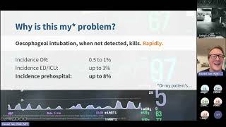
Just a different style of presentation of our systrev. I thought maybe use a bit of AI to generate some images and write a poem. So here goes. Tracy's trace Tracey the Bear would always take exemplary care to conduct herself to the letter of the guidelines and each and every byline written by her college. Yet in spite of all that knowledge, she had been given a scare the last time she induced and filled a pilot balloon with air, for Tracy could not see the trace that she'd learned to rely on each time. A tube had been earned by the patient, now fast asleep and quite still as the aminosteroid, the one starting with R, had exerted its will. Now she was quite certain she tubed the right hole. She's a bloody anesthetist. It's our one and only goal. The bag's a bit off, but the sats are okay. Perhaps if I may. She asks her assistant to pass her a stethoscope while she conscientiously observes the tube. After a breath misting, she exclaims, well, that's a good sign. And although some commotion had started to occur, she listens intently and quips, I'm quite sure that if I hear breath sounds we should be just fine. Yet the cap, no remains eerily flat, a bluish tinge on the lips where the tube is still sat. But Tracy suspects it's because of an error, a faulty connector might be trying to scare her or or perhaps bronchospasm or something like that. Yet slowly but surely a bit of doubt had crept in, and not long thereafter his sats are in his boots, the Pulse Less than 10. Then the crash buzzer hoots, the room fills with bears of all creed, kind and station opinions start saturating everything bar alveolar oration. Now, reader, pause briefly and bear with me, please. What Tracy did next should stay a matter of lore, but for the purpose of this tale I do just implore, allow me a bit of liberty. With an academic tease, as if time stood still, she went on to Google to find any evidence relevant, succinct and frugal, and lo and behold, a systematic review of exactly this question. Who bloody knew? Now quickly, let's see how these findings perform. A false positive rate of 72% and lung auscultation falls on every eighth event. Well, this is outrageous. I've been terribly wrong and I should not have waited this blooming long. Quick now, make haste. As he is on death's door, let's pull the misplaced tube and intubate once more. Now, not to mine or your surprise, but they could not believe their adrenaline fueled eyes as the patient pinked up and even cracked a wee smile with a square cap. No trace Sustained for a mile, she dodged the coroner and so did he. Bless him. That day, Tracy learned an invaluable lesson that when the tube's in the gullet, there is no messing with clinical signs. So repeat after me. They are all inferior to capnography. Now, this had a happy ending and Tracy managed to save the patient and all was well. But unfortunately, when esophageal intubation goes unrecognized, it's quite rapidly fatal. And often when it is unrecognized, it leads to poor outcomes for patients. This is the so called anatomy of death by esophageal intubation that was proposed by Tim Cook and not the Apple CEO Tim Cook. Tim Cook of Nap for Fame, a British anesthetist and professor. So, as you can see here, we sort of go from a situation where we have perhaps an easy intubation to begin with, using a direct laryngoscope or a video laryngoscope followed by a flat capnograph. And this leads to a fixation error. We might think, oh, okay, the capnograph might be malfunctioning, but we also have anchoring bias. We're shorted, we've intubated, or we think, oh, maybe there's anaphylaxis, for example. As things progress, as clinical signs start deteriorating, we're getting into the territory of a clinical crisis. And then we have increasing situational complexity. We might fail to do a proper A to E assessment and we might over rely on clinical signs as well. And sooner or esophageal intubation when it's not detected will lead to cardiac arrest. And again, we have a fixation error. We're thinking, what's causing this cardiac arrest? Now, in the prehospital space, you might encounter patients already in cardiac arrest. And if you then intubate them, you might not reverse their hypoxia if the tube happens to be in the esophagus, but you might might just progress it further and actually worsen the patient's outcome as opposed to if they had a supraglottic airway inserted, for example, rather than a tracheal tube. Now, at the point of cardiac arrest, in theater, at least usually there will be a call for help and the situational complexity will rapidly increase. So you will have difficult crowd control. You're going to have consideration of diagnoses. There's going to be an absence of a structured response. There will be a lot of people flooding that theater or that anesthetic room and a lack of space as well, ultimately all of this leads to chaos. And you can see how we're sort of going down, down this vicious cycle really almost down the vortex in a sense. And we're losing situational awareness with all of this. Nobody keeps control of the whole situation. Now this might be less of an issue in the pre hospital space, but it will become highly complex when you get the patient into ED and we might be over relying on the history. So you can say, oh, I've successfully intubated this patient. It was a grade one view, it was easy, you know, and this just leads to anchoring throughout. And then when finally somebody recognizes this, they say, oh, we have no captography. I'll second guess this and I'll have another look. And then usually the airway is actually easy after recognition. And the reasons for that, which we'll explore later, might vary. This is finally followed in the aftermath by a local apology and usually a local policy change, maybe some local training and governance changes and usually a family plea for it not to happen again to others. Or it might might entail a lawsuit as well, depending on whether negligence was at play. Now you might ask, why is this my problem? I'm shithoted intubating. Well, we all think we are, but esophageal intubations still happen and when it's not detected, it kills rapidly. As we've said, the incidence of esophageal intubation in theater, depending on who is intubating, is from 0.5 to 1%. It can be less than 0.5 in very, very skilled intubators hands. In ED that number increases to up to 3%. And then in the pre hospital space estimates vary, but it can be up to 8%. Now this is a combination of unrecognized and recognized esophageal intubation, obviously, but you can imagine that if it's unrecognized in the prehospital space, this might lead to further worse outcomes downstream.
Now this still leads to mortality worldwide. Now this is a very recent case from San Diego, actually from UC San Diego, where a young woman, 39 years of age, Latricia Tillett, underwent an elective breast reconstruction. And following intubation, or sorry, extubation, she developed laryngospasm and then became hypoxic. And there were attempts to bag mask ventilator and an oropharyngeal airway was used. And apparently she was an easy intubation. But only after 13 minutes or 15 minutes was esophageal intubation Recognized by then she had been in cardiac arrest, anoxic and ultimately then died subsequently. Even though when this was recognized.
The esophageal intubation was recognized, it was very, very easily rectified. Now, in this case, it's unclear from the available records at at present who exactly intubated the patient and what the sequence of events was. But it's worth noting in the, in the 1518. Note here that both the CRNA and anesthesiologist listened with stethoscope and confirmed bilateral coarse breath sounds in left and right axilla. So just worth bearing that in mind. Now, looking at the anesthesia closed claims database from 1995 to 2013, we can see a number of cases and these are available on the PUMA guidelines, sorry, the PUMA website to read. But we can see that 45 deaths occurred over that time period. This is an ongoing issue. People continue to die because we don't recognize tubes in the wrong hole. Now, going back to clinical signs, what about them? Misting, for example, when we first put the tube in, we have a look, we give them a tidal volume and we can see misting of the tube. How good is that? So misting only assessed in three studies admittedly, and historic studies I might add, has a very, very, very poor, poor predictive rate. So it has a false positive rate of 69%, meaning that in almost 70% of cases it will give us the wrong conclusion. Now, if we don't see misting, it's a good predictor of esophageal intubation, actually. But if we do see misting, it's actually worse than a coin toss. Presently, looking at lung auscultation and five point auscultation, we're looking at about a 14 to 18% false positive rate. And here we're looking at long auscultation at either 4 points or adding a 5 point auscultation, listening to the epigastric area as well, not very good. And finally, the esophageal detector device, which we included in our systematic review as well, has reasonable performance characteristics as a test. So this is a low cost device that I will show you the principles of later in a bit. But it's also of somewhat historic significance rather than something that we would use in clinical practice regularly. The main argument being that we should be using waveform capnography, ideally not calorimetric capnography, because that can be misleading as well. Now, if we then apply a Bayesian application of thinking about clinical signs, and this is a worst case scenario and we've taken the likelihood ratios from our study and just map them onto a few fake nomograms in sequence. Now, saying that we have a hypothetical situation where we're confident that we've intubated the trachea. So we're saying 90% confidence, for example, 90, like I'm thinking, okay, this was an easy intubation, you know, get it in. And we don't know the likelihood ratios for chest rise, unfortunately based on the data that we've collected. But for misting, this increases from 90% to 95% and then a cognitive error happens. So once we've seen misting, we're thinking, oh, this is, this is good. Okay, this confirms my prior belief of the tube being in the trachea, where in fact it is in the esophagus. Now then we see a flat or abnormal capnography and then we might think there's something wrong with the capno, or we might disregard the capnography. Okay. And if we then proceed to ignore the capnography and not let that lower our likelihood of tracheal intubation, then we go to auscultation and this further increases our post test probability from 95% to 99% giving us the wrong conclusion. Okay. And even if we map this onto so called naive priors, 50, 50 odds of tracheal or esophageal intubation where I might just not know which hole I'm in. So I'll let the tests do the talking. And again, in the worst case scenario, chest rise positive, don't know the likelihood ratio, so it stays 50% whereas with misting it starts to increase from 50% to 60% and then from 60 odd percent to 95% with auscultation, again if we disregard capnography. So the main thing to remember here is that we need to listen to capnography or we need to take capnography into account. And we shouldn't be relying on clinical signs because they will lead us down the wrong path. Now, speaking of wrong paths, we have a couple of different views here and just for the sake of time, I'm not going to do a straw poll of the room, but these look like adequate targets for maybe a bougie. Right. Now this is what we call glottic impersonation. And this is one of the reasons why esophageal intubation can happen. And we'll explore this in a bit, but you can easily imagine if you ram the laryngoscope far in and you get essentially.
Above the glottic opening, you can just see the esophagus and that's your hole to put the tube in. Okay? So that can give you false confidence that you're in the right hole. Now, the PUMA guidelines or the PUI guidelines by Nick Kremes that we will dissect in a bit more detail, give us some insight into why this happens. And we need to think about the occurrence of esophageal intubation and failure to recognize esophageal intubation and why it happens in the first place. It's usually going to be one of misidentifying the larynx, as we've seen earlier. So glottic impersonation or.
A compromised laryngeal view or some equipment issues, flicker of the light. It might be a delivery issue, the tube or the introducer or bronchoscope not passed into the trachea, but it's displaced when you railroad. Or it might be movement after successful tracheal placement, more common in children, for example. Or if you have CPR that's ongoing, you might see this happen more commonly and it can happen quite easily. Then we also have failure to recognize esophageal intubation. Once it's happened, we can have limited access to capnography, which in certain situations still is an issue, especially in resource limited settings. We might have failure to confirm CO2 detection, so we might have equivocal capnography and we just don't confirm it correctly, or a spurious CO2, so we might have a false positive if we use colorometry, for example, and then misinterpreting the monitor display. So sometimes the airway pressure waveform has been mistaken for the captnography waveform, and there are efforts in place to try to standardize those monitors to be fully filled in, as opposed to having a non filled in filled in trace. And then finally, the failure to acknowledge the potential for an absent CO2 trace. And this might be cognitive biases, knowledge deficits, or any sort of communication deficits. Now, it's worth remembering as well that CPR or cardiac arrest is not a cause of a flat capnography trace. And we usually do see a capnogram during cardiac arrest. So we need to see at least something. And the definition from the POI guidelines goes further on this. Now, again, preventing esophageal intubation, the guidelines from 2022 by Puma recommend routine use of a video laryngoscope, which goes hand in hand with the findings of The Cochrane review from 2022 as well that we conducted the laryngoscopy technique. It recommends doing a stepwise anatomical sequence where you essentially do what's called epiglotoscopy. And I think Rich Lavitan from the United States coined this term and advocates it. Means that we first try to identify the epiglottis and use that as a reference point rather than getting the scope right in then stating the view at laryngoscopy. Very important. That orients everybody around us to what might be happening. So we immediately increase situational awareness for everybody present in the room, then recognizing esophageal intubation, observing tube placement. Again, this can be an exercise in teamwork. When we have a video laryngoscope with a separate dedicated screen, two people can actually look at the screen and exhaled carbon dioxide detection and specifically sustained exhaled carbon dioxide for at least seven breaths. Now, this number is somewhat arbitrary, but it is important that it's sustained because if it's not sustained, this can still be an esophageal intubation potentially. And they provide us with this lovely figure here. And again, the level rises during exhalation and falls during inspiration. It's consistent or increasing amplitude over seven breaths. So that's still criteria satisfied. And the peak amplitude needs to be more than 1kpa above baseline. Now, this was somewhat contentious in the discourse following the publication of the guidelines. And again, arbitrary line that's drawn somewhere because in the context of cardiac arrest it might be somewhat lower, consistent with a very, very poor outcome and a very low cardiac output.
But usually we want to draw that line at 7.5 millimeters of mercury and the reading being clinically appropriate. Now, we can see a couple of examples of where these criteria are not satisfied as well here and some other issues to consider. Cardiac arrest and bronchospasm are common confounders. There was just a case report published yesterday, I think, that I shared on Twitter earlier, and I'd urge you to follow me on Twitter and you can read that as well. And there is sharing of experiences of bronchospasm where you might get a stiff chest high pressures, as you would with esophageal intubation sometimes as well. But you get no capnography back, even though you are in the right place. And then you need to use adjuncts to confirm that you are indeed in the esophagus and not operate on the assumption that, oh, you're probably in the trachea anyway, so it's fine. And Then observing oximetry. So you can have sustained sats, especially if we've preoxygenated a patient, quite well, more common in the OR rather than pre hospital space. But you can have falsely reassuring sats and then verbalizing the checks for sustained exhaled carbon dioxide and adequate oxygen saturations. Again something that you can use a two person check essentially. And that would be I can see sustained exhaled carbon dioxide and adequate oxygen saturation. Do you agree? Yes, I agree. I can see that as well. So it's a two person check. The default position should be that of removing the tube and attempting ventilation before re intubation. And this is highlighted in the graphic displayed here. So we essentially just need to ask ourselves if anything is questionable with the capnography and we should all be using capnography in this day and age in the developed world. Is removing the tube dangerous? And if the answer is no, remove the tube and re intubate followed by sorry, following an attempted ventilation. So we want to confirm that we can still ventilate. But if removing the tube might be dangerous potentially, then we need to ask ourselves, are the sats adequate? If the sats are adequate, then we can have a bit of a play around with some adjuncts that we will go into later.
Then finally we confirm again using capnography, that the tube is in. Again a reminder that we shouldn't be using clinical signs to rule anything out. Then alternative techniques to exclude esophageal intubation. The PUMA guideline provides these valid alternative techniques, including a repeat attempted laryngoscopy, ideally using a video laryngoscope when possible. And the most expert person available should be doing this at this stage. And then finally with one extra supplementary technique, either a bronch ultrasound or an esophageal detector device. And again not using clinical signs. Now thinking about flexible bronchoscopy. It's ubiquitously available in well resourced settings, although not in the pre hospital space unless you have a very very specific arrangement. But you can use a single use.
Device such as an Ambua scope or kaleidoscope, started producing one as well. Or you can use reusable stacks, very easy to confirm. You get it down the tube and you can see the carina and that's that there's no studies evaluating efficacy or effectiveness because it's usually considered a reference standard or the gold standard when comparing to other index tests, but again less relevant for the pre hospital setting. Now the esophageal detector device. The short answer on whether to use it and this is actually a photo of one odd that one of my colleagues had had in her drawer still.
Maybe from a bygone era, really from 20 years ago. And the answer whether to use this or not is depends. If you have access to waveform capnography, and it's very unlikely that you wouldn't in Florida, for example, but it might be likely that you can't get access to capnography in sub Saharan Africa, then the esophageal detector device works well as an adjunct. And I can show you the principle of how it works. So you can either put it into the, attach it onto the tube, inflate it or deflate it, and then if you put it into an airway, it'll reinflate. So it just reinflates easily. Now if I use pursed lips as a simulation of an esophagus, really. So it'll do this.
And it stays deflated. Okay. So that's the basic principle and it's quite reliable based on the data that we have. And then thirdly, we have ultrasound, decent test performance characteristics. We didn't evaluate it because many, many meta analyses have been done looking at ultrasound and it does perform well.
It shouldn't be a replacement for capnography, but it performs adequately. But it requires training and access to equipment. And here we can see the esophagus is usually behind the trachea. And in normal circumstances, when we've intubated the trachea, we just see one barrel. But then if we've intubated the esophagus, it's going to displace it to the side and we're going to see two barrels or the two barrel sign, essentially. Again, just a final reminder, do not use tube misting, do not use chest rise, do not use absence of abdominal distension or any sort of auscultation. A quick check that we can use whilst we're troubleshooting capnography to make sure that other causes of esophageal intubation have been excluded is provided as well. But I'm not going to belabor the point here and I'll allow you to peruse the guidelines yourself when you get a spare moment.
As it provides a very good set of recommendations. So finally, some take home points. Do not trust clinical signs when excluding esophageal intubation. They will. You do so at your own peril or at your patient's peril, unfortunately. And the default position for any clinician that sticks a tube anywhere into the mouth should be you should doubt tracheal intubation initially and assume an esophageal intubation until you've proven otherwise to yourself and someone around you, and follow the 2022 guidelines and use reliable techniques to prove tracheal intubation and exclude esophageal intubation, such as sustained exhaled carbon dioxide. And that's all from me. Please feel free to have a look at the Universal Airway webpage and follow Fiuma on Twitter and myself. And you can scan the QR code for the Twitter link as well. Thank you.
More from Peter

Melissa Olen MSN, FPN-C, CCRN: Sudden Cardiac Arrest Training in Schools (Project ADAM) 4/25/25



Pediatric Hospital Readiness: How Teamwork & CPR Metrics Drive Better Outcomes

Chelsea Kadish, MD & Kyle Goodknight, EMT-P, RT: Pediatric CPAP in EMS (3/14/25)