Jason Mansour, MD: The OMI Movement: A Paradigm Shift in MI (10/11/24)
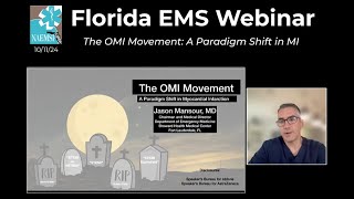
Jason Mansour, MD presents the OMI (Occlusion Myocardial Infarction) framework as an alternative to STEMI/non-STEMI classification. The approach identifies coronary occlusion through ECG patterns like de Winter complex and hyperacute T waves that classic STEMI criteria miss. AI tools like PMCardio are emerging to help hospitals and EMS detect these cases earlier.
Questions this video answers
Friday, October 11, 2024 (Florida EMS Webinar).
2018 (Pendell Myers, Scott Weingart, Steve Smith and others).
1994 (Lancet reperfusion trial cited by speaker).
Approximately 25–30%.
Approximately 15–33%.
Chapters
All right, welcome everybody. It is Friday, October 11th. Welcome to another edition of the Florida EMS Webinar. Myself and Tanner Smita, who we, we have the great privilege of hosting this incredible speaker every single week. I had the privilege of, of hearing Jason talk several months ago and I was floored. It was just such a great talk. And I said, please, you got to come on the EMS Florida EMS webinar. And he agreed. So Dr. Jason Mentour is the chairman and medical director with the Department of Emergency Medicine at Broward Health Medical center right here in Fort Lauderdale. He's been there for 18 years and does an amazing job. He treated some of my family members. So I can, I can vouch for him personally. And it's a great topic. We have a lot of interest in this topic regarding, you know, moving from the STEMI and STEMI to omi. So without any further ado, Jason, thanks for joining us and take it away, my friend.
Thank you, Peter, I appreciate it. And thank you to everyone that's joining for this really important topic. For those that have heard, for those that have heard me speak before, I love talking on these very high impact topics. These things where little subtle subtleties to the way we practice really change and save lives. OMI is one of them. So I'm sure there are some in the audience that already know about this movement. I'm sure there are some that this, they're hearing about this for the first time. So I'm going to kind of just introduce the topic in the early slides, show some examples. But I'll, I'll warn you in advance that this is hard to pack into a 30 minute session. I have some links at the end for some longer videos that you could watch if you want to learn more about this topic.
So our objectives today, we're going to talk about the STEMI paradigm, historically, how we ended up here and where we're going in terms of the OMI movement. You know, what is it? Why is it a better paradigm? And then I'm going to show lots of examples, as many that I can fit in the time that I have. And then I'm going to summarize. We're going to look to the future and talk about AI and how it's going to be playing a role probably in the near future when it comes to diagnosing omi.
So my, I have a question to kick things off. Stemi is STEMI a disease or is STEMI a disease marker? Right. So there are several other things in medicine. For example, if I see a patient with right lower quadrant pain, and they have an increased white blood cell count. Those are disease markers for the disease. Appendicitis. I have a patient with belly pain and an elevated lipase. Those are disease markers for the disease. Pancreatitis. Patient walks in with a cough and a pulmonary infiltrate. Those are disease markers for pneumonia. So is STEMI a disease or a disease marker? If you agree with me, I believe it to be a disease marker. Okay. But over the years, we started using it as a disease. You know what happened to Joe? Oh, he had a stemi. STEMI is not actually a disease. It's a disease marker. And there are several other disease markers out there that represent an obstructive mi. Okay. So if stemi is a disease marker, the disease it's marking is an occluded coronary artery or an obstructive mi. And it is not the only disease marker that points to this diagnosis. And we and people in healthcare should care more about the disease and less about the disease marker.
So if you've heard of this before, you may have heard it on a podcast. There are several videos on YouTube, a very popular website, life in the fast lane. There's a lot of articles on it there. MRAP is a podcast that's popular in emergency medicine, MCrit, and all of these. These. These publications. You'll find some mention of the OMI NOMI paradigm shift, but it all started with the OMI Manifesto, which was published by Pendle Myers and some others like Scott Weingart and Steve Smith. And this was published in 2018. This is a PDF that was published, followed by a podcast on MCrit. And this was the first introduction to the topic. And I'll admit that I took some of the slides from the OMI Manifesto to introduce the topic to you today. So let's start with a historical perspective. If we walk back more than 30 years, we never really had STEMI and non STEMI. We had Q wave MI and non Q wave MI. Q wave MI was a completed infarct. A non Q wave MI was. It was an acute infarct. But back in those days, we didn't have reperfusion options.
We just essentially sat on these patients and dealt with the consequences of the MI, but we couldn't reperfuse them until about 1994. In 1994 is the reperfusion era. When this was published in Lancet, showing that we could actually get fibrolytics and reperfuse these patients if we caught them during an acute mi.
We found a mortality benefit. If you had this one EKG finding called ST elevation, if we look at that with some greater detail, if you took all comers suspected MI and you gave them all lytics, there was a slight benefit. But if you took the. The subset of population that had ST elevation on the EKG, they had a greater benefit is 12.8% versus 10.4 for another number needed to treat a 42 patients. If you had ST depression or you had a normal EKG, so there was no ST segment derangement, you actually caused harm by giving those patients lytics with number needed a harm of 42 and 143. So this was the line we drew in the sand back then and said, if you have this finding ST elevation, we should be sending those patients for reperfusion. And if you had either of these findings, you were actually harming them by reperfusing them. And I'll remind you, this is in the. They're talking about lytics at this time.
And then what have we done since then? Well, we did subtle shifts to the recommendations, but for the most part, this ST elevation finding has not gone away. Right. So if you look in 2000, there were some published guidelines where we increased it to 2 millimeters, then we brought it back to 1 millimeter, and then we had a male and female difference in V2 and V3. So there's these little subtle tweaks to ST elevation. But in general, we just accepted that if you have ST elevation, those patients should go for reperfusion. And if you didn't have ST elevation, those patients, even if we felt they may be dealing with a totally or subtotally occluded coronary artery, we weren't taking them for reperfusion if they don't have this particular finding on ecg. So if we stay with this paradigm, it would imply that every time we have ST elevation, we find a complete occlusion, and every time we lack that finding on ekg, we. We either have incomplete occlusion or no occlusion at all. And in a perfect world, if this was the way we. We should be practicing, this is how things should be. However, that's not reality. Okay? Reality is that, yes, if you have ST elevation, there's a. There's a significant number of those patients that have complete occlusion, but there's also a 15 to 33% false positive rate. Those patients have ST elevation, but the cause is benign early repolarization pericarditis bundle branch blocks, left ventricular aneurysm left, LVH, etc. We would call those false positives. They have the finding, but they don't have the disease. What I'm more worried about is the 25 to 30% that are the false negatives. These patients do not have ST elevation, but we find in the cath lab that they have a complete occlusion. Okay. In fact, less than 25% of the time that you see ST elevation, is it actually an acute MI? Okay, there, there are more of the other diagnoses on this screen. Lvh, bone to branch blocks, early repo, pacer, pericarditis, et cetera. That where you're seeing ST elevation, but those patients actually do not have the occluded coronary. So who are these patients? The 25 to 30% that don't have ST elevation, that have a proven occluded coronary. Those patients have other things on EKG that would, that would suggest they have an occluded coronary Wellen syndrome, the Went de Winter ST wave T waves, Scarbosa criteria in the setting of left bundle branch blocks, pacers or LVH. They have posterior MI, they have hyperacute T waves, etc. These are ECG abnormalities that are not ST elevation, but they are certainly disease markers for a complete, complete coronary occlusion. What does the literature say about these patients? What happens to them? Well, firstly, if they have one of these diagnoses with an occluded coronary without ST elevation, their mortality doubles. I have three papers here, but there's countless papers that show this. We're not taking these patients to the cath lab. In fact, it takes us 10 times longer to get them to cath. Right. If they have ST elevation, on average, they're in the cath lab in 41 minutes. If they don't have ST elevation and they have the occluded coronary, so it's manifesting as one of these non ST elevation markers. It takes us 437 minutes to get them to the cath lab and in doing so, we double their mortality. How do we accept such poor performance? How can we sit here and sleep at night and say we're going to allow these patients to sit when we know that they we have, which is a strong suspicion, or we feel that they have an occluded coronary? Well, we don't in general. Okay. There's lots of really smart physicians out there that have been talking about this for decades. And we use certain terms like STEMI equivalent. Right. We learned that A Welland syndrome or De Winters T wave is a STEMI equivalent. We should be sending those patients to the cath lab. Or sometimes they have some ST elevation, but they don't meet strict criteria, which means they don't have ST elevation. And two contiguous leads. We might call that a subtle stemi when there's lots of lectures out there, publications, you can look in the literature of these subtle STEMI's. Again, we call them a STEMI equivalent. And we try to prompt cath lab activation based on these findings. Sometimes we call it a semi stemi, but we're bending ourselves into a pretzel, trying to figure out ways to draw attention to these ECG findings that should prompt emergent cath lab activation that are outside our current guidelines that say we need two. We need a millimeter of esty elevation and two contiguous leads.
So with the current STEMI guidelines, there is no such thing as a false negative. Okay, take this EKG that we're looking at here.
Most people would look at this and not call this a stemi. Okay, There are some signs of ischemia here. Oh, let me go back. Sorry. There are some signs of ischemia here. There's ST depression and inferior leads. A little SC elevation here in AVL, there's a little SC elevation here in V2, but these are not two contiguous leads. So many folks would look at this and say they don't meet ST STEMI criteria, but there's signs of ischemia here. And if we admit this patient and they end up with a positive troponin ruling in for MI, they go to Cath later on, within 24 hours, we find an occluded coronary, we just change the diagnosis to N stemi and then everyone sleeps well at night. We didn't miss it because this is not a stemi. And the guidelines would not say send this patient to the cath lab because it's not a stemi. And this is why the current STEMI guidelines allow us to sleep on these patients and allow them to sit with an occluded coronary when we know it doubles their mortality. There's no such thing as a false negative, because if they rule in, we just change the diagnosis to nstemi.
So why not make the underlying pathology drive what we do with these patients? Not the disease marker, but the underlying disease? Okay, enough with STEMI and nstemi and instead, let's. Let's replace it with occlusion MI and non occlusion mi. Now, under occlusion, under the umbrella of occlusion mi, we're still going to include stemi. If we see stemi, we're going to call that an omi simple. But there are many other things that are the disease marker for an occluded coronary that represent an occluded MI that present with something other than ST elevation. Okay. And these are lots of examples here on the, on the slide. And I'm going to go through some of the examples during this talk, but I want to make sure we have time at the end for questions. And I'm going to share a link that, where I go through each one of these in great detail. But let me show you some examples. I'm going to start with the subtle STEMI or the ST elevation in one lead. So it's time to bury STEMI and all of his friends. The term STEMI equivalent, semi. Semi stemi or subtle stemi. Let's bury all those terms and stop using the word STEMI to it for. In this context and, and instead ask the question, does this patient have an include coronary occlusion that will benefit from immediate intervention? Okay, so in the OMI manifesto, which I'll give you a link at the end, they clarify a few other EKG findings that should prompt emergent cath lab activation. And over the years, we put them above the line. We call them semi equivalents. Let's look at the literature when they actually look at this and see if it does make a difference. So this is Pendle Myers. He's the author of the OMI manifesto. And he, he took a look at this and said, let's look at patients that meet OMI criteria but fail to meet STEMI criteria. Okay. So I want to. So if you look at this graph, Let me simplify it for you. On this side in the blue, you see all patients if they met STEMI criteria. There you can see their troponin, right? The kind of the dots you see there. But if they met STEMI criteria, more often than not, those patients actually had an occluded MI in gray. You see patients that did not meet STEMI criteria, but all those dots represent their peak troponins. So there are a lot of people there that did not meet STEMI criteria, that ruled in for mi. So we ended up calling them nstemi. But what if you took those patients and you apply only criteria? So the slide that I showed you before with scarbosa and posterior MI and things like that, we apply those criteria. So I'm now looking at patients that are STEMI negative, but only positive. That's what you're seeing in yellow. And in blue, you see patients that don't meet criteria for either one. Right? Sure, there's some false negatives there, but we can do much, much better in diagnosing an occluded coronary by applying OMI criteria.
So what are some of the things in here and in this paper? He. He's specifically outlines many of them. Again, I'll give you a link at the end where I can go through each of these in greater detail. I can't fit it in the talk today, but I'm going to show you some examples of some subtle STEMI or ST elevation in one lead, which by definition would not meet criteria for being a stemi. Take this ekg. This is a low voltage ekg. So already it's going to be very difficult to get 1 millimeter of ST elevation or depression. But what caught my eye here is when I looked at 2, 3 and AVF, I have just the hint of ST elevation. Now, if I want a textbook STEMI here, I need 1 millimeter of ST elevation and two contiguous leads. I don't have 1 millimeter and two contiguous leads here, but if I look at the reciprocal wall in avl, I'm seeing just the whisper of ST depression, and that would represent the reciprocal ST depression of an acute MI. I'm also seeing ST depression in V2 and V3, which would represent posterior extension of my hypothesized inferior wall mi. This patient does not meet STEMI criteria. Your computer would not call this.
It may recognize the st depression in V2 and V3, but it's not going to call this a STEMI. But this person certainly is having an occlusive MI and would benefit from emergent cath lab activation. Let me show you another example from my practice. This is a patient that presented with chest pain. If I zoom in here, what caught my eye was in lead 3, I had 1 millimeter of ST elevation. So I'm really going to scrutinize lead 2 and AVF, which are the contiguous leads, and look to see if I have ST elevation in either1. Now, AVF, I have slight ST elevation, but it's about a half millimeter and would not meet semi criteria. And I'll tell you, the computer did not call this an mi. But when I look at the reciprocal wall, I already see ST depression. And when I look at V2 and V3, I already see the ST depression of what would be posterior extension of my inferior wall mi. So the textbook would not call this an mi, but I would call this a subtle stemi, or ST elevation in one lead. That would represent this patient having an occluded coronary and an omi. This patient would benefit from the cath lab activation. Now, I've been practicing for almost 20 years here in Fort Lauderdale. I've seen a lot of EKGs that look like this, and a lot of times I've sent this to a cardiologist and they say, well, it doesn't meet criteria. Why don't you get some troponins and some serial EKGs, and if you meet criteria on EKG number two or EKG number three, we'll take them to the cath lab. But the argument that I'm trying to make in this talk is I already know this patient has an omi. Why are we sitting and waiting for the disease marker to fully manifest? Why can't I send the patient to cath lab now, based on my belief that this patient has an obstructive coronary, which they turned out that they did. One final example. Okay, almost the same thing. This is a 48 year old Hispanic female male. I'm sorry. Who presented with chest pain. And you notice that there's a PVC here, so we're just going to overlook that for now. But what caught my eye again was there's just a hint of ST elevation and lead 3. I look at the contiguous leads and I do not see ST elevation there, but I do see the reciprocal change, the ST depression in the reciprocal wall, as well as the. The depression I'm seeing in V2 and V3, which would represent posterior extension. Okay. Again, I'm advocating for this patient going straight for reperfusion rather than sitting on them for serial EKGs and seeing if we. If the. The true STEMI by definition manifests itself.
This is the EKG I showed you earlier where I said there's no such thing as a true negative. I'm sorry, as a false negative.
There'S your ST depression. That everyone would see that. And you might want to be tempted to call this diffuse ST depression or inferior ST depression, which would represent ischemia. But there is ST elevation in AVL. There is slight ST elevation in lead 1, which would be a contiguous lead, but it doesn't meet the 1 millimeter criteria that I need. And I have some ST elevation here in V2.
Right.
This is an OMI that should be. Should be called OMI and should be sent to the cath lab, not sat on as acute coronary syndrome with the. With ischemic EKG changes and wait 24 hours for their cath.
So that's one example. I'm going to go through one more example, and I want to leave enough time for questions. This is the Winters STT wave complex. Okay. If you haven't heard about it, I'm going to show you the literature on this, which came out about a decade ago. What do you notice on the EKG is there's some ST depression in V2 that goes immediately into a tall and peaked T wave. This is called the Winters ST T wave complex. It's described in 2008 by de Winter as a STEMI equivalent. That signifies a proximal LED obstruction. It's 1 to 3 millimeters of ST depression shown in the blue arrow that goes immediately upward to a very peaked and tall T wave at the J point. That is, is what. What is called in the literature at the Winter's T wave. And you say, well, I haven't heard about this when it's first described. Here it is. It's a letter to the editor in the New England Journal of Medicine in 2008. Essentially, de Winter writes a letter and says, look, I look back at a lot of my LED proven LAD occlusions from the cath lab. I look back to their EKG, and 2% of them had this particular finding of ST depression going right into a peaked T wave. Since then, there's been other publications. This one by Vidin, that he looked at 1890 of his patients that went for cath, and he found 2% of his patients had this ECG pattern as well. 23 of them had proximal LED and the other ones had distal LAD. And then this is one example of an EMS EKG patient comes in, and you see there's ST depression here. And V2 and V3 goes right into a very peaked and tall T wave. That's a de Winters ST T wave complex. We were calling this for years a STEMI equivalent, but I would like to remove that term and replace it with an omi. De Winter's STT wave is an omi. It's an obstructive mi. Okay.
Here'S another example. Okay? ST depression going right in a very tall and peak T wave. This patient did not go to the cath lab. This was not called an OMI or STEMI equivalent. This was thought to be hyperkalemia because of the peak T waves. The ST depression going into the peak T wave was overlooked or perhaps recognized, but was not recognized as something that needed to go immediately to the cath lab. 90 minutes later, this EKG was repeated and we found now ST elevation with Q waves telling us that this infarct has now evolved into what we would recognize as the classic ST elevation mi. Now with QA is telling you we've completed this infarct. This one presented to Broward North. Chest pain, tachycardic. And we see the ST depression going into a very tall peak T wave. So this is DE Winters ST T wave complex. It's one example of an omi. Okay, now I don't have time to get to more examples. I promised you I would show you, give you some links at the end where you could show some examples. I want to go to, I want to take a look to the future.
So this, I give a talk and I go through a. I'm sorry, I go through a step by step method for how to recognize. Let me go back. I'm sorry, I'm clicking too quick. This is a step by step method on how to recognize omi. And the top half of this step one through five is to make sure it's not a mimic. And then step six through 10 is to look for all the things and I showed you a couple examples that would represent OMI on EKG that does not present with ST elevation. I have a whole talk on this and I'm going to share that the QR code at the end. If you want to watch that talk.
This is a nice pocket guide. Here's the website omg guide.org where they go through many, many examples of going through what I just showed you, a step by step algorithm to make sure that you could recognize what an OMI is and when to prompt immersion cath lab activation. Okay.
Where are we headed? This is Steve Smith, Pendle Myers and Robert Herman who have created an app called PM Cardio. And it's, it's, it's not yet FDA approved, but it's being used overseas and essentially it'll allow you to take a photo of an ecg and it applies the algorithms that I just outlined earlier and it makes a recommendation whether or not it feels this is an omi. So I, I feel like this is going to be a valuable tool that we're going to be using in the future. Right now it's not for widespread use because it's still being looked at.
That's all I have for this talk, this lecture with that stepwise approach, that ten step approach. The one hour version of this talk is available with this QR code. Here.
If you're kind of at the entry level of EKG interpretation and you want something a little simpler, this is a talk that I gave for grand rounds years ago where I just talk about ST elevation and I talk about when you should worry about it representing included coronary versus when it's a stemi mimic that. That's a QR code for that.
I mentioned the OMI manifesto by Pendle Meers and Steve Smith, Scott Weingart, if you'd like to read it in the PDF form, there's a QR code for that. If you want to watch the video lecture on YouTube, there's a. A QR code for that as well.
Amazing. Jason, I will, if, if you wouldn't mind sending me the QR codes, I will forward it to the group. But we do have a couple of minutes left. Amazing talk. And I think that it's very clear, I think, to many of us on this call, that things have to change. My question for you before I pass it over to Tanner, is that in ems, it sounded earlier like you are even having trouble within your own hospital, like many people do within the cardiologist. If we have, on the EMS side, people calling an OMI and they're calling into the emergency department.
There'S always going to be that kickback from either the ED doc or the cardiologist. So how do we bridge the gap from where we've been in STEMI to this new paradigm and then extend it into ems? If you have an answer for that.
That'S a tough question.
So for years, we've recognized several of these EKG patterns as being STEMI equivalents. And so I would present them to the cardiologist as a stem, the equivalent. And, you know, I would use, you know, I, I use the phrase as, you know, quite a bit. You know, I would say to the cardiologist, as you know, this de Winters complex has been shown in New England Journal to represent an occluded lad. And that's why I'm concerned.
Right.
There's some finesse in terms of trying to convince another colleague to have the same urgency that you do when you, when you're looking in ekg. I'll also say that many of these concepts start in the emergency medicine literature and they end up showing up in the cardiology literature. But it lags behind. Right. A lot of this stuff starts with us in em, and then as we validate the studies, then we start to see it show up in the ACC and AHA guidelines, and then it gets adopted more Broadly, I think we're right in that kind of that valley, right where, where we, we've identified something here. We're getting the word out and I, you know, I've given this talk several times now, like on. More like regionally and now.
Correct.
And the word is getting out. But I feel like in the next five years you're going to really see this move to the mainstream and it'll be a lot easier to have these conversations with our colleagues when they end up in the AHA and ACC guidelines.
I completely agree. Great, great comment, Wayne. Before I get to you, let me get to Tanner and then, then we'll go over to Wayne. Go ahead, Tanner.
Yeah, absolutely, yeah. Thank you for the, for the excellent presentation. I think this has intense relevance to the EMS setting, Right. In terms of kind of triaging these patients to PCI capable centers, especially, you know, in rural EMS settings and systems. So I wanted to say a little bit more about the AI model that you mentioned.
So you know, I like, I think.
Many others in the field have been following Stephen Smith, the blog and some of his collaborators for some time now and they have published some validation studies of this AI that I think are really, really impressive. So in one international cohort they found that their AI model achieved an accuracy of over 90% and a sensitivity of about 80% for occlusive MI. And when we're, which is incred, really impressive in comparison to STEMI criteria which only had a sensitivity of about 33%. Right. And even in comparison to, you know, people that they adjudicated as kind of ECG experts. So.
Right.
Cardiologists who are, you know, intensely familiar with the kind of physiology underlying ECG changes that do these analysis, you know, These analyses of ECGs every day have hundreds of thousands of interpretations under their belt. The AI model even outperformed them.
Right.
And so, you know, I, I think that, that, you know, as we move forward with this, I think we're going to get a lot closer to ECG interpretation being more like a, like a blood glucose test. Right. Where we're recording these ECGs. We're getting an AI interpretation that is better than what, you know, the average physician is able to provide and then making decisions in a standardized way kind of based on the, the AI decision. So I was wondering if you had any comments on that. And then also in terms of the prehospital setting, there some pre hospital data on this, the Queen of Hearts AI algorithm that suggested that the use of the algorithm can reduce false positives. And in that study like false positive STEMI activation from the pre hospital setting. And in that study no omis were missed. So. Yeah, just wanted to comment on that data. I'll put those papers in the chat, but wondered if you had any thoughts about AI taking this out of our hands completely.
Yeah, I, I feel like, I, I feel like it's never going to take it out of our hands completely.
Right.
I think it's going to be a very valuable tool that we're going to be able to use to help influence our decisions. But ultimately we are going to be the decision makers on those. I, until I have some experience with it and I can, you know, I have more and more confidence with it, I guess once I've seen it being used and, and I'm confident that it matches what my interpretation would be and I see the outcomes being in a positive direction. I think for right now I'm going to remain, and I think in a healthy way, a little skeptical until I can see it with myself. Right now the only thing I really have compared to is the computer model that's written on the EGKG when it gets printed. And I have so many examples in so many of my lectures of the computer model not picking up something that I show you that, you know, it should, should actually go to the cath lab and what have you. I'm sure AI is exponentially better than the computer models that are being used currently with our EKG machines, but I need a little more experience with it. I know we're headed there and I know it'll be used as a tool. I just don't think it's going to be replacing us completely.
Great, great, great question, Great answer. Let me bring in the, the, the doctor who hired Dr. Mentor. Dr. Lee would love to hear your comment.
You know, obviously that was a good day when I hired Jason. He's all.
Jason, first of all, you know, gee, you've always been impressive in terms of your educational abilities and what you've done and your intellect and you know, this just, you know, shows that I really appreciate it. And you know, you know, my environment today is, is, you know, with the paramedics in ems and I, and you know, I, I sort of feel like Tanner, I think I'm going to have to wait till AI comes along for the paramedics to be able to, you know, make this decision on a reliable basis. And if you think about it and what I'm, my question has to do, it's a practical question. If you think about all the EMS systems in the United States and the hierarchy of those systems from very sophisticated to not so sophisticated. And then even within the very sophisticated systems which we have down here, you, you're, you're, you know, the, the providers, you will have the, you know, the people at the top of the pile and the lower the pile with regard to their ability to interpret the ekg. Now, probably across the country, I'm guessing that probably every EMS system in the country, or 90% of them use a stimulant or something, you know, like that, that, that, you know, gives you a heads up and cath lab and blah, blah, blah. But, but the fact of the matter, based on what you've shown us today, is that we'll be missing a lot of, we'll be missing a lot of stuff. And frankly, I find it very, very hard to train the paramedics on reading EKGs. First of all, I don't think I can read them very well compared to you, but I find that very, very challenging. And you know, not only it's not based on their capabilities or their intelligence, it's based on, we have this dynamic affair where we have people coming and going and leaving and so on and so forth. So my question to is how can we translate? I mean, maybe we ought to get rid of stimulants, you know, and I'm wondering, number one, is there any data that links clinical symptoms to these findings? So for instance, if you look at all the OMIS, would 50% of them have clinical symptoms or 95% of them are so and so forth. But just seems like to me that maybe we should get rid of STEMI's and have a larger umbrella phrase or marker, if you will, that we use in the field. Do you understand my question?
Yeah, you certainly need the clinical context. And you know, in the absence of the clinical context, some of the findings that I'm even outlining here may not have the same relevance if the patient is not presenting with chest pain or some cardiopulmonary symptom. So you really need to take that into account. The ECG is one part of the bigger picture when these patients are presenting to us as clinicians. And that could be pre hospital or in the hospital.
What I'm hoping for is that we empower both pre hospital and hospital personnel to recognize these other manifestations of an obstructive MI and not make it so binary. It's either ST elevation or no ST elevation. ST elevation. We do this, not, it's not there. We don't do this. And Say, here's. Here's a collection of EKGs that would represent an obstructive MI that we should recognize as such. And in the right clinical context, we should increase the urgency of how we manage those patients and not make it seem like this one. Everything hinges on this one particular finding. I think that's the goal here. Now, trying to train all of pre hospital personnel and hospital personnel to recognize every possible omi. You're right, is a tall task. I will say that I feel like we could trim the fat in EKG training. I feel like we bog people down with some information that is less relevant, not life or death information. And I feel like maybe we could shift the focus and, and in EKG training, say, hey, here's ST elevation. Here's a few other things that we think you should know about. You should recognize both pre hospital and in the hospital and rec. And increase the urgency on how we manage them.
It's definitely. Yeah, go ahead, Wayne.
No, I just, you know, I, I guess I don't know. It's. For me, the EKG reading quote is. And I want to be careful not to insult our providers because I don't. I, I think, you know, you probably have the same problem with ER docs, you know, to some degree, but at the same time, I, I find it just very frustrating. And, and we only have a limited amount of time for training. We have a, you know, the arena for ems, the things that we do that we have, our health response for today is expanding on a daily basis, so we have limited time for, you know, everything. And so somehow I just feel like that, you know, we probably should cast a larger net that's not based on the EKG interpretation.
Yeah, Well, I think that just to kind of circle back around once, once the AI model, which sounds like it's very close to being implemented, I think it'll manifest in a way that allow medical directors and other folks who just say, we're going to use this AI tool and we're going to be 90% correct, and we're just going to use that as the basis for it. How far away are we from that? I know, Jason, you have a shift to go to, but how far away are we from that, do you think? Where the Zols and the Life Packs of the world are going to have this model, they're going to buy it from the guys, right? And they're going to implement it into the machine, and the machine is just going to say, percentage of OMI, 87%. And then we're just going to call it in how far away from that?
Two years?
I can't say for sure. I know that I downloaded the AI app to try to just play around with it and it was asking me where I practiced and what have you because I don't think that's ready for mainstream here. But that was probably, I don't know, six or eight months ago. I'm sure things are moving really rapidly. And I, I do think you're right. I think this is going to be another tool that we're using Widespread with Zol or one of the other ones where we'll be able to rely on its interpretation to improve our decision making. Again, I don't know if it's ever going to completely replace, but it's, it's going to make us all better.
Awesome. All right, Well, I know you have a shift to go to. Great talk. We're going to put it up on the YouTube channel. For those of you who are not on the channel, go to the Florida NAEMSP YouTube channel, subscribe and keep coming back here every Friday, 11:00am Jason, thank you so much for a wonderful talk. And we're going to put those links up so everyone can watch the other talks as well. Have a great shift and stay safe, everybody.
Thank you.
Thank you, Jason.
More from Peter
Melissa Olen MSN, FPN-C, CCRN: Sudden Cardiac Arrest Training in Schools (Project ADAM) 4/25/25

Pediatric Hospital Readiness: How Teamwork & CPR Metrics Drive Better Outcomes
Chelsea Kadish, MD & Kyle Goodknight, EMT-P, RT: Pediatric CPAP in EMS (3/14/25)