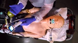
Jeff Mehler demonstrates intubation with a King Vision video laryngoscope on a manikin. He covers patient positioning, oxygenation at 15 L/min, tube prep with lubrication, insertion technique, and securing the tube with a Thomas endolock.
Questions this video answers
External auditory meatus above the level of the sternal notch.
15 liters per minute.
Size 3 channeled blades.
Remove the stylet.
8.0 mm tube depth approximately 23–24 cm.
Chapters
Hey, Jeff Mehler here. Today we're going to talk about our department's new King visions. These are our video assisted laryngoscopes. As you can see of a mannequin here, prepped out as if it were a primary cardiac arrest.
The patient is positioned with the external auditory meatus above the level of the sternal notch. Patient positioning is probably the most important thing that we can do for our patients. Whether we're using the nasal cannula with the NPAS at 15 liters a minute for the no DSAT intubation technique, if we're using that in conjunction with the non rebreather mask and the nasal cannula together in our primary cardiac arrest, that the patient positioning is probably the most important thing that we can do. As we're preparing our equipment, one of our partners can remove the non rebreather mask and go ahead with capnography attached to the bag valve mask device. We would go ahead and administer two full and complete breaths to make sure that the airway is patent Simultaneously, while the airway person is preparing their equipment, we would go ahead and and open up the King vision. And our department currently uses size 3 channeled blades. The blade as if it were to be going into the patient's mouth. The camera would obviously be facing the operator and it slides in and pushes down until you hear a click. There's one button on the reverse side. You push this button and it turns out the camera on.
You can see that there's a light and a camera there and then the channel. You would go ahead and take your endotracheal tube, remove the stylet.
Vigorously lubricate your endotracheal tube. Paying particular attention not to lubricate the distal tip of the endotracheal tube or the Murphy's eye. We would go ahead and then preload.
Preload this tube into.
The King airway and make sure that you've not extended the endotracheal tube past. This will impede your vision as you go ahead and attempt to.
Intubate this patient. One of the tricks that I found is that.
If you grip this King vision much lower than where the.
Rises are on the handle. If you grip this King vision much lower than where the risers are, you will have a tendency to stay shallow. If you grip the King vision up here, way up here, you're going to have a tendency to go deep and you're going to be in the esophagus most of the time. So if you grip the King vision Here and you have a tendency to stay shallow. While we're preparing our patient, we would go ahead and remove the oropharyngeal airway. And then the first piece of equipment that should enter the patient's mouth once they're positioned is a Yankee or suction. I found in my use of this device that if the airway is bloody or has a lot of secretions, the device does not work that well. Another trick of the trade that you can do is stick a piece of tape across the top of the Yankee or suction unit to have it apply continuous suctioning. So.
With my right hand, I would go ahead and insert this Yankee or suction into the patient's mouth and begin suctioning and suction this patient until clear simultaneously. And you can even leave the yankar in. We want to go ahead and watch it enter into the patient's mouth, holding it low.
And immediately rocking that until you have visualization of the vocal cords. You can go ahead and advance the tube.
And you can see that there's no mistake that that endotracheal tube passed through the vocal cords. One of the other tricks that you may want to consider is if.
Your endotracheal tube appears to be deflecting to the right, you can take the camera angle here and rotate it to the 10 o' clock position and go ahead and advance the tube that way. And obviously by the camera alone, you can't miss an intubation on a dry airway if the airway is not dry. If we didn't prepare the patient, if we didn't position the patient, if we didn't suction the patient prior to the use of this, the success ratio on this is not as good as if we prepared the patient as we should and suction the patient as we should. Once we have the ET tube passed, we can remove the king airway.
Shut it off appropriately, dispose of this. We would then go ahead.
And administer a ventilation onto the endotracheal tube. We would listen to epigastric sounds first. We would then auscultate breath sounds. We also have capnography attached, which we had attached when we were using this as a regular bag valve mass device. And then using our standard operating procedures for the use of the Thomas endolock, we would go ahead and and insert the Thomas. Set the Thomas endo lock in there, make sure that the bite block was below the level of the teeth.
Secure the strap first tightly or snugly to the patient's head.
Measure my depth. I'm an 8 millimeter endotracheal tube.
At approximately 23, 20 or 24 centimeters. Go ahead and tighten the tamise endo lock and continue your ventilations at the rate as per prescribed by your protocol. After your endotracheal tube is passed, you can stop the use of the nasal cannula and.
Make sure that you have placement. Make sure that your placement is verified by continuous waveform capnographic monitoring. All other.
Endotracheal tube placement assessment techniques are subjective. Auscultation of gastric sounds is subjective. Auscultation of lung sounds is subjective. The only objective clearly objective proof is in the pudding that the endotracheal tube is in the trachea as continuous waveform capnographic monitoring. Thanks for watching.