Mathew Saab, MD: Graduated Access for Hypotensive Patients (8/23/24)
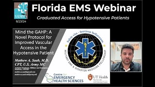
Mathew Saab presents a cadaver study of the Graduated Access Protocol, which uses rapid saline infusion through an intraosseous line to engorge veins for easier cannulation. The cephalic vein expanded 227% in under 30 seconds with less than 100 mL of fluid, but the effect faded quickly once infusion stopped. The technique may help in prolonged field care, though live-animal and human studies are needed before clinical use.
Questions this video answers
August 23, 2024.
23 cadavers.
Cephalic vein, axillary vein (proximal axillary as proxy for subclavian), femoral vein, and saphenous vein.
LifeFlow manual rapid-infusion device (hand‑pumped syringe pump).
Cephalic ~227%; Axillary ~163%; Femoral ~169%; Saphenous reported as effectively infinite percent increase from a pre-infusion circumference of ~0 cm.
Chapters
All right. Welcome everyone. It is Friday, August 23, 2024. This show is going to be a great one. Welcome to the Florida EMS webinar, where myself and Tanner Smita host incredible folks. Every single week, we try to stick to folks who have published research in the EMS field. This came to me because many of us know Scotty Bolliter, Stephen Rahm, David Miramontez, the folks in San Antonio who we all look up to. And Scotty and I were texting and he said, you have to see this paper that we just published. So I read it, I saw the video, and then I reached out to Dr. Saab, who's going to be our speaker today, and he said, yes, I would love to present. And so here he is. What I would like to do is turn it over to you, Matthew, if you can make a further introduction and then take it away. Thank you so much for joining us today.
Thank you, Dr. Anteve. Well, like you said, my name is Matthew Saab. I recently completed the Brooke Army Medical Center Emergency Medical Services Fellowship. There's two years in San Antonio where I worked with Dr. Miramantes and many of his EMS agencies down there. And I also got to participate in this research that I'll be presenting today. And like he said, that was in collaboration with Scotty Bollater over at the center for Emergency Health Sciences. My contact information is here and I will also put it up at the end in case anybody wants to contact me later offline. But today we're going to be talking about the paper that is Mind the Gap, a novel protocol for improved vascular access in a hypotensive patient. And here the logos are just the.
Institutions that we collaborated with on this. I do want to let you guys know, disclaimer up front, everything that I'm going to be talking about are my words and not the words of the army, the Department of Defense, the government, or UT Health, slash utsa. Now, as they joined.
What we'll talk about today's the background about why we did this, what our idea was, why it's important.
What happened, what were the results, what they mean. And then I think we'll have a discussion about what they mean and get further down to the nitty gritty.
What my goal is is that everybody here will be able to understand GAP and the theory behind it. We'll be able to answer all of your questions and you'll have an idea of what I had been doing in my downtime.
As some background. I think everybody who's worked in healthcare and certainly Everybody who's worked in pre hospital medicine can attest to vascular access can be very difficult in certain patients, particularly those who are hypotensive or critically ill. Specifically in trauma patients, when they're experiencing vascular collapse or they have such a high adrenergic state with vasoconstriction, vessels will often get tiny and the target becomes small. So getting access in these patients becomes difficult. One of the things that spurred us to look into how we can better vascular access in these patients was a paper published by Ryan Dumas et al, Moving the Needle. It was an east multicenter study where they compared central IV cannulation, peripheral IV cannulation and intraosseous cannulation at multiple centers, multiple trauma centers, and video recorded the operators. What they found was that placing an IO was just as fast as a peripheral iv, if not faster, and had higher rates of success.
In talking with Scotty Bollater, he had this idea. How can we improve vascular atlas in these patients? Well, as you can see here, what we have is a radial IO and we also have a fluoroscopic image of that IO. You can see that the intraosseous line is innately connected with the vascular beds. So if you infuse something into that entry osseous line, it should go into those vascular beds. And what would happen if we infused a bolus? Could we leverage that bolus to optimize the anatomy of adjacent veins to make them bigger, in a sense, or engorge them to improve vascular access options?
So the idea became for a graduated, we call it gap, the graduated vascular access for hypotensive patients. If we take a distal IO, infuse fluids through it, increase or engorge the veins of the proximal vessels, can we then facilitate and make it easier to establish vascular access? And a lot of people might say, well, why would you do this if you already have the IO, why would you need a vein? And particularly in rural EMS settings where you're participating in prolonged care, or in military settings where you're doing something like that, getting the IO is great. But if you have to sit on the patient for a long time or infuse more rapidly or get a second point of vascular access, this is the idea of where this process could be advantageous. So how did we test about whether or not.
Performing GAP is going to optimize these vessels? We took 23 cadavers. They were going to be used for other research pro, other research and training procedures. And we used three different types of IO needles that were all available to us to see if this would work in all three. And we took the Life Flow device, which I hope everybody's familiar with, but I'll show a picture of it if you're not. It's essentially a hand cranked or hand pumped syringe pump. And we used ultrasound in real time in the cross section to measure.
Blood vessel diameter or, sorry, blood vessel circumference.
This is what it looked like. And up at the top you can see the Life Flow device that has a syringe in it where it pumps fluids through the, through the extension tubing.
And to the right you'll see what it looked like. As we collected data, we were measuring in real time the blood vessel circumference pre and during fluid infusion to see how big it would get or how small it would get, or if there would be no change.
So could we augment blood vessels to better facilitate vascular access by pumping fluid through an IO?
We thought that we would, and we selected four sites to look at. We wanted to look at the axillary vein and the cephalic vein in the upper extremities and the femoral vein and the saphenous vein in the lower extremities. The reason that we looked at these sites where we wanted to do both central and peripheral vessels. And a lot of people will probably say, well, the axillary vein is not a central vein, and that's correct. But the subclavian vein is technically difficult to ultrasound and measure. And the only difference between a proximal axillary vein and a subclavian vein is about a centimeter of difference from whenever it becomes deep to the clavicle. We also looked at both central and peripheral vessels because not everybody is used to or has the ability to cannulate central vessels. So in a prolonged care setting, if you could establish a distal IO in the leg or in the forearm, you could then potentially cannulate a cephalic vein or a saphenous vein.
This is what it looked like when we were measuring our data. If you look at image A, you'll see a radial I O having been inserted. And image C is the corresponding circumference of the cephalic main measured in that extremity. In image B, you'll see that IO while crystalloid was being infused through it. And in image D, you'll see the circumference of the IO pre and post being 0.46 cm and 0.99 cm, respectively. And I bring your attention over to the forearm in image B of this cadaver where you can see that the superficial veins have actually been engorged and are much easier to both see, palpate and potentially cannulate.
So the things we were looking at was one circumference which we talked about. Two was volume. We wanted to know how much fluid would it require to pump up these vessels and three was how long did it take to optimize them. Optimizing was defined as the maximum circumference that they would potentially get while fluid was being infused. We did some statistical analyses, basically went like this.
And after performing these, the output became. Let me move, I need to move the camera or sorry, the.
Need to move the videos of everybody's faces so I can actually see the screen. In our first outcome vessel optimization, defined as the circumference of each vessel. You'll notice I bring your attention to the right column where the percent difference in the cephalic vein was 227%. In the axillary vein it was 163%. In the saphenous, it appeared to be infinity because the.
Pre infusion circumference was typically zero, meaning the vessel was flat. And in the femoral vein the difference was 169%.
I put in the left, the IO site and the corresponding vein site. We did both sides of the body and the the picture that I showed you earlier, or the graphic was just an example of left side upper extremity and right side lower extremity. But we flip flopped each cadaver.
In our secondary outcomes in volume and time. You'll notice that the volume was generally very low, less than 100ml required to achieve optimization. And the time was generally very low too, typically less than 20 seconds.
So what does that mean to us? Well, it looks like the larger vein size was associated with improved direct vascular cannulation attempts in previous studies. So if we could achieve larger vein size, then maybe we could facilitate this vascular access that was not specifically studied in our study. However.
In our study though, every single vein that we targeted had an increase in circumference with a fluid bolus. So it's non discriminatory. It happened in both central and peripheral vessels and potentially could facilitate both central and peripheral access. Step up to central and peripheral axis.
And then the last thing that I wanted to show to you, which I already mentioned, was that the vessel optimization was generally pretty quick, less than 30 seconds. So we may.
Provide safer, less stressful and more stable peripheral and central veins if we perform gap.
That's because typically whenever you have A prolonged transport or you're treating a patient in the field, the IO that's sticking out of the patient is usually less stable, especially in helicopter or, you know, hems transports. If you.
Move the, the arm, it could dislodge a humeral IO if you brush up against the IO, it become, could become dislodged and then you'd end up having extravasation. But if you could get a peripheral access or central access direct into the vein, it may become a more stable vascular access option for prolonged transports and prolonged care. But in our data there wasn't any significant difference between infusion sites and there was only a significant difference amongst the volume of fluid that was required to be bolus to achieve optimization. And that.
That was in the sapenous vein looked like required less. And that may be because it was a smaller vein and you could imagine that a larger vein might require more fluid to optimize. Is this relevant to you? That may depend on where you're working and what type of patient you have. Certainly I would not recommend performing this.
This procedure until we've studied it in a live humans. But also I wouldn't recommend doing it if you have a very short transport time or if your patient isn't critically ill. The idea is that in this critically ill, long transport patients, when you need to step up your vascular access, have more vascular access options, have more stable vascular access options. This is where the GAP procedure may be beneficial. So specifically for the military, where we're looking at probably a lot more prolonged care scenarios in the next conflict and even those that we've already, that we've already needed to undertake.
Sitting on a patient where you're trying to resuscitate them and keep them alive so they could receive medevac. You may want to.
Consider this as an option once we further study it. It could be, you know, perform as a bridge to more definitive vascular access for these patients. But there are of course limitations to what we looked at. One of them being that this was performed in cadavers. We have not studied it in live tissue or in live humans. We have not proven that this is leading to faster vascular direct accumulation of vessels. We have not proven that this is leading to easier direct accumulation. But we are.
Certainly there's some conjecture that this may be a way to achieve those things. And actually my team is looking into, into doing that next.
So we didn't.
We didn't look at time, we didn't look at number of cannulation attempts or ease. We're considering an animal trial for this. And.
The last thing I want to say also on this was that we used normal saline in the study. We did not use blood. Anecdotally, I've used blood on expired blood in this and it did seem to work just the same. I do want to provide a special thanks to my collaborators. We had several of them and some of them may be on this call. But big thanks to them. We wouldn't be able to do it without them. I also wanted to thank those who donated their body to our research and the UT Southwestern Willed Body Program, as well as the center for Emergency Health Sciences for providing their facilities. And thanks to you guys for inviting me to speak. Thank you to Dr. Anteby and Tanner and thank you all for listening. I have several references.
And I will take questions. I do want to say if you have not read the paper and you'd like to, it's open access, so you should be able to get it for free. However, my contact info is at the bottom. If you'd like to email me, I could send it to you or I could send it to Tanner or Dr. Anteven. They could potentially distribute it to you all. I'm not sure.
And I see Tanner just put it into the chat. Matthew, this is an incredible presentation. Yourself and then the list of co authors. You have pretty impressive group of people. I have so many questions and.
I also want to mention some of the commentary that came across on social media because everyone on this call today, obviously we're in the field. We're in the business of mainly critical care. We've all been there where we haven't been able to get vascular access. So this topic I think is especially beneficial and I'm hoping that people on the call today. I see Kim is already raising his hand. I'll get to Kim in a minute. I'm going to go to Tanner first, but my question is he said it took about 25 to 30 seconds to optimize the vein. How long does that vein stay optimized? Or does someone have to start that IV and cannulate as someone is using the life flow in this particular case. Can you comment on that?
Absolutely. Yeah. And we didn't measure that in the study. But I will say, at least in the cadavers, when we're watching, when we were watching on the real time ultrasound, you could think of the, the circumference as well, while it's growing as being kind of undulating where you see like a, an uptick in the size. But then it kind of fell when the pressure fell as the. As the life flow was reloading or recharging with some more fluid.
So I would say, if you're going to be performing it with the life flow and everything, as we did in this study, I can't say what would happen in a live patient, but I can say in the cadavers, the circumference would decrease.
Would decrease rapidly, but not fall immediately to zero. And I can't give you the exact time, unfortunately, because we didn't measure that. But in performing this on the cadavers, yes, the increase in circumference did fall. Once you stopped pumping the fluid.
Can you give us, like, a. Like, would you say on. Just on the cadaver. I get that it's not in humans, but was it within 30 seconds? Was it. Was it within a minute? Because I think that's important as people are thinking through this.
Yeah. Yes. I would say when we were doing this in the cadavers, that maximum circumference would. Would stay for a few seconds, and then it would start to fall rapid. So it would wrap. It would rapidly begin to fall, but not necessarily rapidly fall to zero. And it never really fell to zero when we were doing this. Actually, it didn't at all. But that it would. You could think of like a balloon with a. With a hole in it slow, or like an air mattress kind of deflating would be how I would liken it. And I would say that significantly, within 30 seconds, the circumference would be much lower and would actually, you know, render the whole. The whole process probably ineffective. But if you're able to pump it up with a LIFO with, like, a single operator and then try to perform this, if you could get access within, you know, a minute or, you know, 30 seconds, once you see, if you can see, like, a superficial vein, you could probably perform this as a solo operator, but that peak circumference won't still be present. It'll still be inflated more than baseline, but that peak circumference won't be available.
Got it. Awesome. All right, let me pitch it over to Tanner. Tanner, what do you got for Matthew?
Just one comment about.
More from Peter

Melissa Olen MSN, FPN-C, CCRN: Sudden Cardiac Arrest Training in Schools (Project ADAM) 4/25/25


Pediatric Hospital Readiness: How Teamwork & CPR Metrics Drive Better Outcomes

Chelsea Kadish, MD & Kyle Goodknight, EMT-P, RT: Pediatric CPAP in EMS (3/14/25)