Stroke Triage: The Importance of Pre-Alert
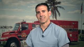
A recorded presentation by Dr. Dennis Reyes (Delray Medical Center) on pre-hospital stroke triage, emphasizing rapid identification, pre-alerting hospitals, and routing suspected large-vessel occlusions (LVOs) to comprehensive stroke centers. Covers stroke types (ischemic, intracranial and subarachnoid hemorrhage), time-sensitive therapies (IV tPA/tenecteplase and mechanical thrombectomy), imaging (CT/CTA/perfusion), field triage scores (BFAS, RACE), and operational benchmarks for door-to-needle and door-to-groin workflows.
Questions this video answers
Within the first 4.5 hours of symptom onset (greatest benefit within first 3 hours).
About 4.5 patients treated (earlier window) to see benefit; at 180–270 minutes NNT ≈14.
Door-to-needle time should be less than 30 minutes.
Example fastest metrics: door-to-imaging 7 minutes; door-to-puncture 45 minutes; puncture-to-revascularization often ~22 minutes (goal sometimes ≈10 minutes in ideal cases).
Standard of care up to 24 hours, with possible extension to 48 hours in select patients.
Chapters
How you doing guys? I'm Dennis Reyes from the Delray Medical Center. I'm a neuro interventionalist at Del Rey Medical Center. Today I'm going to talk about stroke, triage and the importance of pre alerting. I am consulted from panel Brown Stryker. So stroke cases are equally emergent and some of them are ischemic stroke which are highly time sensitive. There are cerebral hemorrhages which you typically present with severe headaches and nausea and vomiting.
You have to treat them with the AVCs on the field. There is also subarana hemorrhage which also present with sudden thunderclap severe worst headache of their life. And it's pretty concerning. It usually relates to brain aneurysms as well. This is a typical scenario of intracranial hemorrhage. This is a CT scan and the bright area is a bleed.
Just wanted to show you a big sample. That's another sample of a bleed related to a brain aneurysm that burst and cause what we call subarachnoral hemorrhage. You see a bright spot on both sides of the brain kind of spread in the surface of the brain scheme. Strokes tend to be very rapidly progressive, especially in the area of the red area, what we call the core and the yellow area is where the ischemic penumbra is happening. There's a risk for having for expanding the stroke from the red area into the yellow area.
As time passes given that the blood flow is so slow in that yellow area. But the red areas typically means that the infarct is going to happen and there's no reverse for that part of the brain. There's TPA trials, we all know that. We try to bring the patient in as quick as possible because we want to give them medication within the first 4.5 hours. Most of the benefit, the best outcome is seen in the first three hours from the symptom onset, the lacinoma with a good 30% of them doing really well. But between 3 to 4.5 hours there is a benefit. It's just a little less compared to those arriving in the first three hours for the clot buster medications. And this is kind of how represented longer you wait. The NNT means that the number needed to be treated the shorter amount of time. You can treat 4.5 patients to see a benefit, but by the time that you get to the range of 180 to 270 minutes, now you need to treat 14 patients in order to see a benefit from giving club buster medication. But good is not Good enough. And the benchmark for stroke, given door to needle time, should be less than 30 minutes. We're trying to do that. It's all about the plumbing, opening the vessels as quick as possible. The big vessels of the brain can shut down, but also.
Whether it's with a clot, with a cholesterol plaque, but at the same time there's something called lacunar stroke where there are tiny vessels of the brain. You see those little branches right there, they can shut down. And that's where the TPA really does a lot of the work, cleaning those small vessels. As there is no really good procedure that we can open those vessels.
At this point. This is again why the clot busting medication is so important to kind of clean up the tiny vessels of the brain.
Moving forward. Lacunar strokes, again, they can be very, very small that appear as a small lagoon over time. Some of them, believe it or not, will present with no symptoms at all. But a lot of times they will present most 100% with significant weakness on one side of the body or loss of sensation alone, or the patient has some weakness and incoordination problems on one side of their body, or they have speech difficulties where they're slurring, but then at the same time they're having issues with their hand. That's what's called dysarthria, clumsy hand syndrome, meaning they're, they're having difficulties coordinating their hand. At the same time, there's learning and there's feet. That could be also including a facial weakness, which is very typical and it looks very small on an mri. One of those tiny vessels just simply shuts down from the changes over time, whether it's related to high blood pressure, high cholesterol, high sugar, it's just shut down. Whether related to, again, risk factors, even age, Age is one of the biggest factors. Unfortunately, we can't do anything about that. But this is the importance of. The only medication benefit that we have for this type of strokes is again TPA or the new medication called tnk.
Which is given as a bolus right away as soon as you get a ct. And you're able to exclude any possible criterias for the medication. But both the small vessels, which again, lacunar strokes or the large vessels can cause weakness, numbness, facial droop, the dysarthria, these are typical for the small vessel disease. Now, when you have a large vessel.
Being occluded, the symptomatology is pretty drastic and it's pretty dramatic as well, meaning they will have a fascia which the small vessel disease will not have. Typically, they will have a neglect. They're paying attention to one side of the body more so than the other one. Or they have a gaze, what we call a gaze deviation. You probably hear that a lot of times. Usually correlates very well when someone has a gaze deviation towards the right side, at the same time they're neglecting the left side. So it kind of goes along hand to hand. But with small vessels, you can have slurred speech again, coordination problems again, balance difficulties, unsteadiness. They can certainly fall, hit the head. And it's pretty confusing because some of them will look like a trauma case, and they'll just show up at the ER thinking this is a fall, this is a trauma patient. But the biggest problem is, was actually.
A stroke happening in the arteries of the brain, not letting the patient control one side of their body. Therefore, they fell to the ground, hit the head. And that's why it kind of looks like a trauma, but it's not to begin with.
So then the big vessels can actually thrombose, especially the carotid, and the neck can build up cholesterol plaque, and they'll eventually shut down to the point that there's not enough blood flow going up to the arteries of the brain. And a little bit of a clot can actually create in the carotid. And all of a sudden, they can fly out to the arteries in the brain and shut it down completely. Kind of looks like this, a tiny clot flying from the carotid up to the outer of the brain. And you have to not only fix now the carotid, but you also have to go after this.
Little clot, shutting down this vessel. These clots also can come from the heart, from a condition very well known by you guys, atrial fibrillation. As you know, that's fine. The rhythm of the heart. The heart is not actually pumping very well, and the blood kind of. Kind of stays. Sits in the area of the heart where it tends to form clots. After 2, 3 minutes, then the clot is already there. So once the heart goes from this finer rhythm back to pumping again, it will shoot up the clot to the arteries of the brain, and that's how they get in trouble. So we follow two different parameters as of. Okay, how do we approach this case is we know that between up to 4.5 hours, we can give TPA. What about how much of brain tissue we can save? And yes, there are scans that we can do and kind of determine what's called a perfusion based algorithm. These scans are very helpful because it gives you the idea of how much blood flow is getting to that part of the brain and how much brain tissue is already damaged, most likely, and how much tissue you can potentially save. And that's what we call the mismatch. So it's just instead of using pure time algorithms, we can use the perfusion based algorithm with the help of the scans. This is kind of what an M1 or right middle cerebral artery looks like. You see the clot completely shutting down the artery on the right side of the brain. There's no flow on the right side of the brain. There's no branches on that side. There's some collateral flow coming across through the top of the head into the right side of the brain.
There's the core where there's not significant blood flow. There's no collateral flow going in there. Therefore most likely that area is going to be permanently damaged. But this area right here is getting blood flow. If you don't open it quickly enough, then it can actually create bigger stroke. If you remember the yellow and red areas that I showed you earlier, retrieval stents is the kind of things, the stents that we use, devices that we use, we also use aspiration catheters to take those clots out of the arteries of the brain. We can go up to the big vessel like the one that says ica, meaning internal carotid artery. But we can go into the M2s, the M1s, the M3, even the pretty distal vessels, M4, and try to aspirate those clots. Sometimes it's very challenging once you get once the clot is in an enforced segment. So but sometimes we try to do so depending on how bad the symptoms of patients are. This is on top of tpa. Obviously the farther the clot is, most likely the higher the benefit from receiving a clot buster medication. But certainly we can push a little bit of this clot buster medication into that tiny vessel through a small catheter in an artery of the brain. And there's many studies and clinical trials have proven the benefit of performing mechanical embolectomy or mechanical thrombectomy, or basically removing the clots from the arteries in the brain. And they should receive endovascular therapy with the stent retriever as well as tpa. Recent studies do approve and suggest that the patient should receive actually both if it's possible clot buster medication as well as the actual surgery. Why is management of acute stroke in the pre hospital arena Kind of complex. And it's basically without special scans, you could literally miss them. You cannot tell what are you dealing with. It's kind of tough to tell. Are you dealing with a bleed? Are you dealing with a big stroke? It's kind of hard in that way. So some hospital have figured out, some institutions figured out how to put a CT scanner inside of an ambulance, but guess how much that will cost. It costs approximately about a million dollars to do so. In order to justify all the trips back and forth and determine is there an actual benefit to you saving time by having a scanner and an ambulance? Some studies suggest it's really not causing effective on the long run. Is it a likelier stroke? Those are the things that you want to answer on the field. Are you dealing with a stroke or not? Is it focal deficit of a global scenario where the patient is having confusion? Maybe it's just weak all over and not necessarily weak on one side of their body or so. Or maybe they're just confused instead of having actual speech difficulties. Were not actually following commands. They don't sound slurred, they just sound confused. Cincinnati score is something that you guys are very well known, but we're not using that anymore. We're trying to keep it as simple as possible for you guys. But using the BFAS score and also the RACE score that we were going to go over. Small vessels are most likely to cause strokes, therefore symptoms should be mild in a way. And then large deficits, meaning a lot of symptoms, most likely going to suggest the big vessel is in trouble. And that could imply again, not being able to move one side of their body along with not being able to speak, not being able to comprehend. The same way they might be looking to the opposite side. You touch them on that side, they're not paying attention to that side either. So the answer is, okay, where is then the most. The closer comprehensive stroke center has availability 247 that not only that patient's going to receive TPA, but also is going to receive potential mechanical thrombectomy. So this is a scale again of how fast the stroke can grow based on a study of the. They looked at the fusion scan and showed that it's very, very useful to have the scans as you can detect those patients that could be fast progressors. Believe it or not, those typically will have a lot of risk factors with high blood pressure, hypertension, diabetes, no matter what you do. They stroked out in three hours or sometimes even less because they don't have any collateral. Their clotters are so poor that the brain will shut down very early in the stroke process and the ischemia will progress very, very quick. Instead of some other patients where they only have, let's say, high blood pressure, then they might have a chance to progress and sustain that brain tissue for a longer period of time and not have as much core infarct as in this case. And this is the bfas. B stands for balance. E stands for eyes. F stands for face. You're looking for a facial asymmetry. A stands for an arm weakness, where there's a drift. Speech. S stands for speech. All three have speech difficulties. They're not comprehending, they're not able to repeat words, they're not able to name words. You can ask them to repeat a sentence, for example. We use a lot. Today is a sunny day, sir. Can you repeat today is a sunny day, or can you name what's in my hand? If they can't do that, then you kind of get a sense that they might have aphasia. You ask them, show me two fingers. If they're not showing two fingers, then you know they have comprehension problems as well. And then next thing you just want to call 911, which you probably guess at that point already. Diagnosis. Enlarged vessel occlusion is very, very tricky. It can be.
Depending on the symptomatology. Usually we try to use the core symptoms. The core symptoms, the most concerning symptoms are gaze deviation aphasia when there's big, big speech difficulties. And again, on the right side of the brain.
You know, they have neglect. They're neglecting their left side of their body. The right side of the brain, remember, controls the opposite side of your body. So they'll be looking like this and the right side of the brain is having a stroke. Then they'll be having weakness on the left side and not paying attention to the left side and then neglecting the left side. And therefore they'll have a gaze preference, the right. So that's kind of what tells you the right side of their brain is in trouble. Most likely there's a large vessel occlusion. This is kind of what a gaze deviation looks like.
And again, so we just want to be apply those scores, including the ray scale.
Which it's important to understand the ray scale not only by the scoring, numerically speaking, but also by adding the plus sign. The plus sign, meaning it means basically that there's a cortical sign. It means the surface of the brain, where the neurons are, is where this important functions are, meaning the speech. The fact that they cannot recognize One side of their body or not, their preference to look one side or the other one, those are on the surface of the brain. And that's again what we call a plus sign. So once you add the plus sign, we know, we understand that this patient is not only having weakness on one side of the body or maybe just facial weakness, but they might be also having speech difficulty that is just not slurred speech, that is comprehension related, unable to name, unable to repeat. Or they're looking to one side. That's the plus sign that it sounds so important as the opposite side where there's not a large vessel occlusion, the small vessel disease, typically they would not have again, that gaze deviation, the neglect, the speech, facial difficulty of naming, repeating. And then mild strokes are usually. When you look at the race score, even though this can fool us sometimes, if it's less than 5, it kind of sounds like it will be a small vessel disease. Anything that is equal to five or more, it will sound that it's going to be a large vessel occlusion. But we have had patients, though, with a race score of less than 5. And then suddenly we find that the big vessel in the brain is shut down. They just happen to have really, really good collaterals in the brain. So.
Race scale of 5 predicts a good 42% likelihood of dealing with a large vessel occlusion. If it's 9, then you close to 65% of dealing with the big vessel being completely shut down. We want to reduce that door to groin time by 40 minutes or more. That's why it's so important that you guys pre alert us that when a race score comes like this, meaning Anything is above 5, even if it's close to 9, we're already pre alerting everybody. We're already letting know the team, techs, nurses to come into the hospital. This seems to be something that is likely to be a large vessel occlusion. And we start mobilizing the team in order to kind of cut down that time and be able to.
Again raise. This is a race against time.
That's why it's so important. The race scale actually is very. It's available if you, if someone doesn't have it through the link, there's an app and you can actually calculate it on your cell phone really quick. If you put that link in your cell phone, it'll be easy to calculate the triage in the field. In EKG is important because you want to see if this patient has atrial fibrillation symptomatology again. Again, Cincinnati, we don't need it as much anymore. We should like you to focus mainly on the BFAs and the race score until we get to the ED and be able to do a scan at least to rule out first the bleed. If there's no bleed, then we keep going trying to find whether it's a large vessel occluded or not. Circulation wise. There is a stratas registry looking at where does the patient should go if you find them with this kind of symptomatology. And it happens that the stratus study in summary basically.
Suggests that we should just go to the highest level of care. Overall, it's felt that transfers are bad. There's 103 minutes door indoor out at a primary stroke center. So patients at a primary stroke center, they're just wasting time. And every minute counts because every minute patients are losing approximately 1 million neurons are dying every minute. So imagine 103 minutes these patients are suffering and they might develop insignificant sequelae as a consequence of waiting on a primary stroke center. So it delays intervention treatment, meaning delays opening the vessel or giving a clot buster medication by 116 minutes. 62% in the strategy summaries were transferred within a ratio of less than 25 miles and approximately 50% were under 14 miles. So it's something that is doable. Transfer patients, they tend to do worse. 8% were less independent and they have more problems, more disability high volume centers. It means that those centers that are capable of doing at least more than 30 strokes a year or usually more than 40. We tend to be faster because we're just so organized. We're into it, we do it almost every day. There is a system in place and everybody's motivated to just go after whatever is going on. Once the code fast, that's what we call it at our institution at the Rey Medical center is triggered, everybody gets alerted and everybody's just running. Including anesthesia in order to get to the clot. And also works with tpa. It's a little bit faster at comprehensive stroke center patients arriving to that comprehensive structure center since again the team is very well organized, they have a better chance to get that clot busting medication in 70 minutes earlier than in actually primary stroke center because they're just not exposed to strokes as in comprehensive stroke centers.
And then hospital triage. Again you want to rate the field severity base bypass two comprehensive center ulcer patients to compress trochenter.
There's a pre alert based on severity. So you want to really see how bad they look like again severity based on the race score and also the BFAs kind of helps you detecting the problem to determine okay, should I take this patient to a comprehensive stroke center or not. But even if it's on the lower again side of the race score.
There has been, I have to be honest with you, there has been cases where even with a low race score, those patients have a clot sitting in the arteries of the brain. We have had some of those really trying to fool us. But at the Ray Medical center, for example, we scan everybody, everybody that comes in stroke like symptoms for 24 to 48 hours, even up to 48 hours. We just go ahead and do what we call a triple scan, which means doing a CT of the head, doing a cta, looking at the vessels and the CT perfusion to see how the blood flow is going in the arteries of the brain. So right there that all happens in approximately 15 minutes and it gives you some much information and that you saving so much time right away.
In order to determine what will be the best next step of treatment. And this is kind of what it looks like. This is a plain CT scan. On the left side the arrow is pointing to what we call a hyperdense MCA signs. It's kind of like increase light up in the vessels when the clot is sitting in there. The clot is sitting so dense that it tends to cause a little bit of a brightness. Then we do the cta, the picture in the middle showing that the vessel is completely shut down on that left side of the brain. Left side of the brain again would be very concerning for I would expect this patient having speech difficulties, unable to speak, probably mute, unable to file commands, not moving the right side, not paying attention to their right side, and probably having a gaze deviation as well to the left side as the whole left side of the brain. We see on the perfusion scan that green area, that green area is the area of the brain that is at risk for having a massive stroke. The red area is more deep in the structure. It's most likely the area that a stroke is going to happen anyways. No matter what you do, it's going to happen in that area.
But the green is the potential brain tissue that you can save and save this patient from significant disability. And this is a big sample of 125cc brain that can be potentially safe. Therefore we took this patient for procedure. You can see that again confirm that the vessel is completely shut down. And we call that M1 segment of the left middle sedative artery. There is good collateral flow.
Through the vessel. It goes up through the surface of the brain into the left side of the brain. But if you don't open the highway, if you don't open the vessel, those collateral eventually is going to shut down. They're not going to resist anymore. So after removing the clot, we were able to restore that blood flow back again on the left side of the brain. The patient did really well. One of our fastest times, the door to imaging has been seven minutes. Door to puncture, 45 minutes, puncture to revascularization. We usually average about 22 minutes. But I've had great cases where even in four minutes we have, I have been able to go through the groin to put a catheter and aspirated clot in the arteries of the brain. We usually try to keep it at. I usually try to keep it at 10 minutes. But it's very challenging on Builders League because it's all case dependent and patient dependent depending on what's going on with the patient. Obviously, time is the primary predictor of a good function outcome. That's why we're just running around trying to get things done as fast as possible. In summary, bypassing to a comprehensive stroke center is now American Heart association national recommendation for patients with suspected large vessel occlusion IV TPA Main impact patients with large vessel occlusions the trials are actually done. It used to be we were concerned about giving TPA before, but not anymore. We want to give the club buster medication even if they have a large vessel occlusion. And then the slower the door to needle times. You'll see those at the primary stroke center, but again, they're just not expected. They don't have the volume. There's not the drive and the motivation that you see at the comprehensive stroke center because we tend to see those patients very, very often. We want to target at least 30 minutes from the door to the time that we put a needle and infuse the medication. And then door in and door out times should be around 50 minutes. For patients who present to a primary stroke center with a large vessel, you want that patient out of the primary stern center as soon as possible so you don't delay treatment if it happens that the patient ended up at the primary stern center. Mechanical thrombectomy is now standard of care up to 24 hours, but it can be extended up to 48 hours. So don't think that it's all depending on the collaterals. Like I mentioned at the beginning, there are the patients that they just stroke dial very very fast within three, four hours because they have so many risk factors. But there are those that have been taking care of themselves, exercising, doing cardiovascular exercise. Maybe they have high blood pressure, but they're going to do well if even at 36 hours later they might be able to keep that brain tissue alive during that period of time. And those two growing times are 20 minutes slower. In low body lymph centers. The median door to groin of 60 minutes or less is now the new normal. We want to get that in less than 60 minutes at our place we try to target 40 minutes processes needed to be adjusted to meet those goals. We need to work with EMS to provide race score and pre alert us. Pre alert the er. The ER will pre alert to us and we will eventually pre alert the team know to get everybody moving.
Always trash the worst report a deficit. If in doubt, just go to go to the highest level of care if you have any doubt. We are only done if we have shown that the arteries are open, you know, and that's what we do with the triple scan in the er. And not every place does this, not every place does a triple scan. Okay, time is brain. Again as a mentioned, every minute delays kill approximately 1.9 million nerve cells in the brain. So that's a lot of disability that the patients will deal with and the families as well. Scoop and run. Again, treatment effect vanishes rapidly. Very helpful if you get a cell phone of a bystander, a family member, so we can learn about their history, whether they have high blood pressure, obviously like I mentioned before, cholesterol of sugar, but also they have afib aphid is a big deal. And whether they're taking an anticoagulant, any blood thinner, not just aspirin, aspirin is not as important. But if they're taking Eliquis, Coumadin, Soarellato, Pradaxa, any of that, that will be just a contraindication to give a clot busting medication.
But if they're not, then definitely we will consider it give it.
So again, more of the race score. It's very, very important to know the last known well time of the patient because that's when we reset the time in order to figure things out. Even though wake up stroke, though we don't know when was the last known. Well, we could still do an MRI of the brain really quickly and determine if the stroke just happened, recent or not based on the two sequences that we can compare. And they're still able to receive clot buster medication. Depending on how the MRI shows. Okay getting a cell phone again medications.
We don't have time for family to drive in. In the news we have really good cases. A man that suffers stroke in I95 we where EMS was involved. May is Stroke awareness month. There's a little video you guys can enjoy and actually May is coming soon. It's very important to let our friends and family members have the typical symptoms and signs of stroke. We also had a patient that suffered a stroke on his birthday and EMS was also involved and saved his life. If you guys have any questions you can feel free to reach me through LinkedIn, Instagram, Twitter or you can call my cell phone or email me any Google reviews. I'll appreciate that as well. Regarding this presentation so we know what needs to be fixed or anything any recommendations regarding the presentations we can fix it later on. I appreciate that. Thank you so much. I hope you enjoy.
More from Peter

Melissa Olen MSN, FPN-C, CCRN: Sudden Cardiac Arrest Training in Schools (Project ADAM) 4/25/25

Pediatric Hospital Readiness: How Teamwork & CPR Metrics Drive Better Outcomes
Chelsea Kadish, MD & Kyle Goodknight, EMT-P, RT: Pediatric CPAP in EMS (3/14/25)