Felipe Oviedo from Microsoft's AI for Good lab describes a deep learning model that helps radiologists spot breast cancer in MRI scans. Trained on over 10,000 exams, it achieved 90% accuracy detecting existing cancer and 80% accuracy predicting five-year risk, while cutting radiologist workload by 80%. The model is designed to assist clinicians, not replace them.
Questions this video answers
Around 300,000 new diagnoses and 40,000 deaths per year.
Data from over 4,000 patients totaling more than 10,000 individual MRI exams.
Around 2% of patients developed cancer.
Over 90% accuracy in detecting existing cancer in patients.
Over 80% accuracy in predicting the five-year risk of cancer.
Chapters
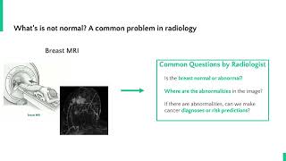
Hello, my name is Felipe Oviedo. I'm a senior research scientist at the Microsoft AI for Good Research Lab. I have a background in applying AI to scientific problems such as computational chemistry, biology and medical imaging, and I'm very excited about the applications of AI in these non traditional domains. I would like to tell you about a medical imaging project I've been working on where we have developed a system that uses deep learning to assist radiologists in the early detection of breast cancer. This project is a partnership between Microsoft AI for Health, the University of Washington and the Fred Hodge Cancer Research Center. Breast cancer is one of the most common types of cancer worldwide. In the US there are around 300,000 new diagnoses and 40,000 deaths per year. Radiologists often use magnetic resonance imaging or MRI scans to perform cancer screening and early detection in high risk women like those who have a family history of cancer. However, even with an MRI scan, small lesions can be missed leading to false negatives and benign lesions can be mistaken for malignant lesions leading to false positives and unnecessary biopsies. AI can be a useful tool to inform decision making of our radiologies to both reduce the interpretation time and increase the accuracy of an MRI exam. Our goal in this project was to develop an explainable machine learning model to assist radiologists in the early detection of breast cancer. We envision this tool helping radiologists by reducing the effort required for the analysis of MRI imaging and quickly identifying areas in the breast that are abnormal and require follow up. A common problem in radiology is distinguishing between normal and abnormal exams. In the case of women with a high risk of breast cancer, an MRI is recommended every few years. This Image shows a 2D projection of an MRI scan. A radiologist analyzes the full MRI scan trying to answer these key questions. Is the breast normal or abnormal? Where are the abnormalities in the image? If there are abnormalities, can we make cancer diagnosis or risk predictions? MRI scans are complex three dimensional spatial measurements and so answering these questions requires a very experienced radiologist and substantial analysis time, particularly in cases that have very subtle abnormalities. This problem can be tackled using machine learning by training a neural network to automatically identify abnormal MRI scans. However, applying machine learning to a problem like this comes with significant challenges. Negative cases, meaning normal breast images, are much more common than positive cases where the cancer is present, especially during the early stages of cancer. To train the neural network, we had access to data from over 4,000 patients totaling more than 10,000 individual MRI exams, but only a fraction of these patients, around 2%, develop cancer. The challenge this presents when it comes to training a machine learning model is that your model could simply learn to predict no cancer for every single image and be correct 98% of the time. In order to effectively identify that critical 2% of positive cases, we need to take a careful approach. The way we did this is by customizing our model for anomaly detection. We which means identifying normal or negative images very well through training on the abundant negative data and then being able to identify abnormal images by looking for features that are absent in the data set or are different compared to the normal images in the data set. This is often called one class classification. And so with this model, for any MRI scan, the model outputs where the image is normal or abnormal. We can take this analysis one step further, going beyond the simple classification of normal versus abnormal and produce maps of anomalous areas in the abnormal images. These maps can then later be used by our radiologists to visualize exactly what a portion of the image was found to be abnormal and inform a more detailed analysis or biopsy. Our final model demonstrated over 90% accuracy in detecting existing cancer in patients and over 80% accuracy in predicting the five year risk of cancer. These results are comparable to the accuracy of a trained radiologist reviewing MRI scans in the context of a patient's complete clinical history. In addition, by automatically analyzing images, our system was able to reduce the workload of radiologists by over 80%. The inputs and outputs from our model look like this, where the top row is a collection of MRI images for multiple patients. The areas in red in the middle row of images are the areas identified as abnormal by the model and the areas in white in the bottom row are those deemed abnormal by a radiologist. With this comparison, we observe a good match between the model predictions and the judgment of our radiologists without any manual annotations. Our next steps for this system are to integrate it into the clinical workflow of radiologists to reduce their workload and help them quickly identify cases that require further analysis by a human. The future of medicine will no doubt include many AI assisted tools like this one, but. But it's important to remember that AI is no replacement for a trained medical professional. Instead, we hope that tools like this can help reduce the burden of tedious work on clinicians and help them provide better outcomes for patients. Thank you.