Watch a Cardiologist Read a Real HEART SCAN: Live
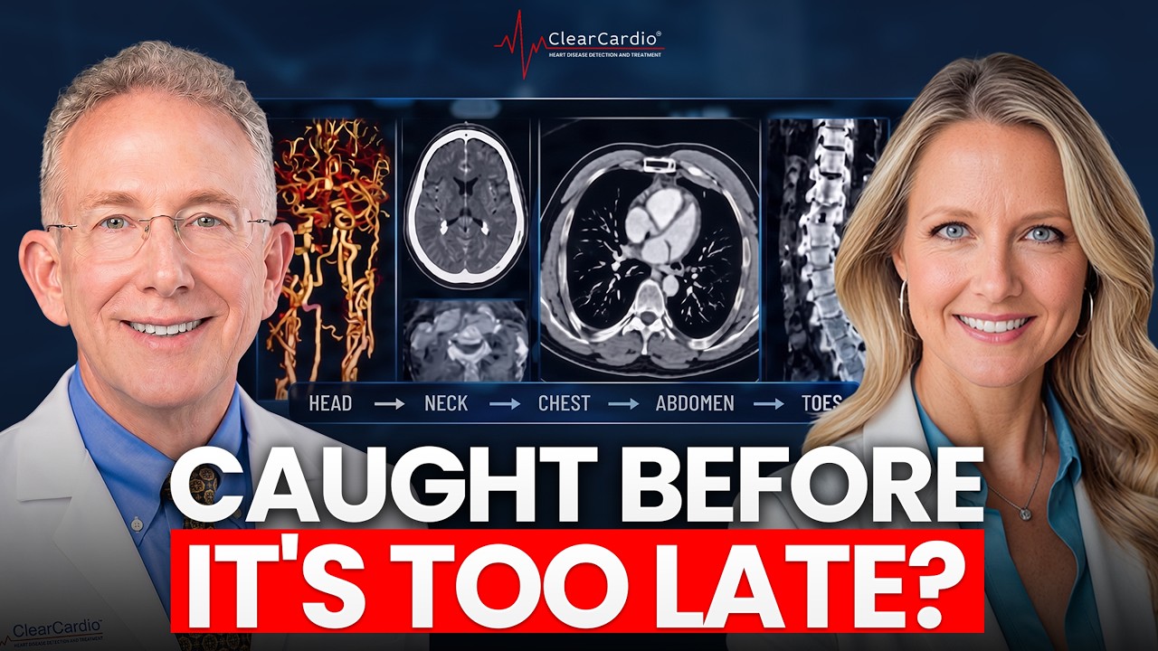
This content was created by John Osborne. This content was uploaded by John Osborne. This is a YouTube video titled "Watch a Cardiologist Read a Real HEART SCAN: Live" (created 2026-10-23) featuring Dr. John Osborne (Chief Medical Officer of Clear Cardio) and Sherry (Clear Cardio COO, volunteer). The clip is a live demonstration/consultation in which Dr. Osborne walks through high-resolution 640-slice cardiac CT images (United Imaging Atlas 960) and AI-based analyses (Cleerly/Cleer Cardio tools). He shows 3D reconstructions, straightened vessel and cross-sectional views of coronary, carotid, vertebral, abdominal and peripheral vasculature; explains AI quantification (plaque volume, percent atheroma volume, chamber sizes, bone mineral density, visceral fat) and clinical thresholds; and reviews incidental findings (tiny gallstone/sludge, a simple kidney cyst, evidence of prior appendectomy). Result: Sherry is described as largely healthy with no significant blockages and only a microscopic speck of plaque. The video discusses clinical implications, screening value of cardiac CT with AI, and upcoming FDA approval for carotid AI analysis.
Chapters
So the AI did detect a tiny little speck in one of the blood vessels here of a heart attack. Now I can see 2,000 shades of gray. I can see not just red, green, and blue. I can see infrared, ultraviolet, microwaves, gamma rays, radar waves. That's what the AI sees. So it literally can see things that we cannot see, that I cannot see. This is Dr. Osborne, the Chief Medical Officer of Clear Cardio. And we're going to use Sherry here, who's our Chief Operating Officer, who's volunteered. She's gotten in the scanner. We've done all these cool scans. She's not actually seen these yet, so it's gonna be all live for her. And we're gonna show you all the additional capabilities that this incredibly special machine can do. This United Imaging Atlas 960 is really the epitome of cardiac CT scanners. It is the fastest scanner out there, which means higher resolution, faster scans. Literally all of the scans that we took, literally head to toe, including the heart, all the vessels in the neck, the carotid arteries, the belly, etc., we did in one scan. One shot of contrast and took about 30 seconds to do all of that imaging I'm going to show you here as well. So let me now show you, we're going to use this now. This is Sherry, believe it or not. So this is what Sherry looks like underneath. I've stripped off the stuff I don't care about. There's no scone, no skin, no muscles. Who cares about that stuff? And this is basically just her blood vessels and her skeleton. So that's a cool view. So this is the resolution that we have with this machine. So we're going to now strip out and just look at her blood vessels alone. So now we go over here and what we're now looking at is just the blood vessels. So we can track— this is the aorta coming out of the heart. We can see the vessels heading off to the right arm here, another big vessel heading off to the left arm, and then coming out of these vessels are the carotid arteries. So they are located right here. That's the one on the right. That's the one on the left. There's also 2 vessels that go up behind called the vertebral arteries, and those actually run through the bones of your spine and your cervical spine. So those are all the blood vessels that go to the brain. All the blood vessels now flaring out in her brain looks beautiful. No aneurysms, no enlargement. I'll show you in a minute. We'll do some microscopic views of the vessels so I can demonstrate no evidence of any buildup or plaque or blockages or whatever. She has a— Completely beautiful vasculature of the head. There's all the vessels in the brain. There's the carotid arteries coming from the front and the vertebral arteries coming from behind. They all come together and loop together in something called the Circle of Willis here in the brain. So this is the level of detail that we can now get. As I love to say about cardiac CT, it's binary. It's there, it's not. It's black, it's white. There is no could be, maybe, possibly. This is the level of resolution that we can see. With the Cleer Cardio technology. So now what we're going to do is get even fancier. We're going to take each of these vessels and now we're going to straighten them out, turn them around, fly up and down them. We've historically done that with the heart. Now we can do this with the carotid arteries as well. So what we're looking at now, again, there's all the blood vessels. We're going to take this vessel right here. This is called the carotid artery, goes to the right, one of the major blood vessels to the heart— to the brain rather. There's 4 of them. And over here, we're going to take that vessel, And now that's the carotid artery. The computer has already used its fancy AI and tracked that vessel, all of this high-resolution, 3-dimensional imaging. Now, interestingly, plaques do have a preference where they like to form. At this location right here, this is a branch point. This is the common carotid artery coming off the aorta. This is the internal carotid heading directly to the brain. And then this is the external carotid artery. What that means is that there's an important branch point, and this is where plaques really like to form and set up shop. And so this is particularly where we look for plaques. But again, we can look anywhere along these vessels as we turn it, spin it, rotate it, look at it in 3 dimensions, do what I like to call the virtual autopsy here. We'll put everything back when we're done, I promise. Mm-hmm. And then we can then take that vessel here and then create cross-sections through that blood vessel. And we can use this to fly in a virtual way up and down anywhere along that blood vessel. So here's that branch point where plaques really like to form. This is now the cross-section. So we're looking down the barrel of her— blood vessel, so to speak. We're going to start at the lower part here, the common carotid artery, and we're just tooling along, flying along, along her carotid artery. There's that branch point, and you can see beautiful donut, as I like to call it. There's the blood vessel wall out here and absolutely no plaque there whatsoever. In addition, we'll be sharing maybe part of this or maybe another little special. We also have AI that we can apply to this as we've applied AI to the coronary arteries, the arteries of the heart as well. We now have AI tools. We'll actually be the first in the nation to offer those as well. That'll be coming here very shortly, but I think we'll be able to do a sneak peek with Sherry in the meantime. So we'll probably either add that onto here or else we'll do a special little show of the first view of AI applied to the carotid arteries. But here, as we fly through those vessels, absolutely beautiful, no plaque whatsoever. So always good to know, nothing here, okay? To put it in a different way, Sherry, you're a big zero. Got no plaque at all. So sorry about that. We can also do the same thing with the left carotid artery. So here we're going to fly up and down that vessel. There's again the cross-section, and we can fly up and down that vessel. Again, looks beautiful. Right here, let's see, where's that little branch point? There's that branch point down here. I'm going to move this up. So this is that branch point, the branching between the common carotid as it branches into the internal and the external carotid here. But the important message about this is this is where plaque really likes to form. And you can see as we fly through that, fly up and down it, look at the cross-section at the same time, Not a speck of plaque in that blood vessel. So, bad news, Sherry, you're not going to get the stroke you were hoping for. Oh
dear.
Yeah, sorry about that. We can also look in the vessels in the back part of the head as well. So there's 4 vessels that supply the brain, the blood flow to the brain. Now we're looking at the right vertebral artery that literally runs through the spine. So if you look carefully here, you can actually see, this is part of the spinal column, and there's a little hole in the spinal column that lets these vertebral arteries pass through them. So they're very highly protected. And here we are again, as we did with the carotid arteries, we're flying up and down those vessels, and they look beautiful. No buildup, no plaque, no aneurysms, no congenital abnormalities, no malformations, all courtesy of this amazing scanner that we have. And then lastly, there's the left vertebral artery as well. But alas, nothing interesting to show you here, Sherry. No buildup, no plaques, no congenital abnormalities, no blockages. So, quite a boring study.
Too healthy.
You're too healthy for me, I love to say. But this just gives you a deeper insight into what we can now do with this incredible scanner, 640-slice scanner, first installation of this device in North America, now expanding well beyond the heart and the coronary arteries in the heart, now expanding to any other blood vessel that we care to look at. So, very exciting stuff. And we will be the first here, it'll be FDA approved next month, we'll be the first to offer carotid AI analysis. Up until this point, we'd use this analysis, which is awesome, but we'll be able to layer on top of that, once it's FDA approved next month, we'll be able to add on AI analysis on top of what we're seeing here with this carotid analysis from the cardiac CT machine. So, thoughts?
It's incredible. It's, you know, it's— and relieving. That's the most important thing, is that when you don't know what's going on underneath the surface, you want to have a good understanding, and this gives you a tremendous amount of relief when you see that there's No plaque there.
Absolutely. And here, what I'm doing now is we're taking those vessels before they're kind of curvy. We've removed all the curves because we can with the magic of computers. And now we've straightened those vessels out nice and beautiful, running all the way from your aorta where they originate, all the way up to the brain up here. And again, just flying, moseying along those vessels, spin them, turn them, rotate them, look at those vessels from any conceivable angle. And they again Look, gorgeous, absolutely beautiful.
Beautiful. So we're going from the head, working with our hands.
So there's the head, there's the head. So there's what you look like in case you ever wondered. There's all my brains. Massive blood vessels, there's the cranial spaghetti as we call it, but all that looks perfect. So what we're going to do now is we're going to go look at some other vessels. So we looked at the heart, we looked at the carotid, so we headed north. Now we're going to head down south.
South, let's
do it. Just for fun, we're pulling up Sheri's heart. This is now rendered in 3 dimensions, and we can then take her heart. And of course, very important to all of us, these little spidery vessels, if you will, that lay on the surface of the heart, those are the coronary arteries. That's where plaque builds up. That's where people can get blockages and narrowings, and we do stents and surgery. Of course, we're out to prevent anyone from needing stents and surgery with early detection at Cleer Cardio. But those are the blood vessels of the heart. And we've done a lot of other examples of this with other people, so I won't bore you with that. On this particular adventure, the Powers Prevention channel, but that's what it looks like in 3 dimensions. So you can get a beautiful view of what your heart looks like, and that's always fun to do. If you like, we can get you electronic versions of these, and you can make a poster of them and put them up in your house.
My heart looks pretty.
Exactly. Now, I will mention, this is probably too much information, but on my watch, you probably can't see it, but that's my wife's heart. We've scanned her. That's my wife's heart that I wear on my watch. So that's such a dweeby nerd. It's very romantic. Very romantic, yeah. Thank you for Cutting me a break on that.
Very romantic.
So this is kind of cool. So here we are, back to the everything including the bones. So now what we're doing is we're gonna go south, as I call it. So now we're looking at everything from basically the heart level all the way down to the hips. So heart-to-hip view. Just to show you here, this is Cherry, at least the inside of her. There's her pelvis, as you can— as you— I'm sure you saw. There's her right femur, the left femur. There's her ribs here. We can spin her around. And there's her spine. So beautiful looking spine. There's her right— sorry, left kidney, right kidney here. Also, by the way, other interesting things here. There's again the right kidney here, the left kidney. This is her liver here. You can actually go all the way out to the degree we want, all the way out to the skin as well. Here, what I'm going to do, of course, being the cardiologist blood vessel guy, I'm going to remove all the bone because who cares about the bone. So now I've removed all the bone. So there's your aorta coming out of the heart. It's going to— we call it bifurcate, but branch at the level of basically the belly button. And then it'll branch into the right side, that's head down to the pelvis, and then down to the right leg, this femoral artery. And then on the left, it's going to branch in the pelvis, and then give off some other branches to the pelvis and such, and then head on down to the legs as well. So what we can now do is we can then take all those vessels, and there's all the vessels of the heart. There's your aorta, the superior mesenteric artery that goes to the— basically the— The stomach. The intestines. There's the kidney artery, the right renal artery. There's your left renal artery. And then here we are back to the aorta and the iliac arteries that head down to the legs. Again, turn them, spin them, rotate them. These are big vessels. So these are almost an inch in diameter. Wow. So quite big vessels. Furthermore, we can then take that vessel and straighten that vessel out. So there's all the vessels we looked at before, now straightened out. The kidney arteries, the splenic artery, Your liver artery, whatever artery we want to look at. There's your aorta here, all nice and straightened out, all the different branches we can see. So no aneurysms, congenital abnormalities, plaque, enlargement, aortic dissections. And then here's the aorta heading down to the iliac arteries, down to the legs. Can turn you, spin you, rotate you, look at that vessel from any conceivable angle. And all of those look beautiful, just like the day you were born.
I've been taking good care of myself. Are we able to see where my surgery was for the appendix?
Sure. So, Sherry, last time she was in Texas, had a little exciting medical thing happen. Her appendix went down on her. So she had her appendix out. So we're going to take a peek at that. What we're looking at now, there's your right kidney, left kidney. There's your spine, pelvis here, legs are down here. And so we're going to take a look at where that appendix used to be. Let me adjust the contrast here. There we go. That'll show up better. So liver here, gallbladder, Now, I'm going to mention this to you, okay? We've not reviewed this before. You have a gallstone in your gallbladder.
Oh, that's interesting.
Look at it right here. Now, note, the gallbladder kind of has a north and south, right? Where the bile duct is that connects to your intestine and all that is up here. That stone is hanging out down here. So as long as it hangs out down here, not a problem, right? So you can see, this is your gallbladder. Right here. So that's all your gallbladder. And then sort of hanging down in the south part of it, you can see this little stone right here.
Is it big?
I'll tell you. We can measure it.
And can you fix that with, like, let's say, lifestyle?
No.
No? It's going to stay with me forever?
I mean, it's not uncommon when you do this. I mean, CT is so— the resolution is so high. Again, if there's a problem, we can see it. We discover these little gallstones all the time. People have no issues, no problems, and probably never, ever going to cause you trouble. But yeah, a little gallstone there. Again, not terribly unusual. Let me just show you all the other cool stuff we can also see with this. Here's a beautiful picture of your abdomen and all the stuff in it. Pelvis here, liver's up here, kidneys here. There's your right kidney, left kidney. Spin you around to the back, your spine. There's your aorta coming out. I took all the other organs out basically. There's your aorta coming down from the heart here, splits right here. By the way, this, with the heart, with the vessels branch, that particularly is areas where plaque likes to form. So even in those sort of high-risk areas for plaque formation, there's nothing there. So little branch there, branches here. Again, plaques like to form at those branch points, but I don't see anything. So here we're cutting through you, front to back. So there's your spine. There's your right kidney here. If you look carefully right here, this little dark spot right there is a little cyst. So these cysts, very, very common. They're just fluid-filled cysts.
Cyst on my kidney.
Yeah, very common. In fact, I only mention these because they're not important. So if you get an ultrasound in the future, or someone does a CAT scan, and they say, yeah, I got a little cyst in the upper poles, we call it, of your left kidney, Saw it, been there, done it, of no consequence, not pathological, nothing to do about it. So these are pretty common. We see them all the time, sometimes in the liver even, sometimes in the spleen, pretty common in the kidneys. But again, not anything of pathology, nothing to do about it or concern about it. Again, I only mention it because it's not important.
And you don't see anything in the chest either?
Yeah, so we did look— I did look at your lungs. We did that cool analysis. I'll just show you here briefly on these images.
You see no cysts in the breasts at all?
So I'm gonna say, for breast stuff, I'm gonna leave that to mammography. Now, I will mention, yeah, we do see the breasts. Resolution's pretty good. I've certainly seen things in women's breasts before, but I would just say, if I see something there, I would say, get a mammogram. That's really the domain for that. But I didn't see anything in yours. But again, it's really more just that, I would say, along the way, all the free data we get from this, you know, if we did see something in the breast tissue, I'd say, get a definitive, yeah, get a Definitive mammogram to follow up on that. But is that good? Is that bad? Is plaque building up? Is it getting deposited? Is it floating right past? You have no idea. Blood work will never ever tell you. Only imaging the heart, only CleerCardio with AI is going to tell you that.
For sure.
Well, he had substantial plaque and even had a stroke. We put him in the scanner, we showed you have plaque, right? Want clarity on your arterial risk? Click now the link in the description to schedule your Clear Cardio patient assessment call. We'll review your risk factors, see if our advanced program fits you, or point you in the right direction. So there's your gallbladder here. I'm cutting you now like this. These are called axial slices. So there's your gallbladder here. We're going to the, kind of the tip end of the gallbladder. See that lighter area there? That's where that little bit of that gallstone is. It's probably not even necessarily a gallstone. It's probably a little bit of what we call sludge. where some of the gall— the bile in the gallbladder just kind of gets a little more dense, a little sludgy. It's more like mud. So it's a little bit of mud or sludge, as we call it, in the gallbladder, but shouldn't cause you any trouble. This is her pancreas here. Beautiful images of the pancreas. And interestingly, I'll say, if you actually look at the data on detecting pancreatic cancer, MR, CT, exactly the same. Hmm. And also in the kidneys too, quite good if there's any kidney issues like cancers or whatever. But again, beautiful kidneys here. Also, by the way, both kidneys are here. Sometimes people are only born with one. There can be a bunch of congenital abnormalities of the kidneys, and you don't have any of those. You could have shrunken kidneys, congenital abnormalities, malformations, polycystic kidney disease, all kinds of crazy stuff I've seen over the years. And of course, kidney stones too, but no evidence of any of those things as well.
Super healthy me. I work out every single day.
And I'll also say, this is one of the things that we'll be introducing here soon. We'll have tools available where we can measure something that's very, very important, okay? Measuring visceral fat. So visceral fat, the fat around the organs of the belly, right? So here's some of the organs in the belly here. The dark stuff is what we call visceral fat. Mm-hmm. This visceral fat is incredibly important. Mm-hmm. If you have a lot of visceral fat, it predicts diabetes, high blood pressure, cholesterol issues, heart disease, fatty liver, on and on and on. So the fat around the organs in the belly, this visceral fat, is very critical. So we'll be rolling that out next week. Or next month, rather, at Clear Cardio as well. So I can say, at least qualitatively, you have very little visceral fat, which is good. So all of your years of working out, eating right, doing all the good stuff— Paid off. Clearly paid out, paid off. And enhance our contrast a little bit here. Not too much. So what we're looking at right here, this is your ascending colon, meaning going up, and where your appendix used to be. See, the appendix is anatomically located in what's called the cecum. That's the very first part of the colon that comes off the ileum for the small intestine. So right, right basically where the small intestine and large intestine And it would have been right here. You don't have one now. So you can verify, yes, you had your surgery, and yes, they did take it out. He is looking at it. Yes, this is quality control.
So if I have that little pain here, could that be from scarring?
Yeah, adhesions, as we call them. Yeah, so.
Okay.
But at least I can tell you everything else looks good, no problems. And again, more importantly for you, no appendix. So all of that looks good as well. This is opportunistic scanning. So we can take the data that's already done in the bag, and then now use AI to measure a couple of important things. One is called chamber sizes. So we'll go back to the heart in a moment, and we can measure all the sizes. sizes of the chambers, the left ventricle, the right ventricle, the left atrium, right atrium. So we can automatically obtain all those chamber sizes. Helpful if someone has heart failure, shortness of breath, congenital abnormalities, atrial fibrillation, high blood pressure actually. So we can get that data for free from the same exact scan. Mm-hmm. And in addition, now we can now look and measure in the thoracic spine. We can now do your bone mineral density, which is essentially the same data we get from a DEXA scan that we use to measure bone density. Bone mineral density with a DEXA scan, but now we can extract all that data, opportunistic scanning we call it, extract all the data that's already there, and then use a different set of AI software, and then come up with your T-scores and Z-scores that we get from your DEXA scan as well. So I'll show you that now.
Let's do it.
So we took your cardiac CT scan, done, fed it up into the cloud, and it can now do this analysis where it can look at all the different chambers of the heart, measure very accurately all those chambers of the heart. If they're too big, if they're too small, we can measure ratios, etc. And so this is just showing all the different chambers that we're measuring, and then we can compare you to everybody else. Mm-hmm. Because people say, how do I compare? So really want the green. You want to be in the green here. So you can see, the good news is all of these chamber sizes are all in the green. So no enlargement of the heart, no enlargement of the left ventricle, the right ventricle, no enlargement of the atria that is very typically seen in patients with atrial fibrillation and other conditions. conditions as well. We can also measure this thing called left ventricular mass. So the heart being a muscle, if it has to work harder, most commonly— but there are other reasons— but most commonly, the most reason— most common reason the heart muscle works harder is high blood pressure. Hmm. Which, by the way, exists in about half of US adults. That's anyone over 18. Many of those people have high blood pressure, do not know it. Many of those people who have high blood pressure may know it, but it's not being treated. So the people who have high blood pressure, which is half of us, that it's identified, that it's treated, and controlled is a very small percentage of people. Right. So one of the nice things with this tool is it gives us a direct measurement of the mass, the weight, basically the mass of the left ventricle. So very helpful in people with high blood pressure. If you see the mass of that left ventricle is high, it says that the heart is really working hard, most commonly due to high blood pressure, and we really need to then Go back and look at the blood pressure and make sure that's well controlled. Interestingly, there are other conditions not related to high blood pressure that can cause the heart to be thick. Certain inherited conditions, congenital conditions, aortic valve stenosis, or narrowing of the aortic valve. Again, in addition to a variety of other more rare congenital and inherited conditions. But if you see that the left ventricular mass is high, you've got to ask the question, why? Look into it and fix it as well. Mm-hmm. So another cool little tool that we now have at Clear Cardio to look at those numbers as well.
And I'm good?
Yep, we'll send you all of the data. This just walks you through all the results and what it means and all of those things. So lots of good educational aspects of this as well. And then we'll go back here and we're going to look at your bone mineral density. So this is a very important thing. A lot of people should be getting bone mineral densities done and they don't. And the other great myth about osteoporosis and loss of calcium in the bones and bone density is that it is an equal opportunity Right. We think of osteoporosis in women. There are lots and lots of guys that have osteoporosis as well. So again, what we've done with the same cardiac CT scan, our CLEAR Cardio scan, we've extracted now the bone mineral density from that. So we extracted that bone mineral density so it can identify the bones of the thoracic spine and then give us the bone density. So that's the bone density in the vertebra. There's the average. This is the data that we get Comparable to DEXA scans, which by the way, this has been compared to and validated against the traditional DEXA scan to look at bone mineral density. So this is your mean bone mineral density of the thoracic spine. There's the Z-score and the T-score. These are the scores we use to determine, are you above or below average? Do you have osteoporosis, osteopenia? And what this shows, this now puts you against everybody else. This is female average bone mineral density. And you can see the middle, the line in the middle, the— 50. Continuous line is your average. So you can see just a little bit above average of other women. So that's great. If that dot ends up being above the— this is your 1 standard deviation, that means you have even better bones. You can see more green here. And then, but if it starts going below this continuous line and below the dotted line, that would mean you're into the osteopenia range. And then further down into the red would be osteoporosis as well. Here we are comparing you to other Young women your age, and you can see you're basically hanging out in the middle. I mean, you're still in the green, maybe a smidge heading into the yellow, but basically all is good there as far as that goes. And so again, another fantastic tool, opportunistic scanning as we call it, being able to extract all of the data that's in that scan, not just the heart, not just all the amazing things we do with the imaging of the blood vessels, detecting plaque, measuring plaque, tracking plaque at Cleer Cardio, but now expanding out to all of the other free data that's part of the scan. And this is the material that we'll be sharing with our patients as well.
Amazing. And I guess for good measure, do we want to show them my very boring Cleerly?
Absolutely. So let's show—
Just to top it off.
We'll show you your Cleerly. So this is your AI analysis here. So there's the coronary tree, the branches of the coronary arteries. What Cleerly will do is detect any speck of plaque in the heart. Now, I will say, the AI is so powerful, so sensitive. I really call it the microscope as we look at the heart. As I looked at all your blood vessels of the heart, they look perfect, right? I won't bother with it because they look perfect, right? Having done 25,000 scans, I would say it's perfect. I will humbly admit, the AI is better than me. Mm-hmm. So when we do AI, when we look at the data coming out of the CT machine, we call that grayscale. We looked at your grayscale images before. That's basically black, white, and we can see or delineate about 50 shades of gray with our eyes between black and white. Mm-hmm. Okay? We looked at yours before. When we do the AI, and I always think of it putting on my AI glasses, when I look at your grayscale images, the blood vessels themselves, the raw data coming out of the CT machine, I can see about 50 shades of gray. When I put on my AI glasses, now I can see 2,000 shades of gray. I can see not just red, green, and blue. I can see infrared, ultraviolet, microwaves, gamma rays, radar waves. As I like to say, analogous to the entire electromagnetic spectrum. That's what the AI sees. So it literally can see things that we cannot see, that I cannot see. And it will then also quantify that very accurately. Here, we can measure this down to a 10th of a cubic millimeter. By the way, a period at the end of the sentence is about 1 cubic millimeter if it were a sphere. We can delineate and measure this down to a 10th of that sphere. A 10th of that. Wow, that's crazy. Okay? So the AI did detect a tiny little speck in one of the blood vessels here. By the way, this right coronary artery here, again, beautiful, nice, great, big, long, open tube, which is what we want to see. Looks perfectly normal. Fortunately, the AI agrees with me, not a speck of plaque here. In the left main LAD, again, beautiful vessel, nice and big. You can see the channel. We can turn it, fly it around, spin it. Again, this is the grayscale, but I'll show you in a moment, it picked up an itty-bitty, teeny-tiny amount of plaque. Okay?
Uh-oh. I'm at risk for a heart attack.
But I will say, of all of the patients we scan, now hundreds of thousands of AI exams later, 1% of us will be completely zero. Only 1%. Okay? Just like somebody scores a perfect score on the SAT, it does occur, not often. That is 1% of us. The rest of the 99% of us mortals, will have detectable plaque with AI.
At least I get into one 1% population somehow.
So I can't say you're absolutely zero, but it is extremely small. And that's the net amount of plaque that you have built up over the last number of years you've been alive. So I never mention numbers as a guy. It's good to know. I'm 50 years old. Well, okay, so you will volunteer that. But it took you 50 years to detect a speck of plaque that is somewhat smaller than a period at the end of a sentence. This is in the whole heart.
It's amazing.
So I think you're good. I'm healthy, yes. I'll also mention, and this is new data, this is pretty cool, I'm sharing this now with my patients as well, just published in the last year or so, we look at another number, this is called percent atheroma volume, just another handle of getting a handle, getting an idea of the total amount of plaque volume in the whole heart. Here's the secret, here's the magic number. What this study showed is if you're below 2.6% atheroma volume, you're less than 0.1, if you're below 2.6%, The risk of a heart attack event like that over the next several years, this is several years out, is basically zero. Okay. With that small, tiny little bit of plaque, it's basically zero. However, if that plaque, if that percent atheroma volume is greater than 2.6%, now your risk goes up 4 to 7-fold.
Wow.
So there's zero, which is zero, and then there's a little bit. As long as that number is less than 2.6% and you're less than 0.1, Eat right, exercise, don't start smoking. We'll see you in a few years, make sure you're still perfect, okay? But if you're above 2.6%, there is a threshold, even though that's not a lot of plaque, that puts you at a whole other different level of risk of a heart attack over the next several years. So this percent atheroma volume, latest, newest, greatest data just over the last several months, is extremely helpful as well. But we, of course, get all that.
And if you were to take, let's say, 100 of our patients, how many out of 100 would be above 2?
Oh, I would say probably 90%, right? So 1% will be zero, 9% will have measurable plaque, but below that, 2.6%. The rest of the 90%, well above 2.6%. And in fact, because we can now quantify plaque so accurately, we have 0, which means no plaque whatsoever. So this is just like oncology and cancer. 0 stage, no plaque. Stage 1, which means that your plaque burden is up to 250 cubic millimeters, right? Mm-hmm. So this would technically be stage 1, but you're just barely there. Up to 250 cubic millimeters is stage 1. Stage 2 is 250 to 750, and above 750, is stage 3, severe, right? What's fascinating is hundreds of thousands of AI exams later, the patient's getting in our scanner using the microscope of AI. A third of our patients getting into the scanner, I feel fine, no symptoms, normal stress test last week, I like to say, will actually be stage 3 the first time we put them in the scanner. Yeah. And if you're stage 3, that translates over the next 10 years to a 40% risk of a heart attack.
Wow.
So that's why we do what we do, right? Helping to eliminate heart disease as we know it. This is what we share with everybody at the end of all this as we walk people through their heart, do their virtual autopsy. So here is the total. You can see it's just in that one vessel called the left main LAD. The right coronary artery, the RCA, zeros. The left circumflex vessel to the left side of the heart, all zeros. Little bit of plaque here, tiny little bit of calcium, tiny little bit of lipid-rich plaque, percent atheroma volume less than 0.1, no evidence of blockages. So one of the other things too, plaque is really what matters. Plaque is what predicts heart attacks, cardiovascular events. Not blockages. However, with the computer, with the AI, we get a very accurate assessment of any narrowings or blockages. This is the right coronary artery, the left main LAD, the left circumflex, and you can see no plaque, no blockages whatsoever. And another thing too, if you've been listening to our channel, plaque always forms in the wall of the vessel, expands outwards. So the last thing to go wrong is when that plaque begins to grow in. That's only at the end-to-end stage. That's the stage 4, if you will, if you think of cancer. Mm-hmm. That's when you have metastatic cancer. That's when the plaque begins to grow inward. So if we wait for symptoms, if we wait to flunk a stress test, we'd never have a little bit of disease. We'd have highly advanced disease, stage 4 in the world of cancer equivalent. But we can detect this not just at stage 3 or stage 2, but even stage 1. If there's the smallest amount of plaque, as we detected in you, we can see it. Right. But importantly, no blockages whatsoever. So if I did an old-fashioned heart catheterization, doing a heart cath on you, putting catheters in your heart, all that kind of crazy stuff that I stopped doing, 20 years ago, because of cardiac CT, it would be absolutely 100% normal. What I'm showing you here would not be detectable on a heart cath. With AI, using the microscope, I can show you a little bit, but nothing to worry about. Eat right, exercise, don't start smoking, we're great. So there's the vessels, there's all the zeros in that right coronary artery, some branches, all zeros, left main LAD, all zeros there. And just in this little branch of that vessel called a diagonal branch, there's that little itty-bitty teeny tiny amount of plaque in the wall, invisible except if we did CT. That's crazy. Well, there are 2 ways to detect this. Cardiac CT at Clear Cardio with AI. But the other alternative, if you're interested, is an autopsy.
No, thank you.
Okay, most people decline that. And then there's the left circumflex vessel going to the left side of the heart, all zeros, a branch there, all zeros, and yet another branch. By the way, this vessel here, here's another little bit of cardiology trivia for you. This is called the ramus intermedius, okay? Only— it's an extra blood vessel. Only 10% of us have this. So you have an extra blood vessel. Hey, lucky me. 90% don't. More blood vessels are always better than less blood vessels. So you're the lucky winner today. The 10% that have that extra ramus intermedius vessel. Thank you, me. And on top of that, no plaque whatsoever.
Amazing. There you go. Head to toe. Give me some high five on the healthiness.
Absolutely. Yeah, so sorry again, bad news. You're too healthy for me. Very boring, nothing exciting to show you. So we'll bring you back in a few years and see if that changes.
10 years. Yeah, exactly. I still plan on being healthy.
Good, good. Well, we'll prove it.
Yeah, we will.
I think you're healthy, but we'll prove you're healthy.
I'll keep the— well, so does it look like I was given the genetics card?
Yes. Yes. Well, and I think too, maybe you want to mention, we have just a couple minutes left. So you did some blood testing recently. Maybe share that and we can relate that to what we're seeing here. If I can, Cheri shared with me, she had Function Health done. They do all these blood markers. I
can pull it up.
Sure, yeah, okay. Yeah, so here we are. So here's on Function Health, they have the heart axis, right? So we have a marker of inflammation called hs-CRP, which we do track and can be helpful. And then a variety of ways of looking at cholesterol and total and non-HDL, et cetera. But what's interesting is, so if you look at her numbers, LDL cholesterol is frankly average, but I'm going to tell you, and I've said this before on the channel, be wary, be careful of average LDL. Because in the US, about 45% of us die of plaque, and we do that at an average LDL of 120. So if you have an average LDL of 120, I love telling people, then I would only expect the average stroke or the average heart attack, because that's 45% of us will have a fatal stroke or heart attack. Hmm. So be careful of being average. You do not want to be average. And if you looked at your cholesterol level, your LDL, It is average, right? But is that good? Is that bad? Is plaque building up? Is it getting deposited? Is it floating right past? You have no idea. Blood work will never, ever tell you, at least I don't see over the near future, will never tell you, is that cholesterol, which is critical, is it getting deposited? Is it sticking? Only imaging the heart, only Cleer Cardio with AI is gonna tell you that.
For sure.
So this is beautiful because we have a lot of people that have, and it works both ways, people whose cholesterol looks perfect. We had a guy, his cholesterol was absolutely perfect. I mean, his LDL was 65. I mean, his triglycerides, HDL, perfect. Well, he had substantial plaque and even had a stroke.
Wow.
With completely normal, I mean, not even normal, well below average cholesterol. His problem was he had an elevated Lp, but more importantly, we put him in the scanner and we showed, you have plaque.
Right.
Right? And people can form plaque even at low levels of circulating cholesterol. Conversely, there's lots of people who are on drugs, taking drugs, maybe getting side effects, paying for drugs, worried about their heart, that they're treating a number, they're not treating a disease. Not plaque. Right? They're not treating plaque because they never looked. Right. Right? So it does work both ways. There's a lot of people we can de-risk, And take off drugs, which is wonderful. Or equally important, we can look at people and go, wow, those numbers look great, but they're not really telling the whole story. You have plaque, we need to treat the plaque. And fortunately, we have a whole bunch of tools to be able to do that, not just things that manage cholesterol, but a whole bunch of other effective and proven tools as well in our toolbox. Absolutely. So the nice thing is we can say whatever these cholesterol numbers are, your small, your particle number, your medium, your non-HDL, just all different ways to look at cholesterol, Bottom line is, who cares? Because it's not sticking. So you don't need drugs. Just keep doing what you're doing. And by the way, you did choose the right answers.
So my little a?
You're 15. So Lp, really bad form of cholesterol, really nasty. 20% of us will have elevated Lp. Causes heart attacks, strokes, blockages in your legs, can cause aortic valve stenosis, so it can cause your aortic valve to get blocked off. Also related now, interestingly, very recent data with Greater likelihood of developing kidney disease. So it's a bad form of cholesterol. It's really sticky. It's really inflammatory, nasty stuff. 20% of us will have it. You cannot tell if you have it unless you order the blood test. It does not show up on a standard lipid profile. The beautiful thing is we check it in everyone at Clear Cardio once. If it's normal, you're in the 80% of the population. Who cares? It'll never become abnormal. It is 100% genetic. So we check this once. That's it.
Right.
If someone checks it again, They've just wasted your money. Wow, okay. 'Cause it will always be normal for you. But we wanna pick up the 20% that genetically are high because we're gonna do some different special things for them and also do what we call cascade screening to find other members of the family, 'cause it's 100% genetically determined, that have Lp too, that can have substantially high risk. In fact, great example just recently, we saw a guy, had a stroke. This is a guy, perfect cholesterol numbers. No one had even talked about being on medicines before. His numbers were great, they're well below average, great cholesterol numbers. He shows up with a stroke.
Wow.
Has almost a 3-week hospitalization. Lots of stuff is done. They don't really come up with anything. They never checked an Lp, even being in the hospital for 3 weeks. It was very high. Perfect cholesterol, high Lp. And he actually— we did his clear cardio, had substantial plaque. So we got him on the right path, did all that. And that was at a very profound, large academic hospital. 3 weeks in the hospital, never checked it.
Wow, crazy.
And then we found out his son, who's 45, has an LDL of 150, which is clearly above average, and has an Lp that's 400.
Wow.
No symptoms, feel fine. And so we encouraged him, come on down, let us put you in the scanner, we need to look under the hood, right? Because that's the classical guy at age 45, healthy, working out, looks great on the outside, does all the right stuff, doesn't smoke, has high LDL plus this nasty high Lp that literally can have a massive sudden acute heart attack and either have a heart attack and hopefully survive, get to the hospital, or else just keel over dead. So pretty worried about him. Hopefully he'll come down and see us.
Hopefully. You know who you are.
Exactly, exactly. You know, I'm the guy below the neck and above the diaphragm. But I will say, you know, being an internal medicine, you know, person as well, they kind of threw the whole panel at you. They threw just shotgun, you know, shotgun screening, right? So they threw the whole kitchen sink at you. So they ordered on part of this profile what's called an ANA, antinuclear antibodies. These are very, very commonly False positives. That is, they'll be very mildly elevated, which is what yours are. That's perfectly fine. They do not indicate a condition or disease. In people who have joint disease and inflammation, particularly something called lupus, that could be a marker for lupus. But those, uh, the titers we use— that's how we measure levels— are not like 1:40, 1:80. That's a false positive, meaning you don't have disease, but the levels are very mildly elevated. But in people who actually have lupus, they'll have joint involvement, lots of other organs involvement, but they'll have titers of You know, 1,200, 1,600, much, much higher. So again, just a false positive. That's why we really don't do these tests just, you know, on everybody, 'cause you have lots and lots of false positives, and people get worried and excited, kind of like your lipids and what we did with cardiac CT.
That's exactly what happened to me is I, you know, a lot of us, right, we wanna take our health into our own hands. So we Dr. Google, we Dr. ChatGPT, which is everyone's new favorite best doctor friend. So I went for a full panel, and of course I get my results before getting the CCTA. Right. And I've spent the last month really worried. I may have called you like 5 times.
I got the phone calls. Times. Look at these numbers. What do you think? What are they gonna do? Am I gonna live? Am I gonna die? Do I need bypass surgery? Do I need drugs?
And so I take these numbers and I put them into our friend Dr. ChatGPT and scared the crap outta me, right? And then you really don't know until you can have the CCTA and do the AI analysis and working with one of the best doctors in the United States. Right. If not the world.
You're very kind. Thank you for talking with my mom.
Well, Dr. Osborne, this is, I mean, wow, what an, it's been an adventure. It's been a real through the body. The happy ending. The happy ending. Looking at my neck and, Nick and me, this has been a lot of fun. Very boring, though, because absolutely no disease.
But the family version certified. It's all good.
Right? So yeah, we've found absolutely nothing. So I'm a very boring case study, but very—
So at Cleer Cardio, here's new news. Yes, we are going to offer you life insurance now.
Oh, great. Isn't that fantastic? I'm expected to have a very long, long life. And yeah, so now we've given you a complete tour of all of our beautiful scans that we now have available at Cleer Cardio. Come on in and see us and Dr. Osborne. So thank you very much.
Well, you know, excitement never stops at Clear Cardio, right? We're constantly looking at the latest, coolest, greatest technology, and I really enjoyed the opportunity with Sherry serving as our wonderful—
Guinea pig?
Guinea pig. Okay, we use the term guinea pig to really explore all the things that we can now do with the imaging available to us. And it's only going to get better and better. So these are exciting times. Exciting times. And we'll keep you up to date with what's happening at Clear Cardio. So thanks, everybody. Be sure to subscribe. Thanks for watching. Check out our website at clearcardio.com. Again, please subscribe to Clear Cardio on YouTube, and we will see you on the next segment. So this is Clear Cardio. You're clear. I'm
clear.
I'm cardio. Don't miss this other episode right here on the screen where we will reveal the missing answers you need to know about true cardiovascular prevention. And if you're ready for absolute clarity on your arterial risk, click the link in the description to schedule your Clear Cardio patient assessment. We'll look at your specific risk factors and see if our advanced program can help you.