An embryologist walks through IVF embryo development and shows how a laser makes a tiny hole in the embryo so five cells can be extracted for genetic testing at a lab in Oregon. She describes watching pronuclei form and cells divide under a time-lapse camera, then shares meeting children born from tested embryos.
Next up, the inseminated eggs go into the incubator.
And if you see those guys right there, those are called pronuclei. So one of those comes from the sperm and one of those comes from the egg. Yes. So when I see that, it's no longer just an egg. Now it's an embryo.
Yeah.
And because we're using a time-lapse incubator, we get to observe the whole process. So it split. It went from two cells to 4 cells.
Holy cow, that is cool.
And now it's at 8 cells, and it's just getting bigger and growing. And essentially it's like, "Where is my uterus?
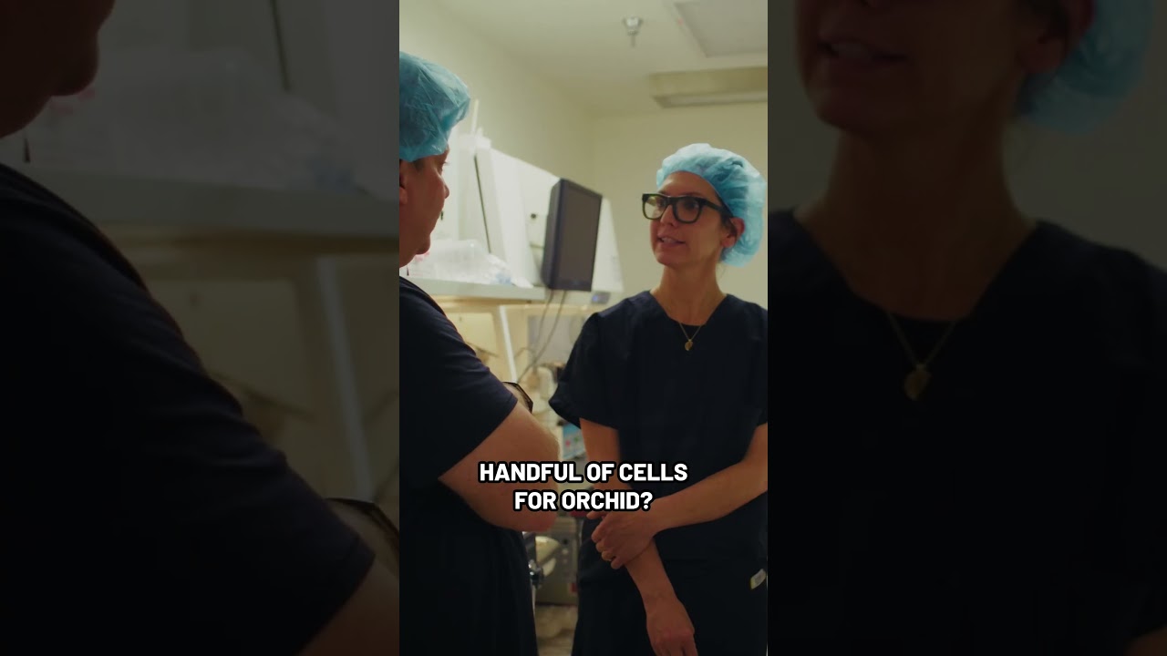
Where's my home?" So we're ready to biopsy it. This is where you would grab a handful of cells for ORCID?
Yes.
Okay.
It's from these cells that ORCID will sequence the DNA.
I break a little hole in the zona and enter in with my biopsy pipette. I have a laser and I'm hitting it a few times I'll take one biopsy of approximately 5 cells, and I want them to kind of be intact and not lysed so that it preserves the DNA quality. If I were to describe it in real life, it's almost like if I had a bubblegum balloon and I'm trying to take a piece off of it. It's very sticky. It's hard to— I can't just cut it. I see. And at that point, it's fully pulled away, and so I'm releasing it from the embryo. And embryos usually survive this process pretty well. And I have the biopsy piece that I'm now ready to send off to Oregon. What's crazy is meeting kids who I literally like froze, I thought, I picked out, I held in my hands, and now she's this amazing little girl who like is— it's, it's wild.