The Best Way To Test How Healthy Your Heart Is: 12-Lead EKG
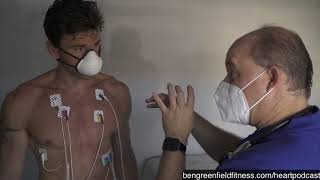
Dr. Dandelaya performs a 12-lead EKG on Ben Greenfield, explaining why 12 different angles of the heart matter for catching arrhythmias, chamber enlargement, and electrolyte problems. A resting EKG paired with a stress echo can detect coronary artery disease through changes in heart wall motion.
Chapters
What are we doing here, Doc?
All right, this is one of our most fundamental tests in cardiology. We're doing a 12 lead electrocardiogram. So Brenda is putting these leads on. So we're going to be recording electrical signals from your heart.
So this is essentially like a resting ekg.
Resting ekg. So we can ascertain your heart rhythm. We can see if there's any kind of athletic heart pattern, chamber enlargement and so forth. So it's just, it's our fundamental test. So we'll get.
This is the one that lets you look at the QRS wave.
Qrs, huh?
That rhythm of the heart.
That's right. We'll look at the QRS waveform and the other waveforms. So.
Absolutely.
Now explain to people who might be wondering why you can't just put an electrode, say, over the heart for this, why the electrodes go in so many locations.
Well, we're looking at vectors. So think about when we talk about a 12 lead EKG, we're looking at the heart from 12 different angles. So we can localize certain disease patterns. As you probably know, there are some consumer devices that are really pretty cool that you can record your own heart rhythm at home. And that attaches to a smart device.
So.
So those are good for.
I use one for that to measure heart rate variability called nature beat.
Yeah.
And so those are good for rhythm analysis. This is good for not only rhythm analysis, but really looking at, again, the heart and all these different leads and localizing any other issues so it's a little bit more detailed.
What would be an example of an issue you might find on an ekg? Electrical abnormality, like an arrhythmia.
Yeah, arrhythmias, tachycardia. You know, in older folks, atrial fibrillation is a common thing that's picked up on an ekg. We could see left ventricular hypertrophy. We could see left atrial issues with the atria. As well. So different chamber abnormalities. Sometimes you can pick up electrolyte abnormalities also. So some really interesting things on the ekg. And then, of course, vagal tone. I suspect you'll have a low resting heart rate. So we'll kind of just see what your resting heart rate is on the ekg. So, yeah, we can pick up all of that.
Will this also pick up heart rate variability?
This one does not. Traditional 12 lead EKG does not.
But my sense on heart rate variability is that you can see that on some of the devices, the OURA rings and so forth, where you can get more of a consistent day to day measure of that. That's probably the most useful to develop a pattern. I think isolated measurements on that can be a little bit hard to interpret. So it hasn't quite made it into the traditional cardiology world. But I think it's going to be fascinating when we have wearables and sensors where we're just gathering data on a continuous basis for folks to be able to aggregate this data and see what this all kind of means.
I'm sorry, what's your date? 12, 20, 1981.
Can you cross your legs for me?
Great, thank you. So as predicted, heart rate of 42. So high vagal tone pretty impressive and otherwise looks like a good ekg. No major red flags here. So we'll review this a little bit more in detail. But this is overall good. So is your resting heart rate usually detracts that?
Yeah, usually if I'm asleep, I'm kind of upper 30s, low 40s.
Yeah, fantastic.
Excellent. So this part looks good.
Depends what I'm up to during the day. You know, whether my mother in law is visiting or not.
Hopefully she doesn't listen to this podcast now.
Okay, excellent.
All right.
All right, so resting heart rate 42.
So that was accurate.
Yeah, yeah.
So we're good.
So all right, on the home stretch here, I think we're gonna get you on the treadmill.
Home stretch. Yeah, perfect.
All right, so, yes, so we've hooked you up like a standard resting electrocardiogram. Again, as we discussed earlier, a little bit different electrodes because again, this is a stress. So you're expecting to be bouncing on the treadmill, exercising, sweating, et cetera. So the electrodes have a little bit bigger surface area and are designed to stay on you during those phases of your test. And then what's cool is we will then use the ultrasound machine again in conjunction with the treadmill. So what we do is we take pictures of your heart at rest. So we look at the regional wall motion. So the heart should look like a bellow. So the heart is going to contract like a bellow with a base that moves up. So that's essentially what the ventricle should look like. Well, each area is around that bellow, essentially are a segment and our coronary arteries feed various segments of those walls. So we do the resting images of the heart. Then we bring you over to the treadmill and exercise. You get that heart rate up. Now what's different from a traditional treadmill is normally, of course, we're at the gym. As soon as you're done. You generally cool down. We don't have a cool down stage. We go from maximum exercise back over to this table as quick as we can, back into our imaging position, and we take those pictures again. And that's so we can see that heart beating extremely fast and obviously looking for any type of blockages along those walls. So back into your Titanic pose. Okay.
That's actually one movie I never watched.
Left arm, left side. Well, that's why you don't know to come over on your side. The most famous scene in the movie is this pose right here. And I think she got a dime or a nickel for that many others.
I never watched Godfather. Oh, I get a hard time for people. I've just never been into movies.
Read a lot of books, though. That's my jam.
Well, that's good. You can get a lot more from a book in most cases than movies.
All right, let's see if we have.
The only time I watch a movie is if I'm stuck on an international flight.
So really, on this test, it's patient preparation. You spend that extra couple minutes getting the electrodes right and getting the right pictures.
It really goes such a long way to get a good quality study. So we don't always get great windows like yours, you know, to look into the heart. So we just have to do everything we can with the patient prep.
Makes sense.
It's going to be fascinating to compare all this, all this data we're getting.
Have it all in one place.
Now, in terms of this workup that we're doing, is it common for, you know, someone, whether it's. It's an executive or an athlete or someone like that, to come into your clinic and for you to do this battery of tests? Yeah, it's called it a full cardiac workup.
It's a full kind of cardiac preventative workup. And again, we're always looking at new technologies and tools to put into the. Into the mix. Part of it is that there's a lot of technology that just hasn't been quite validated with some of the data. But this is a good representative set of things that really does have good data. So we like to look at.
So if someone were interested in doing this same panel, they would just be able to contact Atelier Health and say.
They want the full cardiac workup, preventative cardiac workup.
We'll call it the Ben Greenfield special.
That's right.
Then people will get competitive. They'll start to see if they can beat our results.
See who's got the biggest Heart.
Good.
There we go.
Reading an article the other day about how broken heart syndrome is becoming increasingly common in an era where loneliness and depression is increasing dramatically. But this idea of. Of impaired cardiovascular function due to the emotional and stress component.
Fascinating. Yeah, yeah.
It is fascinating that you can actually have broken heart syndrome. Very similar to some of my experiences. To lose a loved one.
Yeah, yeah. This has.
Or be ditched by a loved one. But you can get the same thing through loneliness, through depression, through losing your job, like Covid.
Yes, you can. The emotional impact, the stress that's created during those types of events. You know, there's varying theories, but can really create a catecholamine. You know, flood the heart with catecholamines, and that can create a stress cardiomyopathy. I think the other term has been a takotsubo cardiomyopathy that they've called before. Yeah. So.
So here we're just getting some good baseline pictures and a couple different angles. This is called a peristernal lung axis view. And then Michael will switch over to a few other views as well.
So just like a film editor or capturing snapshots, and they loop. And the software that. On the machine and plus the software that Dr. Dandelaya uses post test to evaluate all your images, it puts them side by side so you can actually compare the walls as we had discussed earlier. Go ahead and lean back a little bit more. Stop right there.
So these, Michael, are obviously terrific images. So we have a really clear view of all the segments, and we're going to compare these resting to the. What we call the post stress images. And we can see if there's any differences or abnormalities. And if there's coronary artery disease, there would be a segmental wall motion abnormality on stress. So that's the rationale behind doing this. Perfect.
Okay, so we're ready to go ahead and transition to the walking portion of this test.
All right.
More from Ben


The Solution to Aging Might Be Closer Than Anyone Realizes - With Ben Goertzel