Breakthroughs in Proteomics & Single Molecule Protein Sequencing
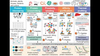
The TDCC/NHGRI genome technology development webinar opened with Mark Adams introducing the series and emphasizing the growing importance of single‑molecule protein sequencing as a complement to DNA/RNA sequencing. Giovanni Maglia (University of Groningen) provided a field overview, highlighting the limitations of traditional antibody/aptamer assays and mass‑spectrometry, and then surveyed commercial‑near‑market single‑molecule approaches: a quantum‑SI device (CLIP‑S‑based N‑terminal recognition), Encodia’s DNA‑barcode ligation strategy, and Nautilus Biotech’s affinity‑sequencing platform that immobilizes full‑length proteins on DNA‑scaffolded microchips. He stressed the need for intact‑protein analysis to capture post‑translational modifications (PTMs) and proteoform diversity. Jeff Duvall (University of Washington) shifted to nanopore‑based sequencing. He explained nanopore sensor fundamentals, the challenge of protein charge heterogeneity and folding, and described four translocation strategies: (1) charged‑tether electrophoretic capture, (2) chemical denaturation with guanidinium chloride, (3) DNA‑motor‑driven peptide ratcheting (showing single‑amino‑acid resolution and rereading), and (4) protein unfoldase motors (ClpXP, proteasome‑nanopore fusions) that can pull or chop proteins. He highlighted recent results achieving single‑amino‑acid sensitivity, the importance of cis‑side motor integration for compatibility with ONT’s MinION, and the critical role of deep‑learning base‑calling to translate noisy ionic‑current squiggles into sequences. Jag Swaminathan (Arizon) introduced the FLORA flow‑cell fluorescence sequencing platform. The method extracts proteins, digests to peptides, chemically labels selected residues (Lys, Cys, Trp, Tyr, phospho‑Ser/Thr) with distinct fluorophores, immobilizes millions of peptides on a glass slide, and performs iterative Edman degradation cycles. The resulting multi‑channel intensity patterns (“floral sequences”) are matched to a reference database using machine‑learning classifiers, yielding peptide identity, PTM position, and absolute counts. He presented two high‑impact applications: (a) ultra‑sensitive detection of HLA‑presented neo‑antigen peptides for cancer vaccine design, and (b) quantitative mapping of phosphorylation stoichiometry across proteoforms. He also discussed overarching proteomics challenges—dynamic range (10¹¹–10¹² molecules per blood sample), solubility losses, and quantitative range—positioning single‑molecule technologies as potential solutions. The session concluded with audience questions about Edman‑cycle limits, photobleaching, chip capacity (~250 M spots), disulfide reduction, amino‑acid discrimination, and motor stepping precision. The panelists emphasized that software advances (ML‑based signal decoding) and biochemical engineering (labeling, motor design) will jointly drive the field toward commercial protein sequencers, likely leveraging existing nanopore hardware platforms. Overall, the webinar mapped the current landscape of single‑molecule proteomics, compared competing technological paradigms, identified key performance metrics, and outlined the scientific and clinical opportunities that will motivate rapid commercialization.
Chapters
Good morning, everyone, or afternoon, as the case may be, and welcome to the second in a series of genome technology development webinars sponsored by the TDCC. I'm Mark Adams. I'm the principal investigator of this grant that is issued from NHGRI. We are a coordinating center that works with genome technology development programs across NHGRI, including sequencing technology development, synthesis technology development, and a broad range of genomic technologies related to understanding the function, structure, and activity of the genome at large scale. We work with over 65 funded programs to enhance coordination and communication, collaboration, and primarily outreach, disseminating the advances from these PIs and their work to the broader scientific community. And today's event is part of that. You can learn more about our group at genometdcc.org. Many of you will have signed up for the whole series. We have 4 events this May. The next 2 next week are going to be on single-cell analyses and genome scale regulatory analysis. If you're interested in those and haven't signed up, please visit our website, genomtedcc.org. With that by way of introduction, today we have a terrific group of speakers in the area of single molecule protein sequencing. This is an area that has really come on very strong in terms of a research area in the last few years. I remember when I was in grad school, protein analysis technologies were way ahead of DNA analysis technologies. I think that's coming back into the fore where it's really important if you want to understand the function of a cell to look at its proteins, not just its DNA or its RNA. So let's get started. Our first speaker is Giovanni Maglia. He's professor at the University of Groningen Biomolecular Sciences and Biotechnology Institute. As a postdoc, he worked on development of what became the Oxford Nanopore Sequencer. And his group is focused on a wide range of applications of nanopores as sensors and activities for understanding biology. Giovanni, take it away.
All right. Thank you very much for the introduction. So I'm very glad to be here to give this web seminar. So I'm going to talk about single-molecule protein sequencing, but I've been asked to give an introduction on the field. And then the focus of the talk will be to give like a brief introduction about the importance of the field, and then focus on the few technologies that are actually already on the market or very close to, to be really part of the markets, and they can already sequence protein at the single molecule level. So it is actually little doubt, and that really probably should not tell to this audience, that the second and third generation or next generation sequencing really has revolutionized the way we see biology. So thanks to this technology, which you can see there here on the left, the cost for sequencing the human genome is dropping in a sort of dramatic sort of way, actually allowed to do certain experiments and understand biology in a way that is really truly revolutionizing. So now the focus is on proteins. And then the reason for proteins is because while DNA is actually a very important molecule, it's really not what is the reality of what we are at the moment right now. So that actually locked into the protein and understanding the protein and the proteome or at the same level that we understand actually the genome, will bring an equal revolution in biology and in medicine. Just to give you a hint, we're going to have about 90% of the drug targets nowadays are indeed proteins. So protein is great. We know a lot about proteins, but actually the proteome is incredibly complex. In fact, we don't really understand that much about the proteome. If you read, you know, reviews and etc., there are even people that think that about 90% of the proteome we don't really see, and then this can be called the dark proteome. So it really depends who you're talking to. But it is actually quite clear that we don't really know the entirety of the proteome. And that's kind of like important because many diseases that we have, they really require understanding the full sample of proteins that is made in cells. One of the reasons why we don't really know much about the proteome is because there is an incredible diversity of proteins. So not only on the shapes and the size of the protein that's diversity, but also on chemical composition. Just to give you an example from one molecule of DNA that we need to sequence for having the genome and perhaps a few epigenetic modifications, we have about 20,000 genes, which the diversity of this gene is amplified by the fact that mRNA can be spliced and can be alternative way that we assemble this part of the genes. But really the major variety comes from the fact that we have post-translational modifications, which creates some say 1 million, some say 5 million different proteins in our proteome. Actually try to understand these proteins is really important to understand who we are, how we're made, and actually all the pathologies that are related to the malfunction of these proteins. Right. So the way that we read proteins nowadays is really using two main techniques. There are other ways, but these are the two main techniques. The one on the left is by, if you know what kind of protein you are after, then there are many kind of antibodies or aptamers or in general binders that you can use. And then you can actually make these assays, which Right now you actually can measure up to almost a million proteins in one, in one go, in which you can actually use in a fluorescent readout, you can tell you what kind of protein you have in a certain sample. You can go in, in real samples, so that's very good. You can almost do quantification. It actually depends who you're talking to. Again, it looks like that's different methods actually quantify protein in different ways, so there's some work to do there, but it's very cheap, it's pretty good. The real problem they have in this method is that you need to know what kind of protein you need to look after. The post-translational modifications are difficult to look. You need to have a binder for the specific modification and really try to understand the nitty-gritty details between, difference between different proteins is really actually quite hard. By far the tool that is used most prominently for de novo sequencing of proteins or discovery of proteins is mass spectrometry. It is an extremely complicated machine. It's been been in development in the last 20+ years for protein sequencing and analysis. And now you can measure about thousands of proteins in one go. It's pretty good, but the sample preparation is extremely difficult. It requires a lot of very skilled people to do that. Not all proteins are detected. Here I put 8% of the protein from blood and 30% from cells. These are numbers that I get from you know, next-generation company that want to displace mass spectrometry. So take it with a pinch of salt. But, you know, it's fair to say that not all the protein can be detected. And the reason for that is because you need to first fragment proteins into peptides. The peptide need to be kind of separated with many different techniques. And a lot of them, they co-elute together. And when you actually go to analyze, because it's an ensemble technique, you actually measure only the most abundant peptide that is co-eluted. At the same time, there's some peptides that don't fly very well in mass spec. PTMs are not very easy to measure and so on and so forth. There are many kinds of difficulties that when you use mass spec, but although it works very well and pretty much most of the information that we have now is from mass spec. One aspect that I really would like to bring your attention is that mass spectrometry needs to break the protein into peptides to be able to sequence them. And, and that actually loses some information. For example, here, if you have a phosphorylation of a protein in 3 different parts, and some that are actually not phosphorylated, it's really not possible to distinguish if you have these kind of things on the left, or actually you have a different kind of combination phosphorylation if you cannot have intact protein modification. Another example here is the tau proteins that can actually be spliced in many different ways, at least 6 are known, and then you have more than 70 different phosphorylation sites in this tau protein, and many other can be phosphorylated and have different PTMs. And it's extremely difficult to actually measure this, and all these kind of PTMs, modification, and splicing are related to disease. So it would be very important and very useful if you could have a methodology that can actually sequence and identify protein as intact proteins. The way that I can explain about these single molecule techniques is that the need that you need to, you know, like that we need to have a single— why do we need to have a single molecule is because mainly protein cannot really be amplified. Like you do with DNA. So you really need to go at the single molecule level if you want to identify the heterogeneity that you have in a sample. All these techniques, they must be massively parallel because there's a huge different range of concentration of proteins. So you need to really measure 1 million or plus kind of these kind of proteins, and better if it's fast. You can do de novo sequencing. That's what the ideal situation is, but also many techniques just are happy to do ID fingerprinting of these proteins. As explained before, it would be great if you can work with intact proteins, but also even fragments would be good because actually can give you all the diversity and heterogeneity that mass spectrometry at the moment cannot really give you. So in the way that actually to divide these different technologies that are popping out more and more these days, there are many different ways that you can categorize generalize these kind of proteins. One way to do it is by the way that actually address single amino acids. So either you, you need to degrade one amino acid at a time and then address the last amino acid that you're actually cutting off, and that's mainly the technology that are more promising and let's say closer to the market. Or actually you pass one of these molecules across the nanopore and then you actually measure these amino acids as they pass a specific part in the nanopore. You can also divide them by the readouts, can be fluorescence, that's the kind of the result of choice for many of these applications, but could also be next-generation sequencing, and give an example, or nanopore current. Perhaps the techniques that is more close to the market, or one that is actually in fact you can already buy right now, is a quantum SI experiment device. You can see here an example of that is this kind of, this kind of like device is basically very similar to what is already in the market for PacificBio, for what I can understand. I don't really want to kind of say something that's not correct, but for what you can see from the paper that they published, You have waveguides, which is similar to a zero-mode waveguide Pacific Biosciences has. You have semiconductor chips with the camera already integrated into the device. And the only difference that I can see is that, like, the substrate here, which is a peptide, is immobilized, must be immobilized by the C-term. And you don't really have an enzymatic kind of like amplification like in Pacific Biosciences, but you actually have the recognition of the N-terminus by protein that actually recognize the N-terminus. The device is already in the market, I was saying, for less than $1,000, and it can provide a massive readout of millions of peptides at the same time. So the recognition happens with proteins that actually recognize the N-terminus. This protein is called CLIP-S. So the description they give in their science paper describe, you know, about 1, 2, 3, 4, 5, 6, 7 kind of amino acids that can be recognized. The recognition happens either because this CLIPase can recognize one individual amino acid, or can recognize a family of amino acids, like in this case leucine, isoleucine, and valine. And the difference between similar amino acids is actually not only in the color, which is attached to the specific enzymes that measure the N-terminal amino acid, but also by the duration of the poles. So if you have slightly different energy in the binding of the N-terminus, then you're going to have a slightly different time that you stay bound. Then you come to one aminopeptidase, which is an enzyme that cuts the last amino acid, and then you have this, you can access the next amino acid. The reading time is actually quite long, is about 8 hours. And also here you can see that you can only see certain amino acids. What is blank here is all the ones that you can see. In the, in a deck that they, they sent for, that you can find on the internet, you can see an update of different families of binders. So now they have about 15 different types of amino acids that can be recognized. That's what they claim. And then you can actually getting closer and closer to actually having a full sequencing of a peptide at a single molecule level. There is another company called Encodia, which for what I can see, they are in stealth mode, so it's very difficult to get information. But for what I can see, it does exactly the same thing, immobilizes the C-terminus. But here there is a piece of DNA that is used as a barcode, and then you, as there is recognized one amino acid, the terminal, you actually ligate the equivalent piece of DNA to the barcode DNA. Then you're going to have a sequence of DNA here, which is elongated, and then you send it for next-generation sequencing, and then you get the sequence of the protein. The advantage is that it's simpler. There is no optoelectronics that is needed and no dyes. That's the main kind of problems that I can see is could be the immobilization and slow runtime, but they probably can be solved. Full-length protein probably cannot be solved because it only can work with peptide. The structure is going to be a problem. They enter amino acid properly there. It's not clear if they all can be identified. And also, if you can have, you know, only one amino acid is identified rather than a group of amino acids that looks like it's the same, at least the last two are identified. And it's not clear if you have enough colour for the fluorescence to do real-time analysis of all of that. There are many kinds of unknown yet, but it's quite impressive. I I might say that you can already sequence about 15 amino acids. For technology that can address full proteins, I can select here something that has been described by Nautilus Biotech. They use also affinity sequencing of antibody adaptamer to actually sequence, or actually better say identify, a full-length protein. They have a fluorescent readout, and they again, they have a massively parallel readout that they can have. The way they immobilize is to take a protein, immobilize it through chemistry into a DNA scaffold, and this DNA scaffold is actually lodged into very well-defined spaces into a microchip. And here they claim to have about 10 billion molecules that can be immobilized and addressed individually. That works again, not like Pacific Biosciences, but as a second-generation sequencing technique in which you can actually send one reagent at a time, wash and send, wash and send. And these reagents are antibodies or aptamers that can bind and recognize a very small, short sequence, amino acid sequence in the full-length protein. You do hundreds of different iterative cycles of binding and washing, and eventually you feed it to a machine learning algorithm. And then at the end, it gives you the identity of this molecule. So the advantage here, you don't actually cut amino acid by amino acid, you recognize intact proteins. So you can address the entire molecule. What they claim is that with 200 or 300 cycles, you have about 90% cover of the whole proteasome. In theory, you can actually identify new proteins that have not been characterized before. In practice, their recognition is only 20%. So if you have every epitope that is recognized, if it's present, only 1 in 5 is recognized because structure most likely have a part of that. But because you have so many cycles of, of, of washing and recognition, then you can end up to actually recognize the full protein. The challenge for this technique probably is that the neighbor residues are going to be influencing the binding. Structure is probably a factor that is there you cannot really remove, and the reagents are a little bit expensive, and the runtime is still 1 day, but probably because you have billions at one time, that's still probably a minor problem. Now I leave the baton to Jeff that's going to talk about nanopore sequencing and how you can sequence protein with nanopores. And thank you very much for your attention.
Wonderful. Thank you very much, Giovanni. So our next speaker is Jeff Duvalla, who's a principal investigator at the Molecular Information Systems Lab of the Allen School of Computer Science and Engineering at University of Washington. Jeff got his PhD as part of one of the biggest Nanopore groups at UC Santa Cruz and then did a postdoc with George Church before joining there. Jeff, welcome.
Thanks, Mark. Thanks for the opportunity. Hello, everyone. Nice to be here today. So as Mark said, I've been working on protein sequencing since I was in grad school, actually, and off and on since then. And so I lead a group right now where we have a big focus is on single molecule protein sequencing with nanopores. And so my goal with the talk today is really to not focus just on the work that my lab is doing, but actually try to give you a general overview of sort of the state of the art in nanopore approaches to single molecule protein sequencing. And in only 15 minutes, this will be incomplete. So I'll just try to give you a flavor of different approaches that people are using to solve these challenges. So Giovanni just gave a great motivation as far as why do we need sort of, you know, think of them as next-generation proteomics technologies, orders of magnitude more complexity at the proteome compared to the genome. Yet the tools that we have to analyze these, it makes it much more challenging. And so I see it sort of twofold where single-molecule proteomics can have impacts. It's the ability to read and sequence proteins, not just sort of at the fundamental proteomic side where, you know, you can actually make— derive new biological insights as Giovanni was talking about, but also on just sort of general technology side where next-gen sequencing is used as a readout for all sorts of molecular assays. As a way to— for different molecular counting applications. And I think if we can extend that to the protein level, we can open up a new swath of different techniques to do protein-based counting to answer all sorts of questions. And so I'll focus here, of course, now on how are we going to develop nanopore sensors for this sort of next-generation proteomics sequencing. So Nanopore Sensors 101 for folks that are not familiar, how does the nanopore sensor work? So I'm going to be focusing mainly on protein-based pores here, which has shown the most promise for actual sequencing. And those are what's commercialized in DNA and RNA sequencing devices right now. But they're also another class of solid-state pores. I'm showing you here a protein pore. And the way this works as a sensor, essentially, you have your pore embedded within an insulating membrane. You can apply a voltage across that pore. And that drives ionic current flow through that nanopore. And that's what generates your signal. So you can see here, your signal— these are nanometer-sized holes, so you're driving— very, very minute amounts of current measured typically in the picoamperes. And you're measuring this over time. And the way that this works as a sensor is when a molecule actually flows through that— a large macromolecule actually flows through that pore, it blocks or occludes some of that current, and essentially that generates your signal. Okay? And you also— people in the field also like to maybe call this a squiggle, is the way we like to refer to this. This type of blockade here. And very early on, this was conceived of as a potential way to do DNA sequencing where if you could thread a single-stranded piece of DNA through that pore, potentially each base could occlude or block the current in a way that would allow you to translate its sequence of bases as that strand moves through the pore, map basically that signal into the sequence of the nucleotides moving. Through the pore. And so very early on, it was discovered that if you're just relying on electrophoresis, right? So one of the nice things about DNA and using nanopores and having this electric field being part of your sensors that you have some ability to exert force on charged molecules. So DNA being having that nice net negative charge along the backbone, you're able to electrophoretically force it through a nanopore just using that electric field. And what you find though, is if this is not a good approach to actually sequencing a strand of DNA because it moves through the pore much too quickly. So showing you down this little plot here, each of these little blips is a long piece of DNA actually moving through the pore, and you really don't have enough resolution in that signal to actually sequence that strand. So the big breakthrough came about a little over a decade ago. Sort of the secret sauce here is having a motor that can actually control the translocation of that DNA strand through the pore. Let's see, by using a helicase or polymerase that can ratchet it through the pore base by base, controlling the movement, and so using that controlled movement with a sensitive pore, that's a pore that has the right structural dimensions such that it makes it sensitive to single nucleotides along that strand. Putting those two things together made the secret sauce that actually was the first demonstration of single molecule DNA sequencing. Using a nanopore, and it came about this, this collaboration for Mark Aikison's lab and Jens Gundlach's lab, where Mark had the, the motor and Jens had the nice pore. And so it was a good collaboration to, to bring this together. And essentially now this is a version of this, is, has been commercialized now in, in by Oxford Nanopore Technologies, where they have these MinION, they're called MinION devices, which essentially is an array now of hundreds of nanopores that can operating in parallel. And the big game changers here is that they deliver direct single molecule reads. So you're getting base modifications. They can read really long strands, millions of base pairs in a single read. They give you real-time data streaming and they're portable. And they're also scalable. So more field-based applications, you can have smaller devices with less throughput all the way up to more genomic size applications. So more like a benchtop type sequencer. And they've already led to a number of breakthroughs in completing the human genome, direct RNA sequencing, as well as some of the field-based surveillance and things of that nature. So it'd be great if we could extend some of these features for reading proteins, right? And so if you look at some of the challenges now of protein sequencing, so it's— the bottom line is really much more difficult for a number of reasons to to analyze proteins or to sequence proteins with nanopores compared to DNA and RNA. So the first thing we can look at is just the charge distribution. As I was mentioning, DNA and RNA have this nice net negative charge on the backbone, which gives you sort of a handle on the molecule just by using that electric field. Proteins, on the other hand, don't have this net charge along the backbone. So they have more heterogeneously charged. And so it's harder for you to sort of get a handle and thread them through the pore in the first place. And then they also have— Proteins are folded, so they have this tertiary structure that you need to somehow deal with to be able to thread them through the pore. Okay, so there's a number of different approaches that people have been looking at over the last decade or so of how do we get, how do we thread, linearly thread these proteins through the pore. So on the left is sort of this general class of using basically chemical methods similar to how you might thread DNA through the pore, which is if you can essentially attach the charge tether to one end of the protein, let's say an oligonucleotide or a charged peptide, it sort of gives you a handle to then be able to hopefully pull that whole molecule, then unfold it and pull it through the pore. So this is sort of this non-enzymatic control. And on the right side are showing you two different approaches where you're actually using molecular motors similar to how you might sequence DNA. We're able to use that molecular motor to actually ratchet strand— full-length proteins or peptides through the pore. So I'll walk you through some different examples of these approaches. So I'll start off here with this non-enzymatic control here. So this was first demonstrated about 10 years ago out of Hagen-Bailey's lab where they attached onto the N-terminal of the protein, they actually attached a DNA oligonucleotide, and what this allowed them to do is actually pull single protein molecules into a pore with electrophoretic force. Okay, and when this happens, basically the protein will become captured at the top of the pore, and then it will eventually unfold underneath that electrophoretic force, where essentially there's this— by mechanically pulling on the end of that protein. And what they're able to observe is is really interesting, basically, this co-translocational unfolding of the protein and observed different unfolding intermediates as that protein went through the pore. Okay. So it was really interesting way to sort of examine different unfolding states, but it really didn't give you much control of the translocation process or the speed. So as far as a sequencing approach, didn't quite hit the mark for them. More recent work, actually, just a few months ago out of Minnie Woon Yoon's lab is using a similar approach, where he's attaching a charged tether to the end of the protein. In this case, it's actually a charged peptide molecule. Okay. And then in the buffer, he's put guanidinium chloride, and it essentially gets rid of all the folded structure of the protein such that now you can electrophoretically thread that protein through the pore and actually get a full-length translocation of that protein through the pore without having to deal with any of that folded structure. And so what they're able to show is actually sort of you're getting a coarse-grained view of this protein as it moves through the pore. It's still going through very, very, very fast. So each of these little spikes, excuse me, in the signal here is actually hundreds of amino acids going through the pore at a time. And so this method was really good at sort of driving full-length translocation of the proteins through the pore, but it sort of lacks that sequence sensitivity to actually get towards a sequencing approach. But they were able to demonstrate sort of this coarse-grained view where you can get some idea of the shape of a protein that you have some ability to to identify or distinguish between different types of proteins. Okay. So moving now towards the enzymatic-based control. So getting, in my view, a little bit more closer to actually sequencing and some of the tools that we'll need to continue to develop to actually get to more single amino acid sensitivity. And so the first route here is actually using a DNA-based motor to pull peptides through a pore. Okay, and so this— so there's a series of papers that came out in 2021 from the Huang lab, Bei lab, and Kay Stecker's lab, all showing generally the same technique, where essentially what they did, they take peptides and they can conjugate a DNA oligo to them, and then they can use a DNA process of motor just like you would use in DNA sequencing, so either helicase or polymerase. And you can use that to ratchet both the DNA strand that the helicase or the polymerase is operating on. And as that is ratcheted through the pore, you're also going to pull along the peptide along with it. And using this approach, you're actually able to get single amino acid resolution in a very controllable way. So I'm showing you traces here. So this is the raw data here. And this sort of translated to a sequence map where you're first, the process starts out, you're actually sequencing that oligotether. Then eventually, that peptide will come up through the pore. And your current changes are actually now dependent upon the sequence of that peptide. And they're able to show, at least in the Decker paper, they're able to distinguish with high accuracy between a small subset of different amino acids. And I think the really cool aspect of this that was in the Brinkerhoff And Decker paper was actually able to show rereading the same molecule many times because as that helicase is ratcheting along that oligo, when it reaches that peptide, it'll slip off. And you can have another motor loaded, already loaded on the oligo. And so essentially, your read will slip back to the previous position where that other helicase is bound. And doing this, you can actually reread the same molecule many, many times and really increase your single molecule sequencing accuracy, which I think will be— rereading will be really key for nanopores. Nanopore-based single molecule protein sequencing. And so some of the limitations to this is that we're not dealing with full-length proteins. So we're still reading peptides here. And so basically, your read length is limited between the distance between that nanopore sensing constriction and the top of the pore. So in their case, it's around 20 amino acids. And the sequences that they showed that this worked for were highly negatively charged. So really amenable to sequencing in this method. So it remains to be seen how well this will generalize to neutral or positively charged peptides, but a super, super promising start here. And actually, last summer, Oxford Nanopore showed some data at their conference where they're actually exploring a similar approach to peptide sequencing using a DNA motor as well. So I think that's going to be a really, really active area for commercialization fairly soon. Okay, so that's what the enzymatic or DNA-based motors. And so now we'll get to my favorite form of approach to nanopore protein sequencing, and that's using protein processive motors or specifically unfoldase motors to pull proteins through the pore. Okay, so what is, what's an unfoldase? So essentially, an unfoldase is an ATP-powered protein unfolding machine. So there's basically, they function in the cell to grab onto protein substrates, unfold them, and then degrade them. So involved in protein turnover. And so ones from E. coli are a common one is ClpXP. Eukaryotes have a version called the 26S proteasome. And so the question is, how do you sort of couple this unfoldase activity to a nanopore sensor? And so there are actually various ways you could do that. So I'm showing you work here from actually from Giovanni's lab, where he made this, engineered this proteasome-nanopore fusion, where you actually take this protein, protein nanopore, and you make a fusion protein such that you put a binding site for the proteasome on top of it. And what that allows you to do is actually assemble this whole proteasome complex and the unfoldase on top of the pore, as it's shown here. And what you're able to do then is capture proteins in solution and then push them down into the pore for analysis and actually do this in two modes. So if you have this, the proteolytic activity of the protein is knocked out, you actually thread and read entire full-length proteins, or you can have that protease activity turned on and you actually end up what they call chop and drop, where you're actually chopping up the proteins as it's— before they're actually threaded down into the pores, you actually start to get reading individual peptides using that method. So really, really cool engineering feat here. And it sort of remains to be seen the ability to distinguish between different proteins using this method and, you know, how single— single amino acid level sensitivity will be achieved using this, but a really interesting approach to coupling this unfoldase activity to the pore. So when I was in grad school, I developed this alternative approach, where you actually have these unfoldase motors in the transside solution, and then you initially thread a protein substrate through the pore using that charged tether, but it gets stuck until that, that that protease, that unfoldase that's present in the trans solution is actually able to grab onto it and then mechanically pull that protein through the pore, as shown in this, this little video here. And what we're able to show, we weren't using a very sensitive pore, but what we're able to show is sort of a coarse-grained sequence-level view that could allow us to discriminate against some subset of proteins. And also using this method, you get some ability to get information built both about the stability because of how long it takes to unfold, as well as the size of different protein domains based on how long it takes to translocate. And this is sort of a method that we've picked up now in my own lab, but we sort of adapt it to— now the motor is not in the trans side, it's actually in the cis side. Okay, so you initially thread the protein through the pore using that charged tether, it gets stuck because we have this blocking domain, and then we load the unfoldase in and it pulls that protein back out of the pore. Okay, and what this is nice, it's completely cis-side only, so this makes it compatible with the commercialized nanopore devices like Oxford Nanopore's MinION array that we're using to develop this approach. And what we've been able to show is actually a single amino acid level sensitivity, though albeit in sort of this synthetic, synthetic context right now. But what it's allowing us to do is sort of get a handle on sensitivity and, and how different amino acids contribute to our protein-based signals. Okay, so this is really exciting work that we're really pushing on right now, and so there's a lot of exciting results that will be coming on that, so stay tuned. Okay, and finally, I'll just wrap up here. I didn't mention anything about signal analysis, right? I'm just really focused on sort of the mechanics of how do you get a protein through the pore. So One of the main challenges in sequencing is how do you sort of translate this noisy signal and actually sequences of bases. And so early DNA sequencing is using, you know, hidden Markov models and lookup tables where now state-of-the-art base callers are really using these machine learning approaches. And I think these deep learning, same deep learning approaches will really be key to protein sequencing as well. But first, we really need a robust reproducible system that works on a wide variety of protein and peptide sequences. And if you can get data at a high enough throughput, I think this ML magic will be able to do some useful decoding there. But as Giovanni said, protein sequencing, you know, can be reduced to a lookup problem. So I think that will be much— will come much sooner. We're essentially using a protein fingerprinting approach for nanopore-based sequencing. And I think even sequencing synthetic proteins will have their applications, as I said, for things like, like barcoding. And with that, I'd like to, yeah, just acknowledge all the folks in my lab and thank Mark again for the opportunity.
Great. Thanks a lot, Jeff. Our next speaker is Jag Swaminathan from Arizion, which Giovanni alluded to. He's the founder and chief technology officer. Before that, he was a PhD and postdoc at UT Austin. Going to talk about a different technology for single molecule protein sequencing. Jag, it's all over to you.
Yeah, thank you, Mark, and the organization committee for the kind invite to speak at the TDCC workshop seminar series. I'm really excited about the field. As you can say, I've been at this field for about a decade, starting with my PhD, and now I'm leading the effort at Erisian to commercialize the technology. You guys have heard about all these new emerging fields from the previous two speakers and and the incredible challenges that they had to encounter. I've been given a little mandate in this talk to try to peel back the layers and ask, what is the single molecule technologies really going to be good for? Like, as you all know, invention— necessity is the mother of all inventions. So I want to look at it and make you guys also reflect on, like, what this technologies could be helpful useful for. So I'm just outlining this talk. I'll be going over a few thoughts on how to think about the applications, and then I will speak to two of the very interesting applications that we are working on. And to do that, I have to give you a short primer on how our technology works, and then finally I'll summarize with what I think the next needs for the field are. So, but before I do that, I just want to acknowledge every single person in our team, both at UT and Erision. We were founded in March of 2018, and we are commercializing the instrumentation, and we want to have a— set the field up for very new and exciting applications. So I don't have to tell you guys why proteomics is important, but I just want you to reflect on the fact that the central question in most proteomics research is the following: which proteins are present in my sample, and at what counts are they present, or what is the concentration at which it is present? These are information that, as you can think about, cannot be got from DNA sequencing technology. So you cannot identify post-translation modifications that appear in disease samples, or a very sensitive detection of antigenic peptides for cancer therapy. The— before I launch into the different applications, there is one thing that I want you all to keep in mind when you think about why proteomics is very much challenging than DNA sequencing, and I kind of like laid out a little comparison, and you can see that we unfortunately don't have polymerases. There are plenty of diversity of the amino acids and hundreds of post-translation modifications that happen. There are two critical aspects of why proteomics remain to be a very challenging area of research, and that is due to the vagaries of solubility of the proteins. They have littered with different types of charged molecules. They have differences in solubility in different solvents, and you tend to end up with loss of samples as you transfer them from your tissue to your instrument. And dealing with that is really a big problem in proteomic research. The second biggest challenge is the wide dynamic range that we have to deal with when compared to the RNA sequencing. A single blood sample could have proteins ranging in abundances from across 10 to the power of 11 to 10 to the power of 12 molecules, and even a single cell is estimated to have a billion order of magnitude of protein concentration differences. What I— so what I tried to frame this application space is to break up the technology features into 3 different components. One is the sensitivity, that is, how many molecules of the proteins can you identify? The second is the coverage question. In my sample, what is the number of proteins that I can identify and like different types of proteins? So in a human sample, you are estimated to have at least 20,000 proteins, and can you identify them all? And the third, and the third dimension, which is sometimes not well understood is what I call as the quantitative range. So what this is, is it's a— it is the estimation of the dynamic range of a proteoform. So people also call these as stoichiometry of modification. So for every 100 proteins, how many of my proteins was modified with a phosphorylation? So this kind of stoichiometry is what is dynamic and and oftentimes a disease indicator. Layering on top— and you can see from this chart that mass spectrometry does a fantastic job of covering a large swath of features.
It is—
it can, as was alluded, there are studies that say we can get to lower femtomole to upper attomole levels of sensitivity. And you can detect more than 10,000 proteins from wide fractionations. But then there is a limitation in terms of quantitation. So you find it difficult to measure the percentage of phosphorylated residue on your favorite protein of interest to the total proteins that is present. So as you can see, there is a large swath of space to play with. And my speculation is that a lot of these technologies, like the full-length nanopore technologies that was described earlier, or the nautilus approaches, or the shotgun methods, they have a place in this application. And we might find different technologies applying different applications. You can think about some of the interesting ones, like the single-cell proteomics, which has been big challenge for the mass spectrometry field. And just like how single-cell RNA sequencing revolutionized a lot of medical research, we can think about single-cell proteomics doing a similar job. Purity of biologics is one other thought that I had, which is that if you have a glycosylated antibody that you have produced, how pure is it in your sample? And now we're not talking about impurities from other proteins. We're actually talking whether the exact same patterns of glycosylation was found in every single biological molecule that you're going to inject into a patient sample. So these are the kind of questions that are currently not tenable to be answered by any of the antibody-based methods or mass spectrometry. I will be talking about two methods that we have focused on in our work, which is the detection of antigenic or the HLA peptides from cells and the determination of mapping the sites of phosphorylation and the stoichiometry of them. So before I give you that, I want to give you a quick, very quick preview of what flow sequencing technology is so that you can look at the data in a different light. Our central concept is that if you know the positional information of a select few amino acid residues, that is sufficient to infer who the protein is. So in this case, if you know the database, which is seldom overdetermined by the genomic and the transcriptomic information— so as it was alluded to before, oftentimes it's just a lookup problem. And it turns out that 4 different amino acid types and knowing the position of the protease is often sufficient to make an inference of who the protein is. What we have done is we assembled a technology to piece— to get this positional information together. And so what I'm going to show you is a short video of us talking of the description of the technology. So we take cells, we take the proteins, extract out the proteins, digest them into peptides, just like how you would do in a traditional mass spectrometer. Experiment. These peptides are then selectively labeled, so the amino acid side chains go through a covalent chemistry of attaching fluorophores to it. And since there is a lot of work that has been done in bioconjugation methods, we have distinct chemistries to label lysines, cysteines, tryptophan, tyrosines, and a number of different amino acids. We immobilize these peptides on the glass slide, and now we end up with millions of these individual spots, fluorescent spots. They look like stars on a dark night. And we take measurements of intensities of these peptides across all the different channels. We then perform Edman degradation chemistry, which is a classic chemistry of the 1950s where you cut one amino acid at a time from the N-terminus, leaving the rest of the peptide back bone intact. So what comes out is that every single spot on this billions of peptide molecules gets a partial sequence of who the position of the different amino acids are. And if there is no change, that is marked as an X, and we move on. And this pattern, which we call the floral sequence, is used to match to a reference database to infer who it is. We can do similar modifications of these fluorophores but we can also modify the side chains of post-translational modified residues. So in this case, we teach how to modify the phosphorylated residues and attach fluorophore on the sites of phosphorylated serine/threonine. And what you can see here are the counts of molecules with the phosphorylated signal at the distinct position where it was labeled. So this gives us a— 2 important types of information. One is the position of the modification, and second, the concentration at which this modification occurred in the sample. A very quick overview of how we look at the data. In this example, we have a peptide, a full peptide mixture labeled with different types of fluorophores, 2 different types of fluorophores labeling the lysines and the cysteines, and then we perform FLORA sequencing experiment.. And here you see some of the raw data where you see drops of intensities corresponding to the different channels at which it— at which the fluorophores were labeled. And this gives us a pattern which is completely different from the next spot on our flow cell. What comes out from this and applying a number of machine learning algorithms to solve our image correction and image analysis and a protein inference question, we end up with a machine learning algorithm that can score our floral sequencing reads to the different peptides. And here you see a classic UMAP plot which distinguishes the 4 different peptides that we had in our sample. Now I want to shift gears. Now that you have a flavor for our technology, I want to shift gears to talk about two applications that I alluded to before. So the first is the neoantigen. You may have seen a recent article from the New York Times which talked about this incredible clinical therapy for prostate cancer where spiking the vaccine, a cancer vaccine, actually helped clear out this tumor. So there's a lot of effort in this field to develop these cancer vaccines, where knowing a few peptides on the surface of proteins is sufficient to make an inference of who they— of determining these cancer vaccines. Turns out that mass spectrometry is extremely challenging to get to the sensitivity, and so we are left with the DNA sequencing information where we predict what are the HLA peptides, that are expressed on the cells of these proteins, and we make an estimation from that. What we did is we took this information of the genomic predictive power and layered on top of it with mass spectrometry by characterizing a monocellular allelic cell line, B-cell line, which produced these HLA peptides, and it turns out that the— there is very small overlap between what is predicted by DNA and what comes out from actual detection of peptides. So there's a huge gap to be solved. We applied this to our method and spiked and wanted to determine the recovery of these peptides in the complex mixture, and it turns out that the sensitivity is where we really can address, and this is where I think one of the one of the applications of our field could be. And now we could layer this top on top of DNA information and we have more confirmation of the target antigenic peptides. All right, one more minute. I just want to quickly point out that the phosphorylations is always a challenge. It has always plagued the field of mass spectrometry, given the limited amounts of phosphorylation events, the poor ionizability, and the inconclusive peptide identification problem. And we tackled this through recombinant proteins that were in vitro phosphorylated and compared them to mass spectrometry. And I just want to take— want you to take home this message that single molecule resolutions provide for a much more highly resolved digital counts of these phosphorylated molecules, and we can measure multiple different sites of phosphorylation that comes out from it. All right, that's my time. So I just want you to leave back with this summary that there is a new technology in the field of, which is the single molecule protein sequencing, that can help to address a range of new applications. And I want you to think more about some of the needs and challenges that can encounter 3 different considerations that we need to overcome is to address the huge dynamic range of protein abundances in any sample, especially because we lack the LC and the electrospray that is there in mass spec, a method to avoid sample losses during sample preparation, and finally think about the sampling biases we get when we try to account for low concentration of and allowed to be observed. With that, I will leave, give back control to the host.
All right. Thank you very much, Jag. Actually, I'm going to take these out of order just a little bit. A couple of questions come in. The first one going to you, which is how many rounds can you read through Edmund degradation and do you have to worry about photobleaching for imaging?
Great question. So we actually routinely perform Edmund chemistry for about 15 rounds without a hiccup, and at single molecule level, the question becomes very interesting, which is that it is now a binary decision that every peptide molecule plays, whether to Edmund or not to Edmund. And so what we end up is a phasing difference. So there are some population of molecules that just shift, and this We adopt borrowed techniques from DNA sequencing and call this an insertion error. And so we have developed a top-down model for estimating all the different parameters, including photobleaching, which turns out to be the least of our problems. But it does occur at a particular rate, and we select for fluorophores that survive those. And then there are other parameters that play in there. Play in the game, and we model them all up front.
And how many peptides does the, does the chip permit analysis of at the same time?
So it's all a question of real estate. The longer you want to image, the more number of reads. Our flow cell, which is roughly the size of a regular slide, takes somewhere about 250 million spots. This is, and then there's filtration. So there is a, yeah, but you know, every cell has 10 to the power of 9 proteins. So yes, you have to think of that as one of the limitations too.
Great, thank you. Jeff, we're going to switch over to you. One question about, do you need to break disulfide bonds, generally speaking, for doing analysis in nanopores? And a second question about distinguishing between similar amino acids like lysine and arginine. What's it going to take to improve that kind of discrimination at the current level? And do you think you can do that from software or is that going to have to be biochemistry as well?
Yeah.
Yeah.
So for the first question, definitely we can do it. We do do a reduction step to reduce the disulfide bonds. And so we don't have to deal with those covalent linkages that would presumably could clog the pore with the narrow enough pores that we're using. And as far as discrimination between similarly charged or similar amino acids, so definitely similar amino acids with similar chemical properties will give you similar signals. But in the majority of cases, we are still able to resolve between these subtle differences, let's say between D and E and K and R. So I think a lot of it comes from software. So I do think we'll be able to discriminate amongst those. But of course, there are different chemical tricks that you could do if you want to resort to labeling. And you could imagine more of a fingerprinting-based approach could increase your resolution there for particular amino acids.
Right. And I think perhaps related to this, and there was a question— I saw you and Giovanni were going back in the chat that our— attendees couldn't see. Maybe you could talk a little bit about the, the Klipex stepping, using that as a motor, and sort of some of the advantages and disadvantages of that, and, and whether— I see you're looking at other motors. What, what do you look for when you're trying to control this process of passing a protein through a pore?
Yeah, yeah. So one thing you want is a really consistent— what you'd like is a consistent step size. So FLIP-X steps in a 1-nanometer distance, so it doesn't step in a particular number of amino acids, so it steps a particular physical distance. And why is this a challenge? Because that could be a variable number of amino acids for each step depending on the backbone conformation of that substrate protein as it's threading through the pore. So that would not be ideal because it sort of complicates your signal analysis because every time you see a change in the current, in your ionic current, it could have— you could have passed a variable number of amino acids, maybe 5, maybe 8, maybe 10 in that single step. So definitely complicates from a sequencing approach. It complicates your analysis. So what we're looking at is alternative motors that perhaps step in units of amino acids. And there's different sort of active fields of research as far as the biophysics is concerned, where people are trying to determine how different motors step along proteins. And there's some classes of unfoldase where they think they actually— some type might actually step in units of amino acids as opposed to distances. And so we're looking at those as well.
All right.
And then back to— this is a— since we only have a minute left, prognostication question. I thought it was cool that you're building on top of the ONT platform. Of course, there's lots of different pieces to all of these technologies. Even in the Nanopore space, there's lots of different pieces. Software might be generalizable, but only in the context of a certain hardware, et cetera. When will a Nanopore protein sequencer be on the market?
I wouldn't want to speak for ONT, but I will say a lot of, from my perspective, a lot of the time that it took ONT to get that platform to market was, as you mentioned, Mark, that hardware and getting a system where you could reproducibly and robustly have a platform where you can collect data from hundreds of nanopore sensors. And so now that I think those platforms are established, that might have been one of the hardest parts. And now that you have those platforms out there, it would be relatively simple to translate a protein analysis system to those platforms. Platforms and as we're doing right now.
Well, one of the things that's really exciting to me about this area, and I think you guys captured this as well, is there's so many different technological approaches that are being applied. Jag, thanks for telling us about Arision's approach and Giovanni for walking us through QuantumSci and Nautilus, some of the things that you're doing. It's, the protein analysis space is huge. The technologies are really interesting and really looking forward to seeing how these things are used in the coming years. Thanks to each of you for presenting today and for your perspectives. Really appreciate it. For those of you who didn't have a chance to get your question answered, please feel free to email us tdcc@jax.org, tdcc@jax.org, and we'll pass those along to the panelists. We'll have these videos available on our YouTube channel, Genome TDCC. Take a look out for that. And for those from the others in the series, Look forward to hopefully seeing some of you next week at our other events. Thanks for joining us today.
Bye-bye. Bye-bye. Thank you.
Bye. Thanks.
More from Joshua







Scientist Stories: Hans Waterham, The value of phenotypic assays in peroxisomal disorders




Scientist Stories: Joel Zlotogora, Genetic carrier screening programs for reproductive purposes