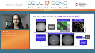
Marina Madrid, co‑founder and VP of Product at Cellino, presents the company’s vision and technology to the Alliance for Regenerative Medicine. Cellino has raised a $16 million seed round led by The Engine and Khosla Ventures to develop an AI‑driven laser editing platform that automates and scales the production of induced pluripotent stem cell (iPSC) therapies. The talk outlines the limitations of current manual iPSC manufacturing, the multidisciplinary team (physics, engineering, biotech), and the integrated software‑hardware stack that combines high‑resolution bubble‑mediated laser processing, imaging, and machine‑learning‑based cell/colony characterization. A custom laser system with sub‑5 µm spot size enables precise cell removal, spatially selective intracellular cargo delivery, and clonal isolation within a closed, robotic workflow. The software records every image and experimental operation as a searchable document, supporting regulatory data collection. Machine‑learning models can generate virtual fluorescence from brightfield images and predict colony quality from time‑series growth data, guiding laser decisions to maintain monoclonality. Cellino envisions a “foundry” where clinical‑grade cell products are manufactured in parallel, closed systems, reducing fixed costs. The platform is applicable to both reprogramming and differentiation stages, and the team seeks collaborations with cell‑therapy partners.
Chapters
Hi, my name is Marina Madrid. I'm a co-founder and VP of product at Celino, and I want to thank the Alliance for Regenerative Medicine for giving us the opportunity to present at this wonderful conference. At the end of last year, we secured $16 million in seed financing co-led by The Engine and Khosla Ventures with contribution from Humboldt and 8VC, and we're using that to build really the first AI-powered laser editing platform that will be used to massively scale the production of stem cell-derived therapies. We're taking a very multidisciplinary approach, combining stem cell biology, laser physics, and software and machine learning. And that is definitely reflected in the makeup of our team. All 3 co-founders, including myself, we have backgrounds in physics and engineering, The entire team has significant experience in industry biotech instrumentation, cell therapy development. And right now we have nearly 25 full-time individuals. I'm sure by the time Meeting on the Mesas is taking place, we'll have grown even more. We are advised by a truly fabulous group of individuals with significant experience in induced pluripotent stem cells and regenerative medicines across academia and industry. And I don't think I need to tell this audience why we are so excited about induced pluripotent stem cells. They really are a regenerative medicine platform in and of themselves. They have the code in them to become any cell type in the human body, so there's almost unlimited potential in terms of the patient suffering that we could alleviate. But the way the iPSCs are manufactured today is using a very manual, very labor-intensive process. It typically involves a highly skilled stem cell scientist looking at the cells during the reprogramming process, deciding based on their own personal experience which cells look promising and which cells don't, and then physically, with a plastic pipette tip, scraping away the unwanted cells. So, you can imagine this process is highly dependent on operator skill and therefore prone to operator bias. And also very difficult to scale. And so what we're doing is we're taking this process and we're automating it. And the way we're doing that is by taking a multidisciplinary approach. So we're combining imaging, image-guided machine learning characterization, and laser processing. And this approach that we're taking is applicable to both reprogramming and differentiation. This is a schematic of our software infrastructure. It allows us to characterize single cells and colonies and to track colonies over time. And what happens, each cell that we image essentially becomes a document in the database, and that document can hold flexible contents. So it can hold the results of image-guided machine learning-based characterization. It can also record every single experimental operation that's ever been performed on that cell, such as automated liquid handling steps. So we're expecting that this level of Data collection and tracking will be very valuable for engaging with regulatory agencies such as the FDA. Our entire platform is enabled by our custom-built laser processing system. My background is in laser physics, so I could talk about this for hours. The only thing I'll say here is that this laser system has a beam spot diameter of a little less than 5 microns, which means we can target individual cells without causing any collateral damage to the neighboring cells, which is very important. And the laser works not via a heat-mediated mechanism, but via a bubble-mediated mechanism. We have a laser-activated surface that sits at the bottom of the cell culture vessel. We can culture cells directly on that surface. In the case of iPSCs, we put down a matrix. We're using laminin, and then we culture the iPSCs on that laminin layer. And what that laser-activated surface does, it's very efficient at absorbing the energy from laser photons and transferring it to the surroundings in a way that generates a bubble. Bubble, and we can precisely control the size of the bubble by the energy in the laser pulse. So the higher the energy of the pulse, the larger the bubble. The smaller the energy of the pulse, the smaller the bubble. We can use large bubbles to remove, kill unwanted cells, for example, residual pluripotent cells in a differentiated batch of cells, and we can use smaller bubbles to poke temporary holes in the cell membrane for cargo delivery. This is us really just showing off with the laser. So this is iPSCs that have been grown to a confluent layer in a 96-well plate. We stain them to make a nice image and then laser-removed individual cells to develop this salino pattern. We can also do spatially selective intracellular delivery. So here what you see from left to right, these are iPSCs, and from left to right, that black stripe on the left We've laser removed all of those cells. The blue stripe is a viability stain, so we haven't done any laser manipulation to those cells. The green stripe are cells to which we've laser delivered green fluorescent cargo, and the red stripe are cells to which we've laser delivered red fluorescent cargo. So we can do spatially selective delivery, and we can do multiplex delivery. We can deliver some cargo to some cells and different cargo to neighboring cells. So you can imagine this could have interesting applications in tissue patterning, for example. And this is a video outlining our automated approach. We image cells in brightfield and fluorescence, use that to train image-guided machine learning algorithms that we can use to characterize individual cells that we've imaged in brightfield, and then we use that characterization to make decisions as to which cells to target with the laser. Here we're doing it manually so that it's slow enough to be seen in a video. We can also use the laser to do clonal isolation in a manner that's compatible with closed manufacturing. We can use the laser to remove individual cells to achieve a more uniform density across the well. We can use the laser to make pretty images for conference presentations like this one. This is a video mockup of our automated development grade system that we have built in collaboration with BioCero. I have images of the real system at the end of this presentation. But we're using this to collect large volumes of data to train machine learning algorithms. And this is a rendering of our foundry. So this is our vision for the future in which clinical-grade cell products are manufactured in closed automated systems. So the cells get to stay in a nice temperature-controlled environment while the imaging and laser processing takes place. And because it's a closed system, this will allow us to process thousands of samples in parallel in the same facility, which significantly reduces the fixed costs of cell therapy manufacturing. This is a workflow for how we're automating the production of clonal induced pluripotent stem cells. And I won't go into every single step in detail, but at a very high level, what we're doing is we're replacing the image-guided— we're replacing the human visual selection part of the process with image-guided machine learning-based characterization. And we're replacing that physical scraping away of unwanted cells with laser-based bubble-mediated removal of unwanted cells. And at the end, the iPSC clone is expanded. We're still doing all of the same standard NQC assays that everyone else in the industry is doing. So in this case, we're not using machine learning here to replace NQC assays. We're using machine learning really to make better in-process decisions to improve our yield of the process. And we can train machine learning algorithms with image type data to do cell-level characterization, which is shown here. We can also train them with non-image type data to do batch-level characterization. Which I'll talk about in the next slide. But here, to train with image type data, we take pairs of images. In this case, we included brightfield and DAPI and Oct-4 stains. And then we fed the untrained neural network enough of these pairs until it could start to build a correlation on its own to where we can give it just a brightfield image and it gives us what are essentially virtual fluorescence images. So what it would have looked like if we had done the DAPI stain and if we had done the Oct-4 stain. So we can image a well in brightfield, not do any staining, but get a prediction of the location of each and every single cell and a prediction of whether that cell is part of an OctoR-positive colony or not. And our next plans on the machine learning development side are to then laser remove all but one colony, allow that colony to grow, take time series images in brightfield of that colony, and then perform standard QC assays on that colony, like the qPCR scorecard from Alexander Meissner's lab or PluriTest, and feed this data encoded as vectors into a more traditional machine learning framework until the machine learning framework can start to make correlations between calculations that are easily made from time series images, like the day the colony emerged, shape, size, circularity of the colony, edge character— characteristics, broken proliferation rate, and whether that colony is likely to perform well on these objective measures of cell quality. So I've talked a bit about how we decide which colony to keep versus which colonies to remove with the laser, but how do we decide when it's time to remove colonies with the laser? Because the truth is that if colonies aren't at risk of colliding, We actually want to give them as much time as possible to grow because the more time series image data we can take of colony growth, the better we get at deciding which colony to keep versus which colony to laser remove. So for that purpose, we've developed the simulation that predicts colony growth and predicts when colonies are at risk of colliding so that we can remove colonies before we lose planality. It's very important for us to maintain monoclonality of these iPSCs that we're generating. And here I've shown how we can use the laser to remove all but one colony. So this is a way of doing clonal isolation in a manner that is compatible with closed manufacturing, which is really unique to our platform. So the image on the left is before, the image on the right is 24 hours after laser removal of all but one colony. You can see the colony that's been left behind has grown out of it over those 24 hours. It looks like these cells are glowing. They're not. That's just the image-guided machine learning algorithm doing its job, marking the location of every cell in a brightfield image. We can also use the laser to remove individual cells in arbitrary patterns. And you can see it again. These are before laser removal and after. You can see it most obviously in the top half of the white square on the right side. We've achieved a more uniform web-like density in the iPSCs. So we can use this to manage density of iPSCs if there are certain differentiation protocols that do best when you start the cell process off on the right foot with a very specific cell density or pattern. That's something we can do with the laser. These are images of our automated development grade system, which is up and running in our lab at the Engine in Cambridge, Massachusetts. We developed the system in collaboration with BioCero. The way it works, we have a robotic arm that transfers well plates from subsystem to subsystem, so from incubator to liquid handler to imager to laser processor. And we're using this in the near term to take the large volumes of data that are necessary to train the machine learning algorithms to do things like connect time series image characteristics of iPSC clones to qPCR scorecard data. But next year, we'll also be using this system to produce preclinical doses of iPSCs for our cell therapy developing partners. I've talked a lot in this presentation about how we're focusing on the first step in this process of making an iPSC-derived cell therapy by automating the reprogramming process, going from patient blood samples to induced pluripotent stem cells. But this process is also applicable to the differentiation side. And we've done work going from, for example, iPSCs to retinal pigment epithelial cells, We have several cell therapy partners who are interested in, say, applying image-guided machine learning algorithms to their differentiation process or developing laser processing protocols to remove unwanted cells that pop up during the differentiation process. So if you're working on an iPSC-derived cell therapy or just interested in talking in general, please don't hesitate to reach out. We're always happy to, to chat and to explore potential collaboration opportunities.