Founder Stories: Uğur Şahin, Co-Founder and CEO of BioNTech
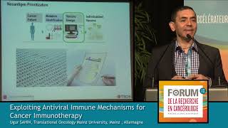
In this talk, Uğur Şahin, co‑founder and CEO of BioNTech, outlines the evolution and translational progress of RNA‑based cancer vaccines developed at the TRON translational cancer center in Mainz. He begins by contrasting immune‑checkpoint blockade with cancer vaccines, emphasizing that vaccines aim to teach the immune system to recognize tumor antigens. Şahin then details the technical optimization of the RNA platform—novel 5' caps, engineered UTRs, and class‑II targeting motifs—that together increase RNA stability and potency by several thousand‑fold. Two delivery strategies are presented: (1) direct intranodal injection of naked RNA, which exploits rapid dendritic‑cell uptake via macropinocytosis and simultaneous TLR‑driven maturation, enabling repetitive vaccinations and high frequencies of antigen‑specific T cells; and (2) an intravenous liposomal formulation that distributes RNA systemically to dendritic cells in spleen, lymph nodes, and bone marrow, inducing strong type‑I interferon responses and robust T‑cell activation. Şahin describes the personalized “polytope” vaccine pipeline: tumor exome sequencing, bioinformatic prioritization of neo‑mutations (including expression level, MHC binding, clonality), selection of ~10 mutations per patient, assembly into a 27‑mer cassette, GMP‑grade RNA production, and ultrasound‑guided intranodal administration. Clinical trial logistics (3‑month manufacturing timeline, sterility testing, safety monitoring board) and early immune‑monitoring results in adjuvant melanoma patients are discussed. Data show amplification of pre‑existing neoantigen T‑cell responses, generation of polyclonal CD4 and CD8 memory cells, and, in one patient, a partial tumor response followed by tumor escape via loss of β2‑microglobulin, highlighting the need for combination with checkpoint blockade. Finally, Şahin introduces the systemic liposomal vaccine now in clinical testing, noting dose escalation (7–29 µg), strong T‑cell responses to both mutated and non‑mutated antigens, and the mechanistic role of interferon‑α‑producing plasmacytoid dendritic cells and macrophages. The talk concludes with acknowledgments to collaborators.
Chapters
So I would like to thank the organizers for the invitation to this wonderful congress. And I'm happy that Ignacio Melero was the first presenter here and described immune checkpoint inhibitors and agonistic antibodies.. And the approach based on immune checkpoint blockade is based on the idea that there are T cells around which are able to recognize the tumor and to make these T cells fit combinations aimed for the idea that somehow immune responses are stimulated. Cancer vaccines have a complementary approach. Cancer vaccines have the idea that we really teach the immune system about relevant tumor antigens and thereby generate this type of T cells which can then be activated by checkpoint blockade. I'm from Mainz. So Mainz is near Frankfurt. University city with a dedication for immunology. There's a long tradition to do translational research in Mainz, and from this translational research, we have created a translational cancer center which is TRON, which deals with the topic of testing ideas, developing ideas until they can be continued by biopharmaceutical companies. And we have also biopharmaceutical companies in mind, spin-offs from the university, taking these concepts, bringing that to patients, and ultimately aiming to get new drugs approved. My group is focusing on developing cancer vaccines and the platform which we are focusing on is RNA vaccines. RNA vaccines have a number of advantages.
For example, you can take just an antigen and encode this antigen by a template, do in vitro transcription, and then you can use the RNA to transfect cells.
And our approach is not transfecting cells but bringing the RNA directly into the body, and so that cells in the body can do the translation. What we did, the main disadvantage of using RNA is the instability of RNA. RNA is degraded in the extracellular space, and it is not stable if it comes to the cells. So we, in the last 15 years, we use several approaches to improve the pharmacodynamic activity, the potency of the RNA. For example, by putting novel caps which inhibit the 5' degradation of the RNA, but using also UTR regions allowing to increase the half-life or the translational capacity of the RNA. Moreover, we included modifications like, for example, MSC class 2 targeting motifs, allowing us to bring the translated protein in the cytoplasm into class II presentation pathways, thereby allowing to increase T cell responses. This is all published and resulted in several thousand-fold increase of the potency of the RNA. The second aspect is which are the right cells to express the RNA. And of course, we all know that dendritic cells crucial for induction of immune responses. And indeed, about 20 years ago, for the first time, dendritic cells have been used for antigen expression via delivery of RNA.
It was Eli Gilboa's group describing the approach, transfecting dendritic cells in vitro and bringing this dendritic cells back to the body.
And actually, this approach, this RNA-based dendritic cells transfection approach, is one of the most powerful vaccine approaches, resulting in strong T cell responses in many, many studies. Our approach is slightly different. We are not transfecting dendritic cells in vitro. We are aiming to transfect dendritic dendritic cells directly in vivo. And we have two approaches.
One approach is injecting of the RNA into the lymph node by direct injection of the lymph node. And the second approach is based on using liposomal RNA, which is injected intravenously. And we developed liposomal approaches allowing us to bring the RNA into dendritic cells in the whole body.
In spleen, lymph nodes, and in the bone marrow.
I will show you later data about this.
Okay, starting with the intranodal approach, this is from mice.
When RNA is injected, labeled RNA is injected into mice, then under certain conditions, if you use buffer systems containing ions allowing to make nanoparticles out of the RNA. The RNA is spontaneously taken up by dendritic cells. This is lymph node after 5 minutes injection of RNA. This is after 30 minutes. The whole RNA is taken up by dendritic cells.
It is the mechanism behind this is, so it is an active mechanism which can be blocked, for example, with mycopinocytosis inhibitor scotlarine. But the nice aspect is that the dendritic cells not only take up and translate the RNA, but the RNA also, by stimulating TLR receptors, induce a maturation of dendritic cells as well as cytokine release. So we get a cytokine milieu in the lymph node which stimulates powerful expansion of T cells. And by this, we are able to do repetitive vaccinations. Since the RNA does not carry any other proteins, we can really repetitively immunize without getting backbone immune responses. And by repetitive vaccination, we are able to get regardless which type of antigen we use, about 20, 30, 40, 50% of antigen-specific T cells in the peripheral blood.
This is, for example, an RNA immunization of HLA-DR mice with tyrosinase RNA. After 5 vaccination cycles, 30 to 70% of T cells are antigen-specific. This is New York Eso-1 vaccination of the same same mice strain resulting in New York iso-specific T cells. And as mentioned, we always immunize with the full-length RNA without taking care which is the right epitope for vaccination.
But of course, the key question is which are the right tumor antigens for vaccination. And we have now, since 25 years, these questions after the first first tumor antigens were cloned, you know the shared tumor antigens like New York iso-1 tyrosinase, the MAGE family members, which are not mutated. On the other side, there are mutated tumor antigens. And we have this debate since 25 years, which are the right tumor antigens. So about 6 years ago, we said we want to systematically test that and want to use mutations for vaccination, but not only single mutation, but the entire set of mutations. The challenge with that is if you are interested in using the entire set of mutations is that every patient has a different set of mutations. So that means you cannot just use the shared ones, then you would just focus on 5% of the mutations. If you really want to use the entire set, you have to do to do next-generation sequencing to determine that. This is exactly what we did. So we developed an approach where we started, take the tumor, sequence the tumor by next-generation sequencing, identified the mutations, and then we tested a number of algorithms to identify mutations which could be immunogenic. What we found is that about 25 to 30% of mutations randomly selected mutations are immunogenic, that most immunogenicity is driven by CD4 recognition of class II neoepitopes and driven by CD4 T cells. And we found that if you take these mutations, yeah, and predict them by bioinformatic tools and put them together, multiple mutations on one cassette, we named this polytope vaccine approach because RNA allows you put multiple mutations into one cassette and immunize mice, then you can get powerful antitumor activity resulting in clearance of established lung metastases or established tumors. So this was a preclinical study which we published about 8 months ago. And the nice aspect about this is even though it is an individual approach, It is universally applicable to all, all, all types of tumors, not only in mice, but we can use the same approach also to treat patients. So we started a clinical trial in beginning 2014 addressing exactly this. Can we use this approach for treatment of human cancers? We did this trial in melanoma patients, and since we knew that we need several months to prepare a vaccine, to do next-generation sequencing, prepare the vaccine, we selected patients who are in the adjuvant stage, who do not have tumors, either being in stage 3 without tumors or being in a situation where a metastasis, single metastasis, was removed prior to vaccination. So the approach works in the clinical setting as follows.
So we get peripheral blood. Tumor tissue, we developed an approach which allows us to use paraffin-embedded clinical routine biopsies. The biopsies are used to extract DNA from the blood and extract DNA from the cancer sample.
So we do exome sequencing of both, thereby allowing us to identify the mutations by comparison between normal and tumor.. And for the identified mutations, we asked the question, how strong are these mutations expressed in the tumor? So what is the expression level? And we can do that exactly by not only looking to the mRNA itself, but to the position which is mutated so that we get really the answer even if 80% of the tumor is contaminated with chromosomes, we can define whether a mutation is expressed or not. And by next-generation sequencing, we get a lot more information. For example, combining that with bioinformatic tools, we can define whether mutation will bind to MHC class I, MHC class II. We can define the allele frequency. We can define whether mutation was generated very early in the tumor development or is a late mutation.. And we use this information and build bioinformatic algorithms, computerized algorithms, allowing the computer not only to identify the mutations, but also make prioritized list of mutations suggesting which antigens should be selected for a vaccine. And we have a target safety monitoring board. These are 5 6 experts sitting together and discussing the suggestions coming from the computer. And if they are approved, we put them into the vaccine. And the vaccine is based on the idea that we want to get CD8 and CD4 T cell responses. So we take about 27-mer with the mutation in the centre. And this would allow, with the mutation in the centre, to get every CD8 epitope and most of the CD4 T cell epitopes. Epitopes. And we not only just take one mutation, but for the first clinical study we were allowed to use 10 mutations for vaccination for every individual patient. We combined this neoepitopes with linkers and put them back into the pharmacologically optimized cassette. Then the RNAs produce under GMP and transported to the center where it is supplied by the doctor by intranasal vaccination. This is the GMP production process. The GMP production itself takes only 2 days, but we need about 3 weeks to get the sterility testing done. And therefore the whole process, everything is complicated by regulatory requirements. If we do that in mice, we have the vaccine available after 2 weeks. Here in patients, we need more than 3 months because of all these controls.
And then, if the vaccine is delivered to the clinic, it is injected by intranodal injection.
This is ultrasound-guided, so this is the nodule region. And I would like to ask someone to start this video. So now the needles come in. This is the lymph node. The needle comes in and then the vaccine is injected and it is— it can be controlled. It's an easy, easy approach with a success rate of more than 96% tested so far. And you can do that repeatedly without getting any changes in the lymph node. So of course, this is a first-in-human study. Important is safety, important is feasibility. The most important for us was immunogenicity, since we did not expect to see any clinical clinical activity since the patients are in the adjuvant stage. So we invested a lot of efforts to, for example, do immune monitoring.
And immune monitoring is done in a way that we get blood before vaccination, the patient until he receives the vaccine gets vaccination with New York-iso-1 and tyrosinase. This is available directly. And when the vaccine arrives, the New York-iso-1 tyrosinase vaccination neuropitopes and the Mutanome vaccination, the vaccine with the neuropitopes does.
And we get blood from different time points.
And if, for patients who are willing to continue and who tolerate the vaccine, we offer continuous vaccination.
So what kind of immune responses we get? We have two different types of immune responses. One is One is amplification of pre-existing T cell responses. So what became clear in the last 2 years is that the success of checkpoint blockade mainly depends on activation of T cells against neoantigens. This is really an exciting field now so that it is now recognized that an immune response against a neoantigen could result in success of of checkpoint blockade. And indeed, if we use the samples from the patient before vaccination, we see that the patients have, for certain antigens, a pre-existing T cell response.
This is the pre-existing T cell response measured with the RNA. And this is the pre-existing T cell response measured with overlapping peptide. And you can see this is mutation number 4. Number 47 in patient 1, there is a pre-existing T cell response.
And what we see is we can amplify this pre-existing T cell response by vaccination. This is the post-vaccine response, a powerful amplification of T cells by the vaccine. And this is— so I see that— OK, go back. Okay, then I will take this. So the slides are mixed, but I can continue with that.
And if we do that, we can check for every mutation in every patient whether there is a pre-existing T cell response. And so these are the mutations in patient number 4, yeah, and you see there are weak immune response against against this mutation, weak immune response against this mutation, and moderate immune response against this mutation.
After vaccination, this patient developed multiple mutation-specific, strong mutation-specific immune responses. And the interesting aspect is, these mutations are against different MHC molecules. So it's a wonderful polyclonal response. And this patient was a patient who developed multiple metastases when waiting for the vaccine. So it allows us to see whether the patient gets—
whether the immune response results in an antitumor response.
And indeed, we have seen that the patient went from progressive disease into stable disease. And then developed a partial, partial response. But after that, the lesions remained for a long time, about 8 months, stable. So the doctors decided to take out these metastatic lesions and analyze these metastatic lesions. This is a metastatic lesion of the patient before vaccination, and this is the metastatic lesion after vaccination. And the pathologist's diagnosis was completely necrotic metastatic lesions. But this was not the case because when we performed a vital staining, we found that still some regions of the metastasis have live tumor cells. So we were even able to analyze, to get a tumor cell line, melanoma cell line established. We were able to get tumor-infiltrating lymphocytes and investigate that.
In this viable lesions, we found a strong infiltration of T cells, and we found also upregulation of PD-L1.
As Ignacio Melero showed, this is a sign for— a good sign for an indication for responsiveness to checkpoint blockade.
Everything is fine here.
And now I I have to continue.
I don't know what happened.
Everything is mixed up. So I'm sorry, I will go back and forth. So this is the story, continuing the story. So we analyzed the tumor-infiltrating lymphocytes, and we investigated the blood of the patient.
What we have seen is there was against this mutations, mutations number 5, there's no pre-existing T cell response in the peripheral blood before vaccination. After vaccination, there is an established immune response.
And if we analyze the tumor-infiltrating lymphocytes, we see that they are infiltrating the tumor. And having blood from different time points, allows us to monitor how fast the immune response is generated.
And you can see from this tetramer analysis that the immune response started to be established after 4 vaccinations. And it's robustly observed about a long period of time.
So we induce after about 20 days a strong a T cell response which remains active and we generate memory T cells in these patients. Analyzing the phenotype of these T cells shows that these T cells are— at least a proportion of these T cells, of antigen-specific T cells, are positive for PD-1. So it's, again, a good sign that this type of treatment can be combined with checkpoint blockade. So we established an approach allowing us not only to analyze T cells, but also to get the T cell receptors out of these T cells.
It is a single T cell receptor cloning approach where we do FACS single cell sorting of these T cells and then perform amplification of T cell receptor alpha and beta.
And then we can reconstitute that and define the specificity of the T cells.
By doing this, we were able to identify T cell receptors which recognize recognizes the mutations. So we have, for example, identified T cell receptors recognizing mutation number 14. And we have identified T cell receptors recognizing mutation 5 in these patients.
And we analyzed this T cells by transfecting that into the autologous PBMC and have seen that these T cells or the T cell receptors confer extra extremely potent cytotoxic activity with high affinities so that peptides could be titrated up to 10^-11. And these are extremely potent T cell receptors. And since we had generated a melanoma cell line in these patients, we investigated the melanoma cell line. Quality controls is to check for MHC class I and MHC class II expression. And it turned out that the melanoma cell line is negative for MHC class I. So we put interferon gamma on these tumor cells, but nothing happens. And then we went back, so MHC is not expressed at all. We sequenced the melanoma cell line, and it turned out that both beta-2-microglobulin alleles both were deleted in this melanoma cell line. And if beta-2-microglobulin is missing, then MHC is not presented on the tumor cell line. So after transfecting beta-2M into the tumor cell line, we observed upregulation of MHC class I presentation. And with this upregulation, we were able to we see potent killing of this melanoma cell line by the T cells, which is not present if MHC class I is missing. We went back to the tumor and investigated and found that indeed in the tumor we have lost the beta-2 microglobulin expression. So this is an indication for an evolution of a tumor Interestingly, it is the only way to escape for this tumor because this patient had multiple T cell responses against different MHC alleles. So that beta-2-microglobulin is some sort of the perfect solution for the tumor. And unfortunately, we did not have this information when we make this decision how the patient should be treated. At this time, we had no allowance to to use combination of PD-1 and vaccine. So we said, OK, we have PD-1-positive T cells. Let's go for PD-1. And under PD-1, after stopping the vaccine, the patient developed multiple metastases and within a few months died from this multiple metastasis. With this information, since we know that we had also CD4 T cell responses, we would perhaps favor to go to continue with the vaccines, hoping that at least the CD4 T-cell response can control this patient. So in the last 5 minutes, I would like to give you also an overview about a totally novel approach. It's a liposomal vaccine. It's actually the idea that instead of reaching a few dendritic cells in this lymph node, gets tried to reach all dendritic cells. The idea is here simple. So the immune system is evolved to deal with infectious disease particles. So if you have an infection just on the arm, you get a limited number of T cells because this immune system is is dealing with economic considerations. So if you have a small infection, you get little number of T cells. If you have a systemic infection, of course, this is life-threatening constellation, and the immune system is activated strongly and generates a lot of T cells. For example, if you have a flu, the immune system is able to get up to 50% of antigen-specific T cells within a few days, yeah. The idea here is to simulate something which is like a systemic viral infection. For this, we try to identify formulations allowing us, after intravenous injection, to bring RNA directly into dendritic cells.
And we found that certain liposomal formulations allows us really to have the signal directly the luciferase signal, which is the indicator for translation, directly in the spleen, exclusively in the spleen. And if we analyse the cells taking up this luciferase signal, it turned out that depletion of CD11c-positive cells resulted in complete loss of the signal. So this is not only the case in the spleen, but we have also transfection of lymph node dendritic cells. With depletion of dendritic cells, completely lost signal.
And in dendritic cells in the bone marrow. So looking which type of cells— oh, okay, pardon me.
Looking which type of cells express this antigen, we found that these are dendritic cells, dendritic cells and plasmacytoid dendritic cells, and to certain aspects macrophages. These are the expression in the lymph nodes. And we have seen that the expression is extremely rapid after 1 hour having already a signal. The mechanism of uptake again is macropinocytosis inhibition with rotlarin completely blocks the uptake. And it is just skip this slide and maturation of the dendritic cells also skips the uptake.. Importantly, these dendritic cells go from the— are transfected in the marginal zone, and the transfection with the RNA results in maturation of these dendritic cells, again, by induction of cytokines. The main cytokine is here interferon alpha. And by this maturation signal, the dendritic cells migrate from the marginal zone to the the T cell zone and provide extremely strong T cell responses. The population producing interferon or the mechanism behind interferon alpha production is mediated by TLS7 recognition. And there are two types of cells involved, plasmacytoid dendritic cells producing early interferon alpha and macrophages producing later stage of interferon alpha. And this results in activation of all cell types, mainly also of T cells. This type of vaccine allows us to get extremely strong T cell responses with just a few vaccinations, allowing us to use this vaccine really in tumor models with large tumors so that we can get rejection of established large tumors. This is an HPV E7 model. But it is also important that even self-antigens can be used in this case. For successful tumor vaccination. This is CT26 and the self-antigen GP70. And the activity of this vaccine strongly depends on interferon alpha, because if we block interferon alpha, the activity of the vaccine is gone.
We have now this systemic vaccine approach in the clinical stage. We started with a dose which is lower than the dose which were used in mice.
So it's 7 micrograms, and we increased that to 29 micrograms. We see the same type of cytokines in the patients in this low dose. And even with this low dose, which is still lower than the dose in mice, we get strong T cell responses here in case for New York-ESO-1 in the first patient, and tyrosinase the immune responses match the activity of immune responses against viral antigens. This is cytomegalovirus, EBV, and influenza immune response. This is tyrosinase and is a response generated by vaccine.
This is the next patient. So we have treated 3 patients so far. And we never have seen such strong T cell responses against against non-mutated tumor antigens in these patients.
Thereby really giving hope that this type of vaccines can be used to translate into highly potent antitumor activity. So this is the mechanism of the vaccine. We have on the one side antigen expression by disease and Type I interferon secretion by other cell populations.
This results in maturation of dendritic cells and activation of T T cells, thereby providing both effector T cells which generate strong T cell responses.
I would like to thank my collaborators, mainly Sebastian Kreiter, Mustafa Dikandlina-Kranz for the RNA work, and our collaborators in the clinic helping us with the clinical studies.
Thank you.
More from Joshua







Scientist Stories: Hans Waterham, The value of phenotypic assays in peroxisomal disorders




Scientist Stories: Joel Zlotogora, Genetic carrier screening programs for reproductive purposes