Founder Stories: Vishva Dixit, VP of Discovery Research at Genentech
In this lecture, Vishva Dixit provides a historical and mechanistic overview of mammalian cell death and inflammation, tracing his own research from the early 1990s to recent discoveries. He begins with death receptors (TNF and Fas), describing the death domain, the recruitment of the adaptor FADD, and the assembly of the caspase‑8 activation complex that drives apoptosis. He links this pathway to cancer immunotherapy, showing how cytotoxic T cells use granzyme B and Fas ligand to trigger the same caspase cascade in tumor cells. Dixit then shifts to the inflammasome, explaining the canonical NLRP3‑ASC‑caspase‑1 complex that processes pro‑IL‑1β and pro‑IL‑18, and how pyroptotic membrane rupture releases these leaderless cytokines. He introduces the non‑canonical inflammasome pathway, revealing that cytosolic LPS directly activates caspase‑11 (mouse) / caspase‑4/5 (human), leading to gasdermin‑D cleavage. Gasdermin‑D p30 forms large membrane pores that allow cytokine release and, eventually, cell lysis. Using ENU mutagenesis, his group identified NINJ1 as a separate protein required for the final membrane rupture that releases large molecules such as LDH, distinguishing membrane damage (pore formation) from membrane rupture. He discusses the implications for sepsis, the failure of TLR4‑targeted therapies, and the therapeutic potential of targeting these newly uncovered pathways. The talk concludes with acknowledgments of collaborators and a brief Q&A invitation.
Chapters
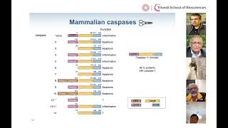
I'll give more of a historical perspective to my work. I'll finish up with some recent exciting data, and the story begins really in the early '90s when we became interested in the mechanism of cell death and especially mammalian cell death. And around this time it was evident that there's a receptor system that if engaged by cognate ligand or by agonist antibodies could induce cell death in the recipient cell. So the receptor system that we worked on was the TNF receptor and the Fas receptor, and these are colloquially referred to as death receptors, because if you engage the Fas receptor, for example, either by agonist antibody or by cognate ligand, then this receptor induces a death signal into the interior of the cell, resulting in the apoptotic demise of the cell. Now, this was quite a mysterious process, but we did know from the pioneering work of Dave Goodell and Shigeyuki Nagata that there was a segment within the cytoplasmic domain of about 80 amino acid residues that if mutated disabled the ability of this receptor to engage the death pathway. So this domain was dubbed the death domain, and I ask you to remember that because it'll be important in a couple of slides. So otherwise, the process was quite mysterious. You engage this receptor and the cells died, and morphologically, phenotypically, under a microscope, it was quite a profound event on engagement of this receptor. Within a few hours, the cells would undergo a dance of death. They would essentially implode. And this is shown in a short movie on the next slide. And it captured my fascination because it said that you engage the receptor and the cells died. And what was the underlying mechanism? So I think you appreciate from this short clip that soon after engagement of this death receptor, all havoc broke loose. And the cell essentially disintegrated. So the question was, how did this happen? And in the early '90s, there was a lot of interest in inflammation, there was interest in cell death and model organisms like the worm, and quite unexpectedly, research from disparate areas seemingly unrelated areas came together and shed important light on the death pathway. So what was this research? The first line of work was being done by investigators at Merck and at Immunex, and they were looking at two cytokines, IL-1 beta and IL-18, These are very pro-inflammatory cytokines, but they occur in an inactive form, the pro-form. And so the question was that this pro-form is converted to the active form, and this conversion happens through a protease, a molecular scissors, which cuts this pro-form to give you the mature cytokine. So the question then that loomed large was what was the nature of the protease? What was this molecular scissors that did this clipping that converted an inert cytokine into an active cytokine that could mediate inflammation? And the subsequent purification and cloning of this protease revealed it to be a very unusual protease. It was a cysteine protease, but what was unusual was that it cleaved off aspartic acid residues. So this protease was termed ICE, or interleukin-1 converting enzyme. I want you to remember that. And its unusual nature was noted. And these workers were working in inflammation in industry. Now, they didn't know that there was a basic research lab at MIT of Bob Horowitz devoted to defining the cell death pathway genetically in worms. And so they had, in particular, they had defined this all-important, centrally important death component that they called Ced-3, and it was mysterious. They didn't know what it did. Then one day they ran it through the databases and lo and behold, it had sequence homology to interleukin-1 converting enzyme, this enzyme that had been identified by workers working in the world of inflammation who had no interest in cell death. And so they realized that Ced-3 must be a protease, must be a cysteine protease, must be a cysteine protease that cleaved after an aspartic acid residue., and it was for this work that Bob Horowitz got the Nobel Prize. So again, seemingly disparate fields coming together. Now we started wondering whether in our system of death receptor-engaged apoptosis, whether similar protease, similar molecular scissors may be related to interleukin-1 converting enzyme or ICE, played a role. Now we had a powerful tool to address that because a colleague of ours was working at Duke, Guy Salvesen, identified a protease inhibitor, an inhibitor of this molecular scissors, from the cowpox virus. It is actually in all pox viruses, and this is termed creme, and it is actually a serpin that inhibits this class of enzymes.. So we said to ourselves, hold on, we have an inhibitor here of ICE and ICE-like enzymes. Why don't we simply ask if it will block receptor-induced cell death? And indeed we did that experiment. The results were very dramatic. If you took cells in culture, treated them with TNF or anti-FAS agonist antibody, the cells promptly died. However, if they expressed Crema, this inhibitor of ICE and ICE-like proteases, then the cells survived. Not only did they survive, they replicated, they basically lived happily ever after in the face of what was previously a noxious stimulus. So what this said to us was that downstream of the death receptor, must be ice or ice-like proteases, and these proteases have since been renamed because it's a family of them, there are about a dozen or so members, as caspases. The C donates it's a cysteine protease and the aspas denotes that it cleaves after an aspartic acid residue. So then the question was, well, how exactly does this receptor using this death domain? Because remember, this death domain was critical because mutations within this death domain disabled the ability of this death receptor to engage death. So how did this receptor, and specifically its death domain, activate these downstream molecular scissors or proteases, caspases in this case? So this we solved in a series of papers in the early '90s while I was still at University of Michigan Ann Arbor, and we showed that quite surprisingly the death domain in the receptor was nothing other than a protein-protein interaction motif. So it allowed for the recruitment of an adaptor molecule that we termed FADD to a death domain-death domain homotypic interaction. Quite surprisingly, FADD had a second death fold that we call the death effector domain, and this death effector domain, even more remarkably, was found in the zymogen form or inactive form of a death protease that is now known as caspase 8. So what we realized was that through a series of these death fold interactions that functioned as a molecular cement, we assembled a signaling complex. And a component of this signaling complex was a protease, a caspase related to the original interleukin-1 converting enzyme, part of that family. And we showed soon thereafter that once it is recruited, it gets activated. And you release into the cell's interior the active protease, the active molecular scissors, the p20 and p10 subunit, and this protease will very precipitously in an amplification manner activate downstream caspases that exist in inactive forms. So their downstream effector caspases 3 and 7 and others that exist as inactive zymogens, but this protease will function to proteolytically cleave them and activate them. So this is very similar to protease cascades you are aware of, like the coagulation cascade or the complement pathway, where you get tremendous signal amplification when an upstream protease cleaves and activates a set of downstream proteases. Regardless, the outcome of this was that very quickly, precipitously, the intracellular protease activity increased and death substrates were cleaved and the cells died. Now, why is this important in real life? Well, this is the foundation for cancer immunotherapy. In cancer immunotherapy, the event that we are all concerned about is the cytotoxic T cell killing a tumor cell. That is what cancer immunotherapy is. And this is a short movie that I'll run which shows a cytotoxic T cell, this purple cell. It's got granules within it. It is adhered to a tumor cell, this large green cell. And it's going to kill it. And you will see it's going to degranulate, it's going to put out its granules, and it's going to kill this cell. And what I'm going to show you is that this killing is due to the activation of caspases. Okay, so let's just run the movie. So that death of this cancer cell is the essence of cancer immunotherapy. This is what it's all about. So what exactly is the killing mechanism? What did this purple cell do to this green cell that it died? So there you see the movie again. The purple cell kills the green cell. Well, the purple cell is the cytotoxic T cell. That's the CD8, that's the cell that does the killing. And this cytotoxic T cell has granules in it which contain a very potent form of molecular scissors termed granzymes. So this granule, I'll show you when I run the movie, will fuse with the cytotoxic T cell and tumor cell membranes. And through these perforin channels, the molecular scissors will be introduced into the tumor cell. And these molecular scissors will cleave these downstream inactive zymogen caspases and activate. Furthermore, the cytotoxic T cell will also express the ligand, the activator of FAS. This is the second punch, so it's a one-two punch. And fast, once this death receptor gets activated, caspase-8, that other set of molecular scissors gets activated, and now the tumor cell is kaput. So let's run this movie. You see the degranulation, you see the perforin channels, the molecular scissors being introduced, these are the granzyme B,. Then you see the ligand expressed, the complex around the receptor happen, caspase-8, this Pac-Man-like structure, also activate zymogen caspases. And so this one-two punch all of a sudden gives the cancer cell an overwhelming amount of protease activity leading to its death. Okay, so that is the story. Now I am going to change gears a bit and talk about the inflammasome for the rest of the talk. Now the inflammasome, you remember I talked about interleukin converting enzyme, that was the first of these proteases that was identified., and this is now termed caspase-1 in recognition that it was the granddaddy. Now this caspase-1, as I mentioned, proteolytically clips proAL-1β and pro-IL-18 to give you the mature cytokines that mediate inflammation. But caspase-1 is all— its activation is regulated. It is regulated by an apparatus, and that apparatus is called the inflammasome. And that apparatus consists of a sensor, an adapter, and an effector. Now the sensor is cytosolic. I show a sensor here, NLRP3. It's a sensor that's part of the NOD family of sensors. It has a protein-protein interaction, death fold, and the pyrin domain, nucleotide binding domain, and leucine-rich repeats. And there are about a dozen or so sensors in mammalian cells, but in plant cells, in plant, in the plant kingdom, this is the mainstay of the plant immune defense mechanism. There are over 200 of these genes. Well, what NLRP3 does in mammalian cells is that it recognizes membrane damage. That membrane damage can be from cholesterol crystals or amylloid crystals, it can be from pore-forming toxins, and once it recognizes membrane damage, the exact mechanism is still debatable, but once it does that, this sensor will now couple to an adapter ASC through a homotypic protein interact, pyrin-pyrin domain interaction. Now this adapter serves as a bridge and allows the sensor to form a complex with the effector caspase-1, interleukin-1 converting enzyme, and through a second set of homotypic CARD interactions. Now, once this is assembled, this is what the late Jörg Schopp called the inflammasome, you get activation of caspase-1. So in my cartoons, the active protease is an open scissors. And this caspase-1 is going to cleave proIL-1 beta and proIL-18 to give you the mature cytokines. Now, I want to tell you something very important about these cytokines is that they don't have a signal peptide. They have no way of getting out of the cell. So these are called leaderless cytokines. The only way they can get out of a cell is if the cell incurs membrane damage and pops open, and then these cytokines will leave the cell. So this popping open of the membrane is a form of cell death termed pyroptosis, really an inflammatory form of death., but it allows for the damaged membranes to leak out mediators of inflammation. So they include IL-1 beta, IL-18 that I talked to you about, they've been processed, but now they're released, IL-1 alpha, HMGB1, so on and so forth. So essentially this says that the death of the cell through this process of pyroptosis is the mechanism that allows for the release of these cytokines. So I will just summarize this part by saying that these are molecular assemblies, whether you think of the death receptor signaling pathway or the inflammasomes, the architecture is much the same. They both have a sensor, the death receptor senses the ligand, the NLRP3 sensor senses membrane damage, They couple to an adapter that has a bipartite architecture, death domain and a death effector domain for FADD, a pyrin domain and a CARD domain for ASC, and this bridging adapter allows the sensor to communicate with the effector. The effector for the death receptor is the death protease caspase-8, and for inflammation is this protease caspase-1 or interleukin-1 converting enzyme. Which is going to convert IL-1 beta and IL-18. So the architecture of these molecular assemblies can be thought of as a sensor-adaptor effect. Now, a few years ago, 2011, we discovered a new pathway, the non-canonical inflammasome pathway, and I am going to tell you about that. So if we look at this family of proteases, there's this family of molecular scissors that are related to interleukin-1 converting enzyme, termed the caspases. There are a dozen or so. And we discussed caspase-1 is involved in inflammation, and we talked about how caspase-8 is involved in cell death, caspase-3 is involved in cell death or apoptosis, caspase-7 is involved in cell death. So many of these function as death proteases, but we were struck by caspase 4 and 5 in humans, that's represented by a single caspase, caspase 11 in the mouse, because these had sequence homology to caspase 1, the inflammatory caspase, and we wondered whether it had a similar function in inflammation. And so we do a lot of mouse work, so we knocked out caspase 11, so that would be the same as 4 and 5 in the humans. And so now we are able to look at the function of caspase 11 in inflammasome activation. The way we measure inflammasome activation is we measure IL-1 beta,, and that's shown here on the y-axis. And here the open bars are macrophages from wild-type mice, red bars are macrophages from Caspase 11 null mice, and the various stimulants on the x-axis are shown. These are pro-inflammatory stimulants, so ATP, cytosolic DNA, infection with Francisella tularensis or Pseudomonas aeruginosa, and disappointingly we did not really see any difference. I mean, it sort of looked the same to the wild type, seemed like Caspase-11 had no discernible function. But then we hit gold when we realized that if we looked at gram-negative enteropathogens, E. coli, Citrobacter rodentium, Vibrio cholerae,. Now all of a sudden there was a massive difference. The generation of IL-1 beta was dependent on caspase 11. So we call this pathway the non-canonical inflammasome pathway, and we published that in 2011. Now what is it in gram-negative organisms that is being recognized in gram-negative pathogens. What is it being recognized? And you probably know that the major constituent of gram-negative walls is LPS, and indeed it was lipopolysaccharide that was being recognized by bone marrow-derived macrophages, cells of the innate immune system, and in response to that, they were activating Caspase-11, and this is shown in this graph here. Again, IL-1 beta on the y-axis, open bars are wild-type macrophages, red bars are macrophages from Caspase-11 null cells, and if we introduce LPS into these macrophages, you see that the wild-type macrophages respond, the Caspase-11 null do not respond.. Now this was again published a couple of years later, and I won't go through the details except to say we were able to make a mutant form of E. coli, because you could ask yourself, how do you know it's only LPS? It could be other things in the gram-negative organism. After all, there are thousands of things that can engage the inflammatory pathway. How do you know it's just LPS? So We made a mutant form of E. coli which was defective in the penultimate step in LPS synthesis, and in live infections we were able to show that this mutant form of E. coli, while it engaged other inflammatory pathways like IL-6, it did not engage the inflammasome pathway. Now this, of course, the work with LPS led us to this question of lethal sepsis, This is an excerpt from The Lancet, which is a plea to the community to continue to work in this area because there are 8 million deaths worldwide. That's more than all the cancers put together. And in countries, in the developing countries, in developed countries, it's responsible for substantial mortality and often in the developing countries in pediatric population. So you would say to yourself, well, hold on, this is a huge opportunity, it's a massive opportunity, so surely the pharmaceutical industry is working on it. But the plea in The Lancet is that the pharmaceutical industry is actually not working on it, not as hard, because they have run 60 failed clinical trials consecutively. That is 6-0. Now, when you run that many clinical trials and you fail, there is not much appetite to study this spectrum, this syndrome. And so it's possible that we are drugging the wrong pathway, or it's possible that the disease is very complex, and there are probably elements of both of them. But the clinical trials are large and expensive. This is the last failure TLR4 antagonist, a 2,000-patient trial where a TLR4 antagonist failed. But you would say, oh, come on, it's such a big problem, obviously people must have studied it, and of course they have. I mean, the 2011 Nobel Prize in Immunology was given to Bruce Beutler because he discovered the signaling pathway, the receptor for LPS, Toll-like receptor 4, He found that there is a mutant strain of mice, C3hej, that doesn't respond to LPS. When he positionally cloned that locus, he found a mutation in toll-like receptor 4, and indeed, if you knock out toll-like receptor 4, mice are very resistant to LPS. This is shown graphically here. If you look at percent survival on the y-axis as a function of time in hours on the x-axis, if you take wild-type mice and inject them with LPS, then they are all going to die within the first 24 hours. But if they are TLR4 knockouts, they lack TLR4, then they all live. So this is very powerful genetic evidence of the importance of TLR4, and it was for the basis of this work that he got the Nobel Prize. And so this has led to the sort of I would say a dogma in immunology textbooks that when a TLR4 is engaged by LPS, you activate downstream signaling pathways that include NF-kappaB, pro-inflammatory pathways. This leads to a massive cytokine storm and lethal septic shock. So I mean, this is fine, but the problem is that there was a pediatric geneticist Jean-Laurent Casanova, who said, you know, I have kids who have genetic defects in this pathway and they are actually not susceptible to gram-negative organisms, so there must be another LPS sensing pathway. So that was a bit of a worry, and then of course there was the failure of the TLR4 antagonist in clinical trials, so it seemed there may be another way to sense LPS. So in 2013, we said indeed there is a sensing system. It doesn't need toll-like receptor 4. It is an intracellular sensing system that is dependent on caspase 11. And this was met with a lot of surprise in the community because they said, Hey, didn't we just give the Nobel Prize for LPS sensing, and now there's another pathway? What's happening? How are these experiments being done? Well, how did we do the experiment? How did we challenge dogma? Well, we said, instead of injecting pure LPS, let's try to mimic an infection, albeit using purified compounds,, but by engaging multiple inflammatory pathways, because when you get infected with a gram-negative enteropathogen, you're engaging multiple pro-inflammatory pathways. So let's try to mimic that. So in this animal experiment, we gave the mice polyIC, which engages another pro-inflammatory receptor, TLR3, but it's well tolerated. So polyIC mice treated with polyIC, if you look at their mortality curve, they do okay. So percent survival as a function of time. So now we took these polyIC primed mice and we gave them LPS because we said, well, now that sort of mimics an infection, more than one receptor system is being engaged, and not surprisingly, they promptly die. But what was surprising was that if we took the TLR4 knockout mice, which had previously been resistant to LPS, and now primed them with poly(I:C) and then gave LPS, kabang, they all died. And the caspase-11 null mice were largely resistant. Remember, the caspase-11 is that that is the one that responds to engage the inflammasome in response to LPS. So we said that in this situation, this priming situation, we are unveiling, exposing another sensing system. It cannot be TLR4 because those mice die. So another LPS sensing system, and that one step or 1,000 steps later talks to caspase-11,, and that gives you the syndrome of lethal shock. So, so why did we do this experiment? Well, we did it because we wanted to mimic an infection by engaging more than just one pathway.. But then what is so special about priming? What did we do in this priming situation now that all of a sudden these TLR4 knockouts that were resistant to LPS became sensitive? What did we do? What is priming? What is this magical thing we did? Well, by 2015, we had figured it out. We realized that what priming was, was essentially arming the weapon. Essentially, if you look at caspase-11 levels in unstimulated cells, then there is very little caspase-11, but if you prime the cells either with LPS or any other pathway agonist, TLR2, TLR3, TLR7, all of a sudden you see now caspase 11 comes up explosively. So what priming does is that it arms the weapon. It's an arming mechanism. So it's all ready to go. And the second signal, what triggers the firing or the engagement of the pathway, is the presence of cytosolic LPS. So that is what priming is. But this still left us with a few questions, which is that whether we talked about the canonical pathway that was dependent on caspase-1 or the non-canonical pathway that was dependent on caspase-11, at the end of the day, you still had to breach the cell membrane to allow for the exit of IL-1 beta and other pro-inflammatory mediators. So how was that happening? Now, in order to do that, we undertook a forward genetic screen using ENU mutagenized mice. I won't go over the details because this is published. It is an ongoing screen. We have screened over 10,000 pedigrees.. And we simply ask from these pedigrees whether we have broken their ability to respond to LPS. So we isolate bone marrow-derived macrophages from each of these pedigrees and we stimulate them with LPS and we ask, is the non-canonical pathway broken? And we can ask that relatively simply by measuring cell death and IL-1 beta generation. There's some more complicated genetics one has to do to identify the mutation of interest, but eventually one is able to do that. And here, let me just show you one hit that we published in 2015 that turned out to be a very important hit for the field, which is shown here in this red dot, but let me first orient you. The Y-axis is IL-1 beta production., and the x-axis is the stimulation with LPS. So most of the, almost all the pedigrees respond very well, they make bucket loads of IL-1 beta when they're stimulated with LPS. The caspase 11 mutant doesn't, but that's how we define the pathway, so that serves as a good control. But here's another one, an unknown, B.11. B11. So what is B11? So we ended up through intercrossing identifying the region and then molecularly cloning it, and it turned out to be GESDERMIN-D. I had never heard of GESDERMIN-D. What is GESDERMIN-D? Went to the library. It is a member of the GESDERMIN family, contains an ill-characterized GESDERMIN domain, no obvious signal sequence or transmembrane segment, functional, no. This is 2015. Like, whoa, that's not very helpful. And the mutation was an isoleucine, one of five asparagine. Now I'll take you through this Western blot because it was very informative. Now here we are Western blotting for GSDMD. And so these are wild-type cells and caspase 11 null cells. And you see the GSDMD band, but you don't see it in these cells from these two knockout strains, GSDMD1 knockout and GSDMD2 knockout. P30 to knockout. So we are looking at the right band. Now we are going to stimulate with LPS and see what happens. When we do that with the wild-type cells, we stimulate with LPS, the intensity of the full-length Gastermin D band diminishes and concomitantly this appearance of this p30 band. But this does not happen when we take caspase 11 in our macrophages and stimulate them with LPS. So from this, we discern that LPS activates caspase 11 and that cleaves GSD11D. And we were able to show that with a purified system. If you take recombinant caspase 11 and incubate it with GSD11D, you get these two prominent fragments, P30 and P20. And if you subject that to sequence analysis, you see that the cleavage site is after a spot at 270. 276. So it is a bona fide caspase cleavage site. But is this cleavage important? And to address that, we made a knock-in mouse where we mutated this aspartic 276 to an alanine. So now GSDMD cannot be cleaved by caspase 11. And the results were very dramatic. So if you look at mice challenged with LPS and you look at IL-1 beta generation, wild-type mice make a lot of R1 beta. GSDermin D null mice don't, but this cleavage mutant, the GSDermin D is there, but it can't be cleaved, that also doesn't make R1 beta. So how is this working? Why do you have to cleave GSDermin D to respond to LPS as evidenced by the generation? Of R1 beta. Why do you have to cleave it? Well, the answer turned out to be relatively simple, is that this fragment p30 that is generated, this is the fragment that disrupts the membrane. So we were able to show that by overexpressing either the full-length protein and measuring cellular viability, so if cells are dying, they are going to take up this die, yo-yo die. And cells tolerated full-length quite well. They tolerated overexpression of the C-terminus P20 quite well. But as soon as you overexpressed P30, kabang, they died. So P30 was causing cell death. P30 was ninja. P30 was what was killing the cells. And p30 is highly cell death inducing. You cannot express p30 in any living cell, bacteria, yeast, plant cells, nothing will tolerate p30. It will disrupt their membranes. So then how is p30 working? I mean, how is it killing these cells so quickly?. So we hypothesized, because the events were so rapid, that p30 must, once it is generated, must oligomerize and form pores in the membrane, and this is what collapses the electrochemical gradient leading to the death of the cell. And so we were able to show that in using liposomes and P30 protein, when you put the two together and looked with negative stain electron microscopy, it was very clear that you were seeing these pore-like structures. And we calculated it that it was probably 2 dozen, 24 or so P30s coming together to form this pore. And we estimated 15 nanometer internal diameter. It's a fairly large pore. And the field just exploded after this. So this was our work with negative stain electron microscopy, but much higher resolution structures were produced by Feng Xiao, who's at Beijing, and Hao Wu at Harvard. And they showed quite clearly at— with atomic-level resolution that in the full-length GESDERMIN-D, they solved the structure of a related family member, but the details aren't important, is that the p30, the death-inducing fragment, is sheathed, it's protected by p20. But once you remove p20, p30 is unsheathed and these beta-barrel domains sort of splays out and inserts itself into the membrane., and this is a structure from Hao Wu which is 27 p30s put together to form this pore with an 18-nanometer diameter. So what they said was that, what was clear was that p30 kills, it kills by oligomerizing and inserting into membranes and forming pores. So the story we had by about this time, maybe a couple of years ago, is that once GESDERMIN-D got activated, you formed pores and membranes. This of course began to collapse the electrochemical gradient, but these pores, 80 nanometers, were large enough that AL1-beta, which is 4.5 nanometers, can go through. So that we thought is the way the cytokines are allowed to leave the cell. But eventually you get rupture of the cell membrane, and this rupture of the cell membrane allows now much larger molecules to come out, and we measure cell death by the release of large molecules like LDH, which is about 140 KB. So this seemed like a very satisfying picture of the events that happened. So I'm going to leave you with a story we just published last month in Nature, which we are still trying to get to grips with. It was a very startling finding. And the ramifications of the finding, I think, will take some time to figure out. Let me tell you what the finding was, which was, again, we are looking at our pedigrees and we treat with LPS and we measure IL-1 beta. And in this instance, hundreds of pedigrees, they all make tons of IL-1 beta. That's no problem. Now, if you make IL-1 beta, measurable IL-1 beta, then you have made these guess-dermin-D pores, right, 80-nanometer pores, and the cell is essentially broken apart with time. And that's— we measure cell death, and we measure cell death with the release of LDH, which is a large molecule of 140 kD, but it's a standard measure of cell death. If you go to the clinical lab, they will use LDH release to measure cell death. If you go to the research lab, they will use LDH release to measure cell death, because you have to have a lot of mega pores, huge membrane damage for LDH release. But now notice here, this strain shown in red, it releases IL-1 beta fine, so it means it must have made the gestamine B pores. Those 18-nanometer poles, but it's not releasing LDH. I'm like, this is impossible. It's not possible, but that's the data, that's the genetics. And so if you look at LDH release here again on the y-axis, if you look at Open bars are wild-type macrophages, red bars are macrophages from this mutant mouse strain which we call NINJ1, and you will see that over a period of a day or so there is some basal leakage but not much difference between wild-type and NINJ1 knockouts, but as soon as you stimulate with a death stimulus, LPS, nigerosin, which is a very potent ionophore, Salmonella typhimurium, that the wild-type cells die, of course they die, they release LDH, but these cells in red from the strain, this NING1 strain, they also die because you've got holes in them, but they're not releasing LDH, so they're not, the membrane is not disintegrating. So what is this strain? This NINGE1 is a protein. It has two transmembrane domains. It's on the plasma membrane. Both the N-terminus and C-terminus are to the outside. And what everybody assumes is that when you damage membranes and you collapse the electrochemical gradient, then osmotic forces passive forces lead to the rupture of the cell and the release of large molecules. But our work says, hold on, that may not be the case because we can genetically dissect membrane damage from membrane rupture. Membrane damage being defined by the release of alpha-1 beta, membrane rupture being defined by the release release of much larger molecules like LDH. And we can do so genetically if we mutate this transmembrane protein, NINJ1. So as they say, look at this movie here. This is wild-type cells treated with Nigerasin. They'll balloon, and then they will implode. So they balloon out, and then they implode, which is what you see when cells are dying. They become larger and then they collapse on themselves. But this is what is fascinating with these NingXuan knockouts, they continue to become large and large and large and then 17 hours, 20 hours, 24 hours, they haven't collapsed, they stayed large and ballooned. So now we have a lot of work cut out for us. I mean, this family of molecules, the NINJ family, is conserved all the way down to the fly, Drosophila, highly conserved. And we can show that if you overexpress it, you get these very large holes in membranes. But I think what While the details need to be worked out, and there's a lot of work ahead of us, I think what the really startling finding is that we were able to genetically dissociate membrane damage from membrane rupture. And in this particular case, the membrane damage doesn't have to be damaged by guest dermin depores. We even used chemotherapy. To kill the cells. They are still dependent on NINJ1 to release LDH. Okay, so this is quite a surprise, and why this mechanism would be conserved, because again I emphasize this membrane damage is lethal, the cells are dead, but they have a mechanism to very actively, very promptly, in a greatly accelerated manner, disintegrate the cell. And our preliminary data suggests both in the fly and in mammalian cells that that may be the need to get rid of intracellular pathogens, is you have a very acute need to get rid of intracellular pathogens into the toxic external milieu, so to maintain homeostasis. Now it will be a lot of work, but that's a working hypothesis. So let me with that finish up, thank the people who did the work. So Nobuhiko Kayagaki is a colleague of mine in the department who working with me discovered the non-canonical inflammasome pathway, works with Ermasto, Bettina Lee, and Ofer Kornfeld who are research associates in the group. And the ENU mutagenesis is a very large collaboration with the Australian Phenomics Facility in Canberra, and Chris Goodnow in Sydney. So with that, I will stop the share and be happy to take questions.
More from Joshua







Scientist Stories: Hans Waterham, The value of phenotypic assays in peroxisomal disorders




Scientist Stories: Joel Zlotogora, Genetic carrier screening programs for reproductive purposes