Neuroimmunology, the Immune and Nervous Systems
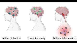
The second half of the eLife Neuroimmunology symposium consisted of an extensive Q&A session that linked the presented research to broader questions about neuroimmune memory, aging, and peripheral‑central communication. Attendees asked about the duration and purpose of immune‑related memory in the central nervous system, the role of contextual encoding (e.g., insular cortex) in immune responses, and whether immune states are altered during insular seizures or strokes. The discussion then shifted to aging, where Saul Villeda described how exposure to young blood or plasma rejuvenates cognition in aged mice, emphasizing the discovery that platelet‑enriched plasma fractions contain the chemokine platelet factor 4 (PF4/CXCL4). PF4 was shown to remodel the peripheral immune landscape (reducing inflammatory signatures, normalizing myeloid‑to‑lymphoid ratios, restoring naïve T‑cell pools) and to improve hippocampal‑dependent learning. Mechanistic probing revealed limited brain penetration, suggesting a peripheral mode of action, and raised questions about PF4 receptors (CXCR3, CCR1), blood‑brain‑barrier effects, and the need for lipidomic/metabolomic profiling. The final segment, presented by Isaac Chiu, explored how nociceptor sensory fibers act as a gut‑brain immune interface. Nociceptors detect microbial products (e.g., Salmonella, commensal bacteria) and, via the neuropeptide CGRP acting on the goblet‑cell receptor RAMP1, rapidly thicken mucus, regulate M‑cell differentiation, and protect against bacterial invasion and chemically‑induced colitis. The panel discussed sex differences, circadian influences, the impact of anti‑CGRP migraine therapies, and the broader relevance of pain fibers to other barrier sites. Throughout, the audience highlighted methodological gaps (e.g., BBB measurements, lipidomics, cross‑species validation) and proposed future experiments to dissect central versus peripheral pathways.
Chapters
Okay, I see that people are logging in. I am Carl Roslin, and I am absolutely delighted to welcome all of you to this eLife Neuroimmunology Symposium, From Evolution to Function in Health and Disease. I'm also delighted to be here today with my colleague and also editor at eLife, Dr. Beth Stevens. So as I can see, we have many participants logging in. I would like to introduce you a little bit to eLife today. So, uh, you probably are familiar with this, but I would like to remind you that in eLife we are an open access journal that publishes research from across the life and biomedical sciences. Now we are a broad range of editors, so I really encourage you to go to the eLife website and look into the section that you're interested and look into the editors that are specialists in all these different fields. And we want to remind you that we really welcome and encourage interdisciplinary research, which I think you will see today in this symposium. So thanks for joining us. I see that many more participants continue to join us, and I very much look forward to this symposium with you today.
Thanks, Carla. I also want to welcome everybody. This has been an incredible opportunity to bring together our community. In fact, I was just told we have 665 registered across the globe, representing many different countries and levels of training. And, and I just want to also remind you of some of the other goals and missions of eLife. And one of the, I think, special things about eLife is is really all of the editorial decisions are handled by active researchers in these fields. And the real ultimate goal is to make peer review fast, fair, and open. And this is actually, I think, we hope and we can encourage everybody to start to think more about contributing their work to eLife. So today's topic, as you just heard from Carla, is neuroimmune interactions. And we've brought together researchers from— that are going to tackle this topic across different perspectives and areas of science ranging from development to within the brain, neural immune communication, from the periphery to the brain, and in different contexts. So, here we have Irene Salinas from University of New Mexico, Michelle Monge from Stanford University, Asya— I'm sorry, Asya Rolles from Israel, and from Salva Leda from University of California, and Isaac Chu from Harvard. I'm now, without further ado, going to pass it over to Carla to introduce our first speaker, who is Irene Salinas.
So just before we get into the main talks today, I just wanted to let you know that we are— this symposium, we want everyone to enjoy it. And so to ensure that everyone has the opportunity to contribute, we ask all participants and everyone watching the symposium abides by eLife's code of conduct. So examples of behaviours that contribute positively to our communities include showing empathy and kindness towards each other, being respectful of differing opinions and viewpoints, and unacceptable behaviour would include making it difficult for others to speak or participate. If you have any questions, so each speaker will have 10 minutes for a Q&A after their talk, please post your questions in the chat and then we'll be able to read them out. During that Q&A section. The event is being recorded and will be available to view after the event, and live transcription can be accessed, which is provided by otter.ai. If you have any technical issues at any point, please just let us know. You can send a chat message on Zoom to Anya Stairs directly, who's a member of the ULI staff who will be able to provide you with any technical assistance. With that, I'll pass you over to Carla.
Thank you, Emma. And it is my real pleasure to introduce the first speaker of our symposium today, Dr. Irene Salinas. Irene is an associate professor and associate chair in biology at the University of New Mexico, and she is originally from Spain. Her lab, it's, I think, doing really cool stuff investigating neuroimmune interactions in organisms such as fish. Fish. And so I think today we're going to really enjoy her perspective on the evolution of neuroimmunology. So Irene, looking forward to your talk.
Oh, thank you, Carla, and thank you everyone for being here. Good morning from my end. Thank you to eLife for putting this amazing program together. And I'm probably the one with the most weird talk, of course, because most people haven't even thought about fish immunology, and now I'm talking to you about is neuroimmunology, but there you go. And first of all, I want to give you a little bit of a broader perspective on the field because I am quite new to the field. I started doing this maybe 7 years ago. I'm a mucosal immunologist by training. And so when I was really welcomed to this field, I really noticed how much diversity of neuroimmune interactions we were kind of missing. And so we wrote recently this review paper where we really wanted to highlight these important process called coevolution. And coevolution overall, it always refers to this reciprocal evolutionary change that occurs between pairs of species and they're interacting with one another or even groups of the species. But we can also think about coevolution in terms of interactions between physiological systems that occur within one organism or a group of organisms or species. And this kind of different scale of coevolution. And in this particular case, the coevolution between the nervous system and the immune system, we think it's really important in the tree of life. So why? Of course, we think that neuroimmune communication is vital for survival of the species, of all species, populations, and individuals. And overall, this communication, what it does is optimization of danger detection, which ultimately is actually the same thing as cooperating with living with symbiosis, with bugs and microbes that are beneficial to the host. In our recent review paper, we thought hard about how perhaps this coevolution had happened early on at the base of the metazoan tree of life, and even perhaps earlier than that, as I'll show you in a second. But we think that this is based on very essential blocks such as ion channels that you guys can see here in this ancestral metazoan cell, or acquisition of pattern recognition receptors or NF-kappaB signaling pathways, or perhaps a combination of the two under microbial pressures that led to the production of perhaps originally molecules that were supposed to regulate neuronal responses but quickly adopted or co-opted functions to perhaps have antimicrobial functions, just to say or give a few examples. So why do we think it's very amazing that the defense game is not just carried out by immune cells? Well, having neurons and other cells such as glia in this game brings a lot of benefits to, of course, detection and elimination of danger. And we can think about this from the inception of the immune response all the way to the resolution of the immune response, starting by very simple avoidance behaviors which have been really well characterized in animal models, C. elegans, avoiding bacterial pathogens, for instance. One that we are really particularly interested in my lab is the velocity and the distance of immune responses. So within milliseconds of encountering a pathogen or a pathogen product, neurons are gonna be able to fire really quick signals. And that has potent and powerful consequences for the entire organism because we are gonna allow detection of danger to happen really quickly, but also to alert far distant parts of the body. During the immune response, of course, here in C, we are going to see how neurons can basically regulate the immune response to either dampen it or potentiate it, depending on what mediators they are making. And we know many people are working on these aspects coming from the periphery to the brain and the brain to the periphery, which I think is really exciting. And then lastly, of course, something that we talk about less, I think, in general as immunologists, is how we shut this down. And I think shutting it down, of course, and the contribution of neurons to these last steps of the immune response is also really important. And I'm hoping that there will be more and more of those works coming up soon. So we put together this phylogenetic tree where what we basically did was looking at major innovations, key steps in evolution about things that happen in both the nervous system and the immune system to basically see in parallel when exciting things were happening. One of the things that was of course very clear is that even before metazoans, which emerged 800 million years ago, we may even think about unicellular organisms already having key aspects of an immune response like glutamate transporters, as well as key aspects of an immune response like complement or PRRs or other important transcription factors. So I want you guys to open your minds about what we know about neuroimmunology, which obviously is right here at the very end of this phylogenetic tree where we are always so human-centric, so mouse-centric, so perhaps C. elegans or Drosophila or zebrafish-centric. But there is all this incredible diversity here. And of course, as a fish person, I'm still only down here in this bottom section of the tree because we We study at the end of the day vertebrates, we study early vertebrates where we already have B and T cell-based immunity and there's already a central nervous system. And we almost can think of a fish that has almost every single aspect of what a mammalian immune system and nervous system would have, but with still some interesting caveats and interesting, I guess, unique adaptations. So in my lab particularly, we are interested in the neuroimmune communication in the olfactory brain axis. We study fish because this axis is actually really clean in teleost fish. Why? Because there's no connection with any other part of the body. Here on the right-hand side, you can see a rosette. The fish will have two of these, they form two flowers like this. They will have two olfactory nerves here. You guys can see one that we dissected out connected to the entire brain, the olfactory bulb, the telencephalon, the optic bulb, and the cerebellum. This allows us to dissect and look at very quick immune responses that happen from this peripheral olfactory system into the CNS without any connection with any other respiratory surface or any other organ. Of course, there's connection to the circulatory system. A few years ago, we started to develop our rainbow trout model. In the rainbow trout, as well as in other teleosts, olfactory organ is composed by the same olfactory sensory neurons that we have, but they also have these unique subset of neurons called CRIB neurons. And CRIB neurons are characterized by expression of Ora4, which is the homologue for the vomeronasal receptor 1 in mammals. And so apart from expressing Ora4, which is a vomeronasal receptor, these neurons also express another receptor called TRACA. And we reported in our work that this TRACA receptor directly interacts with viruses. In our case, we were using a rhabdovirus called IHNV, which is a really well-known virus that kills fish all over the world. We reported, of course, that there were very, very fast activation of neurons in the olfactory periphery, as well as activation of neurons in the CNS, in the olfactory bulb, within minutes of having contacted this virus. The other thing that we noticed is that immune response happened really, really quick in this axis. We were able to detect innate immune responses in the olfactory epithelium as well as in the olfactory bulb. These were not just recorded at the gene level, but we were able to record these really fast migration of CD8 T cells living somewhere, and we still don't know where they are located in these areas around the olfactory bulb and entering very quickly infiltrating the olfactory epithelium. This is where we left it at. Of course, we are still following this up in depth because we have a lot of questions. This original work by Ali was taken over by Pankaj Das, who was a master's student in the lab and now he's at Emory University, as well as Aurora, who is about to defend her PhD, and she's been taking this model to zebrafish because we have a lot more tools in zebrafish to look at the neuronal part of this model. And so for the last few slides, what I'm going to do is I'm going to give you an update of where we are at with this model in rainbow trout, because I wanted to really highlight non-model organisms rather than the zebrafish work, although of course we have really exciting results for both. And the first thing that I wanted to follow up, or we did, we did was to, of course, understand more, is this response happening just to the virus, to this rhabdovirus, or do we have similar responses when we give other microbes into the nose? And to, of course, surprise but no surprise, because these cells were showing this innate-like behavior, we were able to record very quick infiltrations of CD8 T cells in olfactory epithelium, not only in response to the virus but also in response to two different types of bacteria, Edwardsiella ektaluri and Yersinia ruckeri. And in the case of Yersinia ruckeri, we actually killed the bacterium. So this was a heat-kill bacterium, and yet we still recorded this infiltration of CD8 T cells. So this really highlighted an innate behavior, of course. And the next thing that we did was to look at whether danger signals or DAMPs and PAMPs would also recapitulate the response. So we delivered poly(I:C) into the nasal cavity or LPS. We applied a DSS model to damage the nasal cavity, and we gave acetic acid, which is a really well-known irritant for fish olfactory systems. And in none of these cases, at least 15 minutes after we do the stimulus, in none of these cases we were able to record that infiltration of the CD8 T cells. So this was not enough to give us that response. And then, of course, we wanted to check, is this just happening in the nose? And based on our hypothesis, it should be because we are proposing that this happens controlled by olfactory sensory neurons. So we did similar experiments using intraanal delivery of either a Darcella ecteroluri or the virus, HHMB. And again, 15 minutes later, we collected immune cells in the colon epithelium to look at whether or not there was any quick infiltration, and we saw not. So again, this highlights that this is unique to the olfactory-brain axis. So, our next experiment that was really important to us was trying to understand whether these were antigen-specific or not and whether or not we could detect any— some sort of invariant immune response. Because of course there were CD8 T cells, but we could maybe have some sort of NK T cell subset in there. And that innate-like behavior would suggest that that was the case. Our preliminary data had really shown that this was a TCR-alpha, TCR-beta positive CD8 T cell population. So we sequenced the repertoire of sorted CD8 T cells either in control animals or animals that had just received IHMV or a varicella. And we sorted the CD8 T cells from the nose and then we performed repertoire sequencing of the TCR-alpha using next-generation sequencing. And to our surprise, there was no evidence for a clonotype expansion or an invariant population that would come into the nose. Because as you can see here, if we look at the diversity of the CDR3s that come in into the nose, actually when we gave the virus or we gave Edwardzilla, we had way more diversity than when we didn't. And so that increased diversity, of course, suggests or indicates that this is a polyclonal response that is happening. And the last couple of experiments were aimed to dissect more what happens, what are the neuronal signals that are involved. And in fish, the transduction of olfactory signals can happen either through cyclic AMP or inositol triphosphate. And so we first wanted to know, are any of these two involved in our model? And we were lucky because we were first checking cyclic AMP. And to do this experiment, what we do is that we gave a drug called MK-801, H477 intranasally, which basically activates adenyl cyclase and will deplete all of the cyclic AMP in the system prior to the exposure to the virus. And you guys can see we only give the virus, of course we have our usual infiltration, but if we deplete with this drug to get rid of any cyclic AMP prior to exposure to the virus, we then abrogate the response. So this response is for sure cyclic AMP dependent. We don't know whether inositol triphosphate is implicated. And the last question that we had so far that I will tell you about is related to memory. And this was interesting to us because of course, if you think about our noses, we're exposed to viruses all the time, right? You can have an ACEL vaccine for whatever COVID or flu or whatever, and then a few weeks later you can be exposed to the same virus or to another virus. And then the next flu season you can have another ACEL vaccine that is slightly different. So what happens? Do we still have those quick innate immune responses coming to the olfactory epithelium when you have seen the virus once or when you have seen the virus twice. And we've done this in an experiment where we separated viral exposures one month apart. And the first exposure, as you can see, we have two control animals. If we give the virus just once and we detect it 15 minutes later, we know that we have our usual infiltration of CD8 T cells. But if we wait 30 days later, by which time we know 30 days later there should be already onset of adaptive immune responses in our— in rainbow trout because they're kind of slower than in mammals. So we wait a month for the adaptive immunity to be present and then we expose these animals twice. So they've seen IHNV twice or they only see the virus one month ago. And you guys can see that if you see the virus twice, you no longer have that infiltration of CD8 T cells coming into the nose. So this is also making us think a lot about memory and it doesn't necessarily need to be immunological memory. Perhaps there's something going on about the neurons that no longer respond to this virus, and we are really interested in following further here what is the neuroimmune implications of this observation. And so I want to just conclude my talk by just highlighting all of the immense diversity and unique lifestyles of organisms in this planet and how little we know about neuroimmune communication other than model organisms or humans. And I think very extreme environments and lifestyles are really going to give us and push what we find about neuroimmune communication in very cool ways. And we are real advocates of further exploring these non-model organisms. And now with CRISPR and single-cell RNA-seq sequencing, all of these different omic tools, spatial transcriptomics, we do not depend on specific antibodies to do a lot of this work. So I'm really excited for the field and what it's going to bring in the next 5, 10 years. I'm convinced that it's going to be exciting. And with that, I want to thank again the organizers and eLife. I want to thank my collaborator, Mark Huertas, Texas State, NSF for the funding for this work, the Center for Evolutionary and Theoretical Immunology here at UNM because I'm a part of this. And we recently celebrated the 10-year anniversary of my lab and we had a really wonderful retreat. And so I want to thank everybody in my lab as well. And with that, I'll take questions.
Thank you, Irene. That was wonderful. We have some questions already in the chat. Please, anybody that has questions, feel free to write them in the chat. Before starting, I'm really curious, what happens if you disable the receptor for this virus? Can you do that in those neurons? Or if you disable the neurons, what's the contribution of this to the overall immune response?
So in our 2019 paper, we reported that if we block TRACA, which is a tyrosine kinase A receptor, we block completely everything. We block any immune immune response, we block cell death of the neurons, we block everything. It's interesting because no one had ever reported TRACA as a receptor for that virus, but there were others that had been reported. Of course, there's not just one receptor for this virus. So it actually may explain a lot neurotropism that had been reported for this virus in the past. So there are isolates of this virus that have been recovered from the brain. And so that neurotropism those neurotropic isolates may actually use TRAKA receptors in neurons in the brain as well, not just in the olfactory periphery.
Okay, great. Okay, so let's see, we have Sunima Singh. Sunima is wondering, why are you looking at CD8 T cells only? Have you looked into other immune cells?
Yeah, good question. So we looked in our model at IgM and IgT B cells, the two main subsets of B cells that we have reagents to look at. We actually didn't see any equivalent fast immune response going on. We haven't looked at CD4 T cells because at the time we didn't have an antibody. We do have one now, so we can look further there. What we have done recently in zebrafish is basically remove any need for antibodies and go the single-cell way. So we have now kind of maps of what happens in the neurons as well as in the immune cells, both in the nose and in the olfactory bulb. So what we have been doing in the zebrafish is basically looking together periphery and olfactory bulb and then getting rid of any need for reagents.
Wonderful.
I think people are thinking a lot about those neurons and how are they firing. We have a question from Naomi Hamilton. Have you looked at calcium pulses in neurons following nasal infection?
For our zebrafish work again, in this original paper, our electrical activation of neurons was based on two readouts. One was electro-olfatto-grams, which are extremely hard to do electrophysiology in fish, but we did them and we also phosphorylated ERK stainings both in the nose and the brain. For the zebrafish, we can take better approaches, right? So what we've done is to use the GCaMP6s transgenic larvae. I did that during my sabbatical at Harvard at the Enger lab using larvae. And then after that, what we've been doing is the validation of these maps, which is phosphorylated ERK over total ERK in whole mount olfactory olfactory bulbs to kind of like identify which glomeruli are being activated. So for the zebrafish paper, we are going to have both the calcium signaling as well as the whole mounts of the olfactory bulb. Wonderful.
We have at least two more questions. So from C. Yang Li, thank you for your interesting talk. Do you have any suggestions or insight about what type of neurons are triggering the infiltration of CD8 T cells?
Yeah. So we know in our system that the Crib neurons, the ones that I showed you that are similar to orbital melanocyte neurons, are dying really quickly through apoptosis. And we show that in the paper. But we saw neuronal activation was not only in Crib neurons. There was downstream neuronal activation of other neuronal subsets. And so we think perhaps that not— the HHMV virus, of course, is not just one antigen. You're going to have many things going on in their epitopes. So there are probably multiple subsets of neurons from the olfactory bulb whole mounts where we can see that this is not just one glomeruli, although we do see preferential lighting up of the ciliated or the area in the olfactory bulb where the ciliated neurons are projecting to, which is the anteromedial region. So we still don't know. We think that is more of an overall or many glomeruli are being activated because there's many things going on. So there's cell death of one specific subset of neurons, but there's activation of other subsets of neurons But yeah, we are hoping to go deeper into the next stage with the zebrafish because we are moving on to the adults. The downside of doing it into the zebrafish larvae is that olfactory epithelium is not developed as in the adult. So we really need to get into the imaging of adults to get a handle, a better handle to this question.
Right.
Great.
And okay, last question actually from one of our speakers, from Asia Rolls. Great talk. Are there specific immune features that are more conserved, for example, monocytes, and their phagocytic activity. And also, I think Asia is wondering that these fish have maybe unique immune skin cells?
Many unique aspects, of course. Yeah, so monocytes, macrophages, they may be a little bit more conserved. One of the cool things about teleost fish that we are really interested in exploring further in our neuroimmunology concept is phagocytic B cells. So 80% of the B cells are phagocytic. And we are really excited to continue on to this. Another exciting aspect is lack of TLR4 signaling. So they don't have a functional TLR4 or they have lost TLR4 in their genomes. So we are again really interested, for instance, how microbiota signaling is happening in this system. Skin cells. Yeah, I mean, there are really cool aspects of immune— immunity in fish that I could talk for hours because that's what I do. So, but yeah, I guess macrophages are my favorite.
Maybe for another symposium. Wonderful, thank you so much.
Thank you.
Yes, that was great. I'll let now Beth introduce our second speaker. Thanks a lot.
Thanks, Irina. Okay, great. My pleasure now to introduce our second speaker, Dr. Michelle Monge, who's a professor at neurology at Stanford University and a newly minted HHMI investigator and MacArthur Fellow. Michelle is a really creative neuroscientist and neuro-oncologist who's been investigating the growth and development of both healthy and cancerous cells in the brain. She discovered that neuronal activity regulates the myelinating glia of the brain and how this impacts neural circuits. And more recently also discovered that activity also can regulate glial cancer cells in the brain that can drive both glial progression through a host of neuroimmune signaling mechanisms that I think we're going to hear more about today. So over to you, Michelle. Thanks so much for joining us.
Today.
Thank you so much for having me. And I'm going to talk about neural immune interactions in cancer therapy and also in long COVID. So this is very new work, unpublished, and also a new talk. And in the spirit of a lightning talk, I'm going to try to cover a lot of ground that hopefully we can dig into the details of in the Q&A. And so today, I'm hoping that by the end of this 15 minutes or so, that I've convinced you that neuroinflammation, especially reactive microglia and other myeloid cells, can cause multilineage neural cell dysregulation that contributes to cognitive impairment in multiple different disease contexts, including after cancer chemotherapy, after even relatively mild respiratory SARS-CoV-2 infection, and after CAR T-cell immunotherapy. And then I'm going to shift gears a little bit at the very end and discuss how CAR T-cell immunotherapy for lethal CNS cancer is enormously promising, but may be limited by some of the very same immune suppressive myeloid cells. So as this audience knows well, healthy cognitive function really depends upon intact mechanisms of neuroplasticity, and those include plasticity of synaptic structure, function, and connectivity, myelin plasticity and homeostasis, and the generation of new cells in the hippocampus, particularly in early life. And each of these mechanisms can be impaired in the context of traditional cancer therapies, including radiation and chemotherapy. And over the last 20 years or so, we've come to understand that reactive microglia and other myeloid cells are really quite central to this cancer therapy-related cognitive impairment. Cancer therapy-related cognitive impairment is, is a really debilitating syndrome that's characterized by impairment in attention, concentration, memory, speed of information processing, multitasking, and other executive functions. It unfortunately affects a large proportion of cancer survivors to a variable extent depending on the age at which they were treated for their cancer and the exact kind of therapy that was used.
Used.
And in many cases, the persistent cognitive dysfunction is somewhat mysterious. There's no obvious structural damage. Standard neuroimaging is unremarkable, but advanced neuroimaging does reveal subtle white matter aberrations and decreased volume, particularly of the hippocampus. And so these— what has become clear is that central to this are reactive microglia, which can in turn induce a neurotoxic state of astrocyte reactivity, as we know from beautiful work from the Baron lab and Shane Lidlow's group. These neurotoxic astrocytes together with microglia can impair myelination, both homeostatic and myelin plasticity, and reactive microglia and associated cytokine production can also impair the process of neural stem cells making new neurons in the hippocampus. Focusing on that, that myelin component, my laboratory recently showed, or relatively recently, pre-pandemic, so perhaps it feels fairly long ago, that cancer chemotherapy can directly particularly agents such as those commonly used for treatment of leukemia and other diseases like methotrexate, can directly activate microglia. These in turn induce a neurotoxic reactive state of astrocytes, and together they dysregulate the oligodendroglial lineage that forms myelin, so important for really tuning circuit function in terms of speed and metabolic support. So what's really interesting is that systemic methotrexate chemotherapy directly activates specifically white matter microglia. Methotrexate given to a mouse, for example, evokes a very, very particular pattern of white matter selective microglial reactivity. And that makes some sense based on the really elegant work from the Stevens lab and others that has defined a subpopulation of microglia called axon-trusting microglia. Tract microglia. And this distinct population really does seem to be exquisitely sensitive to systemic toxic and other inflammatory insults. We find that myelin homeostasis and plasticity are disrupted again, particularly in the white matter tracts, and that depleting microglia using a CSF1R antagonist restores myelination, partially normalizes astrocyte reactivity, and rescues cognition after methotrexate exposure, at least in mice. And so, you know, noting this, this really reproducible syndrome of cognitive impairment that occurs after chemotherapy, sometimes called chemo fog, there are really stark parallels between that and the cognitive impairment that's now being reported after even relatively mild COVID. This so-called COVID fog, or the brain fog associated with Long COVID is characterized by impairment in attention, concentration, memory, speed of information processing, and multitasking. Really very clinically similar to what we see after cancer therapies. This unfortunately affects a large proportion of COVID survivors, particularly those who contracted COVID prior to availability of the vaccine. And a meta-analysis of nearly 10,000 subjects found that about 1 in 4 individuals who were who were infected with COVID early in the pandemic now experience persistent cognitive dysfunction. This risk of cognitive dysfunction is more prominent with more severe COVID, but it is still quite significant in people who had relatively mild COVID. It's not yet clear what this is going to look like now that vaccines are available and with new variants. And I want to point out encouragingly that recent reports indicate that breakthrough infections in vaccinated people may carry lower risk for long COVID in general. But we wanted to understand the extent to which the systemic inflammation evoked by the respiratory component of SARS-CoV-2 infection might be translating to neuroinflammation and evoking some of the very same pathophysiological dysregulation that we see after cancer chemotherapy. And so, we collaborated with Akiko Iwasaki at Yale, who has a very elegant mouse model in which only the respiratory tract of the mouse is infected. Her lab first introduces the human ACE2 receptor required for SARS-CoV-2 infection just intratracheally. Then after that, SARS-CoV-2 infection intranasally happens, but the infection is limited to the region of the body that has the human ACE2 receptor, which is just the respiratory tract. These mice don't exhibit any sickness behavior. They don't lose weight. They're apparently asymptomatic. And there's no evidence of SARS-CoV-2 infection in their brains. But despite that lack of direct neural invasion, there's really quite prominent neuroinflammation. So this mild respiratory SARS-CoV-2 infection persistently elevates cytokine levels, both in CSF as well as in serum, both at 7 days after infection and persisting to 7 weeks after infection. Together with that, we see that same pattern of white matter microglial activation after mild respiratory SARS-CoV-2 that we expected to see and have seen after cancer therapies of various types. Examining human brains of people who died with COVID these were not mild cases of COVID because the people did die, but they didn't die from the necrotizing pneumonia that puts people in the intensive care intensive care unit. Instead, they died outside of the hospital suddenly, in some cases from things like clots and heart attacks and probably associated with COVID So I want to point out that these aren't necessarily mild cases, but they're also not severe pulmonary infections. What we find in these human cases, which were all from New York City in the spring of 2020, is that there's a stark increase in microglial reactivity again, specifically, or at least selectively, within the white matter. And this is in comparison to age and sex match control human tissue in which individuals died for other reasons. You don't appreciate that same white matter selective or white matter enriched microglial reactivity. Together with that microglial reactivity, we see an inhibition of hippocampal neurogenesis, as has been as I described previously in the context of neuroinflammation. And the degree of inhibition of hippocampal neurogenesis correlates with the degree of microglial reactivity within the white matter of hippocampus. Together with that inhibition of hippocampal neurogenesis, we want— we noted one particular cytokine that was prominently elevated after respiratory SARS-CoV-2 in the mouse that actually became even more prominently dysregulated regulated in the CSF by 7 weeks post-infection. And this cytokine, this chemokine, CCL11, was found in humans who have Long COVID to be elevated in those individuals who experience the cognitive symptoms compared to those who don't experience these cognitive symptoms. And this was just such an intriguing finding to us. Because Salvaleda, who we're going to hear from later in the morning, has previously described the chemokine CCL11 as correlated with impaired cognition during aging and showed that it's necessary and sufficient to decrease hippocampal neurogenesis and cause memory impairment in mice. And so, we'd really like to better understand how this chemokine is playing a role in the cognitive symptoms of long COVID. Together with this neuroinflammation, decreased hippocampal neurogenesis, we see the same loss of oligodendrocytes that we observe in the context of cancer chemotherapy in the context of mild SARS-CoV-2 infection in mice. This decrease in white matter oligodendrocytes persists for at least 7 weeks after clearance of infection. And together with this decrease in myelin-forming oligodendroglial cells, there's also a decrease in myelinated axons. So, this kind of paradigm of white matter-selective microglial reactivity, neuroinflammation, and dysregulated hippocampal neurogenesis and oligodendrogenesis, you know, is present both in cancer therapy, it's present in, you know, systemic inflammatory syndromes like COVID? What about cancer therapies that depend upon inducing systemic inflammatory response like CAR T-cell immunotherapy? So CAR T-cell immunotherapy, which depends upon engineering a patient's own T-cells to express a chimeric antigen receptor that then very specifically targets that patient's cancer, has really been transformative for refractory leukemias and is showing enormous promise for other lethal solid tumors, particularly those that occur in the CNS. And I'm going to share a little bit more about that at the end of the talk. This kind of immunotherapy is associated with really prominent cytokine release and frequently causes cytokine release syndrome. There's a high rate of acute and transient neurological symptoms, including sort of encephalopathy, neuropathy that can happen during the acute treatment phase. The long-term effects of CAR T-cell therapy on cognition are understudied, but a couple of early reports are fairly concerning. And my personal clinical experience makes me very concerned that we're going to see a syndrome of cognitive impairment that follows this immunotherapy, much as we do after more traditional cancer therapies. And so we wondered whether neuroinflammation from systemic or CNS-targeting CAR T-cell therapy might cause a similar lineage dysregulation and impair cognition. And so, we examined this in a mouse model, a mouse xenograft model of acute lymphoblastic leukemia, in which mice are xenografted with leukemia, then treated with a leukemia-targeting CD19-targeting CAR-T cell, and then assessed for cognitive function in the novel object recognition test and analyzed histologically and molecularly. And just like I recently described for SARS-CoV-2 infection, we see really prominent neuroinflammation in these mice in the CSF, elevated CSF cytokines following the clearance of the tumor. And I want to point out that leukemia is a fairly inflammatory state to begin with, and CSF cytokines are elevated at baseline, then further elevated following clearance of the tumors by CAR T cells, including— just highlighting it again— CCL11. CAR T-cell therapy of acute lymphoblastic leukemia activates white matter microglia selectively, just as we've seen again and again in other disease contexts. This decreases white matter oligodendrocytes, impairs hippocampal neurogenesis.. And we find that there is an impairment in cognitive performance in the novel object recognition test that's rescued when we deplete microglia again using a CSF1R inhibitor. So a CSF1R inhibitor put into the mouse chow decreases, decreases the microglial density within the brain by about 80%. That partially rescues the oligodendrocyte density and rescues cognition, at least as measured in the novel object recognition test. And I've been, you know, personally very interested in understanding the long-term effects of CAR T-cell therapy because I, in the last couple of years, have been leading a clinical trial of GD2 CAR T-cell therapy for a particularly lethal form of brain and spinal cord cancer that occurs in children and young adults. This is a tumor that diffusely infiltrates the brainstem and diffusely infiltrates the spinal cord called H3K27M mutant diffuse midline glioma. We have been giving CAR T-cell therapy. We started initially by giving it intravenously and then later by giving repeated doses directly into the central nervous system. Into the ventricular system. And this was based on preliminary, you know, preclinical data from my laboratory in collaboration with Crystal Makel's laboratory showing that this particular kind of cancer has an antigen called GD2 on its surface, and we can successfully target that with GD2-targeting chimeric antigen receptor T cells that clears the tumor in mice bearing tumors either in their brainstem or in their spinal cord. In our early clinical work, we found that really excitingly for a disease that has no therapeutic options, that subjects experienced both radiographic tumor shrinkage, as well as really encouraging clinical improvement. We saw that not only in the brainstem, but also in the spinal cord. And so that was the first time in my career I've been able to give good news to a patient. But it didn't last. These responses were robust but transient after a single dose of CAR T cells. And what we found is that over the course of about a month, as tumors shrank, as if we let the patient then go untreated for a subsequent month or two, the tumors regrew very rapidly. But excitingly, they were— we found that giving a second dose, particularly directly into the nervous system, could elicit a repeat repeated response. And so trying to understand what was going on there, we find in the CSF of these patients that there are really prominent myeloid cell populations. We find greater immune activating myeloid populations after intracranial administration than after IV administration. And immune suppressive myeloid populations that increase later in the post-infusion month, and interestingly overlap with signatures of disease-associated microglia and axon tract. Applying these lessons and giving repeated doses monthly now to patients of GD2 CAR T-cell therapy, we're seeing even more promising results. And sharing, you know, the best response I've seen to this disease in my career, 5 repeated monthly, every 4 to 6-week infusions has resulted for one of our patients in the trial in near-complete resolution of this diffusely infiltrative tumor in the medulla, pons, and in the midbrain. We don't yet know how durable this response will be, but it's enormously encouraging. And so to conclude kind of that overview, systemic immune challenges can cause multiple multilineage neural cell dysregulation. This is associated with elevations in pro-inflammatory cytokines in the CSF and in the serum, especially CCL11 is of interest mechanistically. We see white matter selective or white matter enriched microglial reactivity, inhibition of hippocampal neurogenesis, loss of white matter oligodendrocytes.
Myelin.
And I want to point out that SARS-CoV-2 is particularly immunogenic. It causes a profound immune response that's quite broad. But the extent to which other systemic infections may be causing similar neuroinflammatory pathologies remains to be carefully determined and studied and compared to SARS-CoV-2. And I think it's pressing that that happen. Finally, myeloid cells may contribute both to the long-term cognitive symptoms and limitations in CAR T-cell efficacy for CNS tumors. Hopefully, strategies to address myeloid states may prove beneficial in the context of CAR T-cell immunotherapy for these otherwise lethal cancers of the brain and spinal cord. Many, many people to thank. I want to really point out Anna Gearty, Anthony Fernandez-Castaneda, Lehigh Acosta, Aaron Gibson, and Chris Mount, as well as our critically important collaborators. Hopefully we have some time now for questions.
Thanks so much, Michelle. That was a fantastic talk, and actually you covered quite an arc of a lot of related threads throughout, which I think is one of the things that's most exciting about your work. There's a lot of questions, and I have some of my own, but I'm gonna like make sure that everybody else gets a chance here. So let me start, uh, with the first one. Um, actually, Jeremy, uh, Borninger says, hi Michelle, great Great talk. I missed this, but what do you think is special about the white matter microglia that make them especially vulnerable or sensitive to these changes? Could you comment on that?
Yeah, I mean, Beth, I'm going to refer to your work in the Hammond et al. paper and in others that, you know, single-cell transcriptomics shows that the axon tract microglia are unique. You have shown, Beth, that they contribute to developmental myelination. Why they're so sensitive to these inflammatory responses is an open question that I think we need to understand. And why that change appears to be relatively persistent, right? After methotrexate chemotherapy, that change in state from, you know, more neurotrophic to more neurotoxic persists for at least 6 months in mice. And we see it in patients years after chemotherapy. So, we, you know, really need to understand that. At the molecular level, at the epigenetic level. And I think there's a lot more work to do.
So, yeah, great question. It's exciting. And it's also be interesting to know about how they're signaling to other cells there that might be actually mediating some of the responses. So whether they're the vulnerable cell or whether they're the ones actually mediating the changes. Okay, great. So there's a couple other questions here. Michelle, is microglia depletion sufficient to restore white matter and cognition? Cognitive and cognition in the long COVID in the mouse models? This is from [INAUDIBLE].
So that's a fantastic question. And Kiko Iwasaki's lab and mine are working together to do that experiment right now. If you can imagine working with SARS-CoV-2 in the bunny suit in the BSL-3 facility, these are really challenging experiments. We can't do behavior there. But we are giving them this CSF1R inhibitor in their food and will at least be able to determine whether that rescues the cellular deficits.
Okay, great. And then the question that came up from a few people, which cell types produces the CCL11 and what triggers its long-term production? And obviously, maybe you can comment also on its relevance for a biomarker.
Yeah.
So we— these are such fantastic questions. So CCL11, its receptors, CCL11R, CCR2, 3, and 5 are expressed very prominently in microglia. I actually have some hot-off-the-press facts data from my postdoc showing that they're also expressed in oligodendrocytes and astrocytes. And so, you know, there are multiple cell types that could be responsive to CCL11. And we are presently trying to actually replicate some of the experiments that Saul Villeda did in his really beautiful, beautiful Nature paper in 2011. To then look at microglial reactivity after CCL11 serum elevations and the effect on oligodendroglial cells. So more to come. It's not entirely clear.
Yeah, it's a really interesting connection there. Hopefully we'll hear more from Saul on that as well. Another question that came up a few times, thinking about, again, the characterization of these different myeloid cell states, the DAM phenotype has been observed in immunotherapy-naive glioma patients. Do you believe the DAM phenotype phenotype is expanded by CAR-T therapy and/or that DAM-like macrophages are present during CAR-T treatment? Could they be limiting their efficacy? And this is one of a couple—
this came up a couple of times. Yeah. So the overlap between the DAM signature, the axon tract microglial signature, and the myeloid-derived suppressive cell sort of signature in the same sort of part of the UMAP space I thought was really fascinating. We see very prominent myeloid cell infiltration of these tumors. We don't yet know— so there's definitely more in the mice after CAR T-cell immunotherapy, in part because there's also a lot of cellular debris to clear. And the extent to which they contribute to tumor progression, as well as to inhibiting CAR T-cell efficacy, I think needs further study. But it's a flaming gun, or a smoking gun. Whatever the right metaphor is. And something that, you know, right now we're looking at depleting those myeloid cells in the context of CAR T-cell therapy in mice, at least. And what we're finding actually is that that dramatically increases the efficacy of the CAR T-cell. We can clear the tumors with a much lower dose of CAR T-cells if the myeloid cells are depleted, which really does suggest that they're functioning to suppress the CAR T-cell action. How to how to best achieve that in patients is something we have to sort out. But hopefully, as we move forward to achieve, you know, that amazing response I showed you at the end of the talk in all of our patients, you know, I think we're going to have to figure out how to combine myeloid targeting therapy together with CAR T-cell therapy.
Right.
Great. And a question from Luciana Veneziani basically asked, again, congratulating you for this amazing talk, wondering if during cancer treatment, the elimination of immune populations such as meningeal gamma delta T cells. I knew we had to have a T cell question mechanism here. Could this be related to the microglia activation? And then a related question is about the relationship to the COVID whether or not— I can get to that secondly. Why don't you start with the T cell question, the gamma delta T cell question?
Yeah, it's a great question. And we don't know. What we're hoping to do— we've used relatively old-fashioned methods so far to look at histological changes and immunohistochemical assessment of microglial state and other immune cell state. We're now doing NUC-seq after cancer therapy in mice and hope to have a more granular sense of what immune subsets are present and being changed in that context.
Yeah. Yeah, I think it definitely introduces this need to understand the borders and that communication that's going on in these different— both in the case of cancer and tumors as well as COVID, potentially.
Right? Absolutely.
Absolutely. And then another question there was related to your COVID project and research. When you were characterizing both in the patient brain tissue and mouse model, do you think there's a sort of— are the changes in the microglia and mechanisms more broad in the parenchyma or more restricted to the white matter in these models?
Yeah.
So after I'm going to define severe COVID as— I think you could argue that since the patients died, they had severe COVID. But I'm going to define severe COVID as necrotizing pulmonary infection. In the context of necrotizing pulmonary infection that puts somebody in an intensive care unit unable to oxygenate their blood, there is both cortical as well as white matter microglial reactivity. So in that severe state, the whole brain is clearly inflamed. And in selective gray matter areas, particularly in the brainstem, microglial reactivity has been described even in less than severe pulmonary COVID. In this case, we really saw an enrichment of microglial reactivity, at least as defined by CD68 immunoreactivity, quite specifically in the white matter.
OK, interesting. Thank you, Michelle. Isaac Chu asked a question. Exciting work on long COVID and CAR T cells. Do you detect evidence of immune cells in the CSF or changes in the border regions? Kind of what I just mentioned earlier, i.e., cord plexus or meninges in these models. Like, do you have any early hints of changes that are happening there?
Yes, I would love to understand in both of these contexts what's happening at the brain borders. And we don't have that. And, you know, all we have is the CSF cell— you know, the cells that we can detect in the CSF of the mice and of the patients. And we have half a million single cells we've recently sequenced to analyze. So hopefully we'll have more data. We have a lot of data and we need to look at it carefully. In analyzing the CSF of the first 4 patients over, you know, their disease course and with multiple treatments, you know, we clearly see an increase in myeloid cells over the course of, you know, the 4 weeks after giving CAR T cells. But there's a lot. There's so much that we need to still understand.
Absolutely. Great.
Well, how are we doing on time, Emma? Are we—
should we—
do we have time for a few more questions?
There's 1 more minute. So if you can fit in a lightning question to go with your lightning talk.
OK. OK, great. I will ask 1 last quick question. I guess, is back to this question of trying to better understand those tumor-associated macrophages. Do you see— I know it's early days for your understanding and characterizing them, but do you see certain similar populations across the pediatric and other glioma cases? In other words, are there some common subtypes that might be relevant across these? Or do you think it's disease-specific or even developmental stage-specific?
Specific?
Yeah, no, that's a great question. We looked at— we isolated microglia and other myeloid cells from pediatric diffuse midline gliomas, diffuse intrinsic pontine gliomas, as well as from adult glioblastoma and compared them. And they're markedly different. And part of that reflects probably differential cytokine and chemokine expression by the tumor cells themselves. The GBM cell— adult GBM cells express many, many cytokines and chemokines and the pediatric version is very sort of silent from that perspective. So there are real differences in the immune microenvironment.
Okay, great.
Thank you.
Well, we ended perfectly on time, which is pretty amazing considering the number of questions you had. Thanks so much, Michelle. I'm now going to turn this back over to Carla who will introduce our next speaker.
Thank you.
Thank you, Michelle. That was wonderful. Thanks, everybody, for the great questions and also the lovely messages congratulating our speakers. If we didn't get to answer your question, we are really sorry, but we'll try to get back to you. We'll probably get back to you by email. Now, I'm really delighted to introduce our next speaker, Dr. Asya Rawls. So, Asya is an associate professor at the Technion in Israel. She's joining us today from Israel, and she's also an international Howard Hughes Medical Institute welcome investigator. As you will see, her laboratory is interested in understanding the neural basis of how emotions and thoughts affect physical health. I think you'll see today she employs different approaches from chemogenetics to behavior to address those key questions, and I think we're going to be really blown away by some groundbreaking discovery that her lab has done on how we remember immunity. So, Asya, we very much look forward to your talk.
Thank you very much, Carla. Thank you very much, both of you, for inviting me to this session and for organizing this amazing event. I'm enjoying it very much, and I hope that soon it will be also— we are back to real life. So I would like to share with you some of our recent discoveries and the way we started thinking of the whole concept on how the brain can communicate with the immune system and how we remember or represent the immune response in the brain. Classically, the way we think of immune memory, we are— we now, everyone in the crowd, even if non-immunologists, know it comes in a bottle, in a vaccine form. This is immune memory and it changes our immune system, right? So it's represented in the immune system in antibodies, in memory B cells, but in fact, there are some occasions going back all the way through the 1800s showing, for example, that people that have allergy to pollen, if you show them an artificial flower, they will respond with allergy. So this cannot be explained by a response that it's solely by the immune system, right? It has to be something else. There are also studies going to the 20th of the last century showing that the immune system can be conditioned in a classical Pavlovian conditioning. And the idea there is quite amazing. These were works that were done in Russia by Metalnikov and Korin, and they were kind of lost from the history, rediscovered by Adler and Cohen in the US in the '70s. And I think most of the recent work was done by Manfred Chodosky in the field. And the idea is the following. What they showed is that you can give drug, an immunosuppressive drug, together with saccharin. If you give them, couple them together, like classical conditioning, after a while, it's sufficient that you give them saccharine, they will respond with immune suppression. Classical memory, right? Like the classical kind of memory that we store in the brain for any other type of conditioning. Where should this memory be stored in the brain? We decided to focus on the insular cortex. The reason for that was for several folds. First, conceptually, the insula is this tight hidden behind of the cortex is responsible to generate interoception, namely representation of the state of the body. So it almost doesn't make sense that this kind of representation will not include information about the immune system, which is really informative about the state of the body. Anatomically, this area receives inputs from sensory innervation from the periphery, and it can send inputs to the sympathetic and parasympathetic pathways. And functionally, Martin Przydowski showed the disruption of the insula can disrupt immune conditioning. So we focused on this area. And this was the work mainly by Tamar Koren in the lab together with Kobi Rosenblum from Haifa University. So what we did, we wanted to see if immune activity in the periphery will be reflected in the insula. And we chose as an immune concept, site, the gut-brain axis. And of course, because this is a very active neuronal environment and active immune environment. And as a challenge, we chose DSS-induced colitis. When we give them in the drinking water DSS, this causes damage to the epithelial layer, and the ensuing immune response is modeled for colitis or for Crohn's or colon localized inflammation. To capture the to label neurons, we use trap mice. And trap mice allow us to capture neurons that were active at a given time point, because when there is tamoxifen in the system that we inject, and then we determine when we are going to label neurons, and neurons that are active, they express immediately genes. So this system allows us to capture neurons that were active when we injected tamoxifen, and then capture meaning that we can express Cre in these neurons. Express Cre, now we can control these neurons. That's what we did. We targeted the insula so we can express fluorescent reporter in neurons that are going to be trapped. Mice recovered completely. We gave them DSS to induce the inflammation, and then we injected tamoxifen in the peak of the inflammation to capture the neurons that were active during the inflammation. Then we waited for 3 weeks and we sacrificed the mice and visualized the brain. And you can see here that indeed, we can see activity labeling of neurons in the insular cortex, and there are more neurons that are active during colon inflammation. Okay, so it seems that there is some activity in the insula which was reflecting that. But is it meaningful in any way for immune regulation? Now, instead of just expressing a fluorescent reporter in these neurons, now we express their DREADDs. And DREADDs are muscarinic receptors that are mutated, and now we can control the activity of these specific specific neurons. So now we will capture the neurons that were active in the peak of the inflammation, and when we want, we can control them. Control them, namely, we are going to reactivate them. So after the mice recover from the initial inflammation, we'll reactivate the neurons that were active in the original inflammation and see what happens. So that's what we did. We activated, reactivated this neuronal ensemble, look in the column, and what you can see that we actually can generate the inflammatory response in the colon just by reactivating these neurons. And you can see we also compared it to the original response. So it recapitulates many of the original features of the DSS-induced colitis. And you can see it here, even in staining, the red cells are CD45 cells, and they infiltrated there following the activation of the brain. So yes, it seems that there is something meaningful in this immune— in this representation that is happening in this neuronal ensembles. But what I didn't mention here is that what is my control? My control is that I'm using a control virus. So it's either almost like activating the neurons or not activating them. So maybe it's a general thing if I'm going to activate them. So what we did now, we used 3 groups of mice. In all of them, I'm going to express the DREADDs. So I'm going to reactivate neurons that were active at a given time point. The difference is when I'm going to capture them, when I'm going to inject tamoxifen. In one group, I'm going to inject it before I'm giving them the DSS. So just kind of baseline activity in the insula. In the second group, the peak of the inflammation, the same group that we had before. And the third one is that after they recover. We wait for 4 weeks, they recover completely, we give them CNO, which will activate the these neuronal samples and we sacrificed the mice. What you can see here that only the group that we reactivated the neurons active in the peak of the inflammation actually could recapitulate the inflammatory response. You can see it here again in the staining. You can see that there are more immune cells only in the during group. It seems that it's meaningful. It's kind of a unique activity, but maybe it's innate response. What do I mean? Maybe it's a matter of how many neurons I'm activating. Because as I told you, when in the peak of the inflammation, there are more neurons that are active. So maybe it's a matter of how many neurons I'm activating. So what we did now, we just injected GQ, which activates the neurons, but in non-specific manner. So it will capture, you can see here for comparison, comparison. The green is the neurons that we are going to activate randomly, and in red is the number of neurons we will activate if we are going to do the trapping. So it's— we're activating about twice the number of neurons, and still you can see here in the column, nothing is affected. So there is something specific. It's not that generally once I'm going to activate the insula, I'm going to get the effect. It's something specific in this representation. So it's not an innate response of the insulin, but maybe just specific to the gut-brain axis. We know it's very active. So now we chose a completely different model of inflammation, which was the peritonitis, which has a different dynamics. The site is different. It's the peritoneum. And we inject zymosine, which activates TLR2. And now we capture the neurons in the peak of this inflammation. And now we are already smarter. So we have control group and the activation and the experimental group.— both expressing GQ. It's just a matter of when we are capturing the neurons. And when we're reactivating them and look in the peritoneum, you can see that activation of these neurons generates inflammation in the peritoneum. Again, representative in many features to the original inflammation. But maybe it also has an effect in the colon.
Right?
But no, there is no effect in the colon. This was site-specific. So there is also anatomical information that is encoded by this neuronal representation. So it's not specific to the gut-brain axis. Okay, so how is the signal mediated from the brain, from this insula, to the periphery? To address this question, we collaborated with Oren Kobiler from Tel Aviv University, at the university, we gave a pseudorabies, which will go retrogradely. We injected the virus to the colon and followed where it goes in the brain. And you can see that it reaches several brain areas, including the insular cortex. Makes sense, meaning that neurons in the insular cortex can deliver information to the colon. But are they the same neuron that we captured? So now what we did, we strapped mice again. We trapped the neurons that were active in the peak of the inflammation and now injected the PRV. And you can see that indeed there is big overlap between the neurons that we trapped and the neurons that can deliver messages to the colon. And then we wanted to do the same thing with the colon, with the peritoneum. We injected the virus to the peritoneum, but mice did not hold on. So they just died before we got— we could see any trace in the brain. And this was difficult. And maybe because they need to go through more synapses, we don't know, maybe it's something in the inflammation. So now we decided that maybe we'll just— because we need to keep them to shorten the duration that is required to see a signal in the brain. So what we did, we injected in the trapped mice instead of a regular virus, we injected AV1, which also has some anterograde activity. So we said, okay, this will go one sinus forward, and then maybe we'll see when they— where they meet with the input coming from the, from the periphery. And it was really nice to see where they met, because they met in the— basically in two sites: the DMV, which controls the vagus nerve, and in the RVLN, which controls the sympathetic nervous system. So it seems that these neurons coming from the entorhinal cortex can control both the peripheral parasympathetic nervous system and the sympathetic nervous system. Of course, we don't know what is the proportion, what are they doing there. It's just that there's anatomical connectivity that allows this communication. So it tends to be mediated by the autonomic nervous system.. And finally, so is there any clinical potential to this kind of manipulation? If the brain has this representation, maybe, and maybe it kind of exacerbates the immune response, it can initiate on its own this immune response. Maybe if we stop the activity there, maybe we can somehow attenuate the inflammation. And we did that. So what we did now, we inhibited, used GI, which is a different form of bread, which inhibits neuronal activity. We injected it just entirely to the insular cortex. It's not targeting specifically the representation. It's something that we are trying to do. It's much more difficult to achieve instead of a general inhibition. But already by inhibiting overall, we could see that there was less shortening of the column. So in terms of clinical sign, the column colon length, because the colon shrinks during DSS colitis, so it was less intense. And we also saw that the spleen size was reduced, so less inflammation, as well as more specific effects on cytokines and immune cells. It did not completely eliminate the disease, it's important to note, but it did affect many parameters in terms of clinical and immune inflammatory parameters. So I think kind of to summarize this concept, what we what we see is that immune-related information is stored in the brain almost in mnemonic-like representation. These are reminiscent of memories. But when I'm saying this immune-related information, I think it's very hard to say what— to understand what I mean. And that's what we're trying to understand. What kind of information is stored there? Is it information about the immune response or maybe information tissue? Does the brain store information in terms of anatomy or some sort of pathways? And all of these questions, I think we need to remember that they are completely kind of unknown. What we did show is that the brain can initiate immune reactions. And I think it's especially relevant to entire field of psychosomatics because we know that there are many conditions, actually many gut-related disorders are characterized characterized by an emotional trigger that initiates the flare of the disease. And maybe in a way, it's almost like— I tend to think of that almost as PTSD of the immune system or kind of overwhelming memory that is kind of repeated and can cause, instead of being something beneficial, actually can cause the disease. And I think— and we know that there's anatomical connectivity that can support this potential. And maybe the biggest The question is why? Why should the brain waste power on remembering immune reactions? And I think maybe in a way the answer is how not, why not? It's a, we remember information, we are built or designed or supposed to act in a way that our immune, our immune reactions will, or we will predict what's going to happen and initiate this response. For that, we remember everything around us. It doesn't make sense that we will not remember how our body reacted if eventually we survived and it worked. So I think this is actually fundamental information for us to know. And of course there are many questions, but we are still working on that. I want to thank the people that were involved in this work, especially also our lab manager, and Rui, who contributed a lot to this work, and of course our funding, and thank you for listening.
I'll be happy to answer the questions. Thank you, Asia. I, I really want to congratulate you for your elegant work and really asking very important and fascinating questions. I think we're all like, you know, so many questions. Again, I will make sure that attendees can ask their questions. So let's see. So we have quite a few. So we have one from one of our speakers from Irene, so she says, so excited. So she's wondering what happens to the microbiota composition after the neuronal activation in the DSS model? Does it follow similar changes as when you give the DSS first time? I don't know if you had a chance to explore that.
Yeah, yeah, we actually tried at some point. So we had the collaboration. I'm sorry, we had the collaboration. I have a crazy dog here.
Sorry.
We had a collaboration with Nama Gebazetowski, she is an expert on microbiome, and we tried to see changes, but there was nothing dramatic that we could identify. Actually, after many types of stimulation that we did in the column, we were surprised that we could not see this kind of very dramatic effects in terms of microbiota.
So, I think we are missing something something, but I don't know why we don't see it. Yeah, great. So we have a question from Jeremy Borniger. I think Jeremy must have been very excited when you were presenting your results. So he was curious on how the signal gets to the insula, which you address, but he's also wondering, he has a more speculative question. What happens with repeated conditioning? Does the immune response to insula stimulation get stronger and stronger?
It's actually a great question. In terms of sensory inputs, we are still looking into it. I don't have a definitive answer. We're working on that. But I think in terms of does it get stronger, I think it does. We are doing some experiments in which we are just inhibiting neurons. We are capturing during just just the conditioning. So we give them just saccharine, immunosuppressive drugs, saccharine, immunosuppressive drugs. And then when we give them the saccharine alone, we are asking what happens. We capture the neurons there and see if this can initiate. And we see that the more we do of these representations, the capturing is stronger and the output is stronger. So I think in this sense I can answer that probably Yes, it somehow accumulates or gets strengthened.
Wonderful.
We have a question from Bill Dower. Is there any evidence to suggest an overlap or interplay between the insular pathway you're studying and known neuroimmune pathways targeting the spleen? For example, will the insular activation responses be blunted or prevented by cutting splenic nerves?
It's a great question. I think it should be. Because the way— well, OK, maybe it's a mixture. On the one hand, I think there must be something localized. There is something in the anatomy that it goes to the colon. It goes to the peritoneum. So there is something specific that's happening there. And so maybe there is all these localized anatomical connections. And I think it's a huge work, and we recently showed that local sympathetic inputs can change the inflammatory response locally in the site. But I'm pretty sure that there should be also more systemic effects like going through the spleen or going through the vagus nerve, for example.
Of course. Maybe you can clarify also, there's a question from Walter Koroschetz. He's guessing that lesion, if you would make a lesion in the vagus, would this block the activation in the brain and the brain trigger inflammation in the gut?
It's a great question. We don't know. We're looking into that. I think most also in the context of the sensory input.
Yes, I don't know. Okay, great question. Okay, continuing more with the same topic, I think from Isaac too, he also congratulates your elegant and important finding in the brain-body connection. And he's wondering whether the autonomic signaling to the gut immune system in colitis is via specific neurotransmitters or cytokines?
No idea.
Great question. All of them.
Yeah, great questions. Important findings always give rise to very important new questions to answer. That's the beauty of science. Okay, so we have from Qi Chunglin, how long is such immune-related memory? Is immune response in the central nervous system also stored? I mean, I guess, like, for how long is it stored?
How long is it? We don't know. I think maybe we can kind of— because what we are doing is very artificial, of course. So I'm reactivating these neurons in a way that maybe they will not be reactivated otherwise. I know that if we wait for too long, after a while it's not effective anymore. So we are waiting for these 4 weeks, but if we wait for several months, like happened during COVID because we couldn't get the demand to the lab. So we actually know that it doesn't work anymore. So we lose, even in our very intense activation, we somehow lose the signal. But from all these immune conditioning studies, I know that at least for several, it lasts naturally, it can last for at least several months.
Very interesting. Now from Brittany Smith, She read the other day that the brain is meant to keep you alive, not to make you happy. So do you think this immune memory is a survival mechanism that may sometimes be counterproductive to mental emotional health?
Yes, I think it probably is. The big question of whether we are meant to be— yes, I think that there is an endogenous— it's a natural mechanism that is an evolutionary conserved probably and has a very important function because it's anticipatory immune response. So you say, okay, I'm anticipating that I'm going to have this trouble. Every time I was there, I got this bacteria, I got this gut infection, so I'm already kind of anticipating what's going to happen and I'm generating the response and producing the cells.
And I think—
and sometimes it's just the wrong memory. I think in this sense, I think I'm I meant that it's almost like PTSD, that it's the association are wrong and it's overwhelming response that ends up being counterintuitive, counterproductive.
Yeah, very interesting.
I think many of the questions reflect the diversity of interest of our attendees, which is wonderful. We have a question from Si Yan Li. It also thanks you for the very exciting talk. And the question is, this study resembles engrams cell studies in memory field. So, Xian Li is wondering whether the context, such as a hospital, is also remembered in the insular cortex.
Yeah, it's true. We actually followed the ngroup studies and all their tools. I think that context will probably be important. I don't know if it's mainly an outcome of the way we capture these neurons because we capture everything. I don't know how natural this is encoded. I think from the literature of Manfred Chudovsky and all of these studies, I think we can learn that yes, definitely context has implications. In our case, we ended up to realize that we have to do all the experiments on the weekends. We never trapped neurons in the weekday because you don't know someone is coming in and you smell and it can change everything. At some point we realized that it all has to be done when there is no one there. So yes, I think context is probably very relevant in this sense.
Very interesting. I think at least we can get one more question, one from Michelle Monge. So again, what a beautiful talk and beautiful work. She has many questions, but we'll ask one. Has anyone measured immune state during insular seizures? Insular strokes and seizures can sometimes cause sudden death. This is thought of as mediated by insulin effects on the heart, but I wonder if anyone has ever looked at immune states in these events, as it could be more like cytokine release syndrome.
It's a very cool question. I never— I was actually not aware of this situation. I know that strokes to the insula can cause changes in existing disease or like inflammatory autoimmune disease, existing disease, the stroke can affect that. There's even lateralization. But what you were saying is even more interesting concept and I actually never thought of that and it's interesting to consider.
I will look into it. I am really happy to introduce Sol Valaida, who's Associate Professor at UCSF and Associate Director of the Bakar Aging Research Institute, where his labs for the last number of years has been investigating the mechanisms by which peripheral factors and signals help rejuvenate the aging brain. So we're going to now move to aging. And I can remember back to when I was a trainee at Stanford, you know, Saul really did discover something really quite exciting and important when he was a trainee with Tony Weiss-Coray. Discovered that young blood can reverse some age-related changes and impairments in the brain, in part through regulating neurogenesis. And over the last many years in his own lab, he's been uncovering the specific mechanisms by which these peripheral signals are impacting both the cells and circuits of the brain. And we really look forward to hearing more about some of your most recent work, Saul.
So over to you, looking forward to it. Thank you so much, Beth, and thank you so much for the invitation to the organizers. This has been really fantastic. Fantastic just to hear all these talks.
Great.
So today I'd like to share one particular vignette. I thought it would be sort of good. And it's really one that's taking me more and more into sort of the neuroimmune field, both within the brain as well as outside in the blood. So really broadly, my lab is really focused on the process of aging. So rather than individual diseases, we're really focusing on kind of the biology of aging and saying, well, if we can understand what makes us older, maybe we can understand the susceptibility to age-related diseases. Is, for example, Alzheimer's disease or dementia. And the question really is not just the biology of aging, but whether we can take an organism or a brain, let's say, that's already undergone the process of aging and somehow bring back function. People call this rejuvenation. So somehow restore some of the function that's been lost. Now, over the last more now than 15 years, one of the findings first, I think, first really put out there by Tom Rando's group and Irina Conboy in the context muscle was that exposure to young blood through models such as heterochronic parabiosis, which I'm showing here, is just two animals physically connected to one another, that exposure to blood can actually then rejuvenate or improve, for example, repair after injury in muscle. We showed it in neurogenesis. It's been shown in bone and over the years in more and more tissues. We then went one step further, and this is also, you know, work that we and others now have done really around the world, where it's not just blood, but you can actually take just plasma. And rather than connecting or sewing two animals together, you can just give administration of blood plasma. And that's sufficient to improve function, particularly within the hippocampus, which is really the most vulnerable brain region to the effects of aging and so integral for learning and memory. So here I'm just showing you that injections really over around a month that we just give intravenously continuously. And then when we test cognitive function— here I'm just showing you a version of a water maze where we just divide the arms, divide the pool into a series of arms. We can just ask, how many times do you go into the wrong arm before finding the arm that actually has the escape platform? And here I'll show you just some, some data that we first demonstrated that old animals that received old blood here, just in gray, they continue to make a lot of errors. By the time that we actually do the testing on the second day they're still averaging 2 to 3 errors. So they're going into the wrong arm multiple times before finding the correct arm. But in these old animals that were given this young plasma, you can actually see that they get down to about 1 error. They don't get truly down to like, if we were to look at a very young animal, like a 2-month-old animal, the equivalent of a 20-something-year-old, you get less than 1 error on average. These are performing more around someone that would maybe be in their 40s, early 40s, as opposed to an old animal, which is closer to 2 years, which would be someone more in their 70s. So we're taking cognitive impairments and restoring them to a more youthful state. And that was really exciting because this is just under normal physiological aging. We can actually restore some function. So something is innate within the brain that you can recover. And there's some signals that are sort of being communicated from the periphery to the brain that can promote this function. So one of the very first things when I set up my lab that we wanted to understand was sort of the cellular cellular and molecular mechanisms that are really promoting or mediating this rejuvenation. It's actually been one of the questions that's been the hardest for my lab to tackle. So, you know, oftentimes you start with the first question, it's almost the last one that you answer. And that's sort of what's happened with this. You know, since then, we've gone on to look at other interventions, what we call lifestyle interventions like caloric restriction and exercise, each one with its ability to sort of restore function in the old brain, each one doing it through different mechanisms. So we see that exposure to young blood versus exercise versus caloric restriction, while they can all improve cognition, they're all doing so through very distinct mechanisms. So our idea is, once we start understanding the different mechanisms, can they become additive? Is this one approach we can take to really sort of try and improve function to the, to the most that we can? How close can we get that function back to a youthful state? And this is really work spearheaded by Adam as well as Britt, two postdocs in my lab who really took on this challenge. And the way that they started is first we just took an unbiased approach. We just did unlabeled mass spectrometry, so really a proteomics approach to just look at the plasma and just ask, you know, with age, what are those sort of age-related, you know, proteomic changes that are happening? And when we looked at the different factors that were both decreasing and increasing with age, what we saw is sort of the top biological biological processes was actually coagulation and sort of platelet degranulation that really pointed to sort of these wound healing responses. So we found that really interesting. What we decided to do is, you know, plasma is really devoid of most cell types, but the preparation that we all use in the field actually contains these platelets. So we thought, okay, let's further then, you know, let's further sort of segregate the soluble from that platelet fraction, and let's investigate whether this is just sort of factors that are changing or whether it could be physiologically meaningful that these sort of platelets are within this plasma fraction. So at this point, we really focus on just the platelet-enriched fraction of plasma, and then just repeated the young blood experiment. So if we were to administer this platelet fraction, what is the impact that it has then on the brain? And for this, we focused again on the hippocampus. We just did RNA sequencing bulk RNA sequencing of either the control, the young plasma, or the young platelets. And then we focused on around the 200 genes that were conserved, that changed in the same way between both interventions. And what was interesting was we found things such as a lot of chemokine productions. We saw these immune-related processes as being sort of overrepresented in terms of the biological processes. And up to this point, you know, we focused the sort of the role of young blood on synaptic plasticity, on neurogenesis, but not really looking more at sort of the neuroimmune component. So we decided to really delve in a little bit more and first ask, do the effects of young plasma then extend into sort of more neuroinflammation context? And are they recapitulated by this platelet fraction? And here I'm just showing you just qPCR looking for things such as TNF, C1q, CD11b, and you can see that they're decreased by both the young plasma as well as the platelet fraction. We then looked at EBO1-positive microglia and CD68 within the lysosomes. And again, we're just looking at the colocalization. And consistent with what people see with age, you see this increase in sort of CD68 expression, this difference in sort of the shape of the microglia themselves. But we actually see a reduction of that CD68 in the presence of either a young blood administration, young plasma administration, or that platelet fraction administration. Administration. So we got really excited, right? Now the effects of sort of blood are— they seem to be quite wide-ranging, not just plasticity and sort of neurodevelopmental processes, but really even impacting the microglia themselves. And we wanted to delve a bit deeper, which is, okay, can we start understanding potential factors that underlie this? Now for this, what we did is we focused on that platelet fraction, and because it's so small, in order to do sort of proteomic type analysis, we had to pull together, like, probably 40 to 50 animals' worth to really get enough samples to just look at the mass spectrometry between two groups. So this is really just two groups that we're comparing. And we focused on factors that are elevated in that young platelet fraction. And we focused on one chemokine known as platelet factor 4, also known as CXCL4. And the reason we focused on it is that it's actually been implicated in the effects of exercising young animals under jet lag. Progenesis. In other systemic rejuvenating interventions like neutral blood exchange, where you actually get rid of a lot of those aging factors such as CCL11, which Michelle mentioned earlier today, you see an upregulation of the same factor. So we kept thinking, okay, it's popping up in a lot of contexts, seems like a good candidate to pursue. So here I'm just showing you that we do indeed validate there's more within that platelet fraction in the young platelets. In plasma, you can see that there's higher levels in the young plasma versus the old. So it really is sort of this enriched factor in young blood. So then we just went on and just injected them, just administered systemically intravenously just this platelet factor. And this is 8 times over 24 days. These are in about 20-month-old animals. So again, the equivalent of someone in their late 60s to early 70s. And now we really focused our analysis on the hippocampus and really looking again at either both the microglia as well as some neuroinflammatory markers. So here again, I'm just showing you TNF, C1q, CD11b, IL-1 beta, as well as NF-kappa-B. And you can see that administration of PF4 decreased the levels of all of these markers. And it also decreased the expression of sort of CD68 within lysosomes of these EVA1-positive cells. And then we went one step further, and we actually then treated the animals in vivo and then isolated the microglia and then did RNA sequencing on just those bulk microglia from either Pf4-treated animals. And then when we look then at the RNA sequencing and we look for sort of biological processes, it was really this TNF signaling pathway that was really being decreased across the board when we were looking at the effects of Pf4, really sort of pointing towards these sort of immune-related sort of signaling cascades as being attenuated. So we think that somehow platelet factor 4 in the periphery is able to attenuate some of these signaling signaling cascades. Of course, that brings up then the idea of, you know, how, how does that then work? We're putting in a factor that's in the periphery, we're seeing an effect in the brain. Is it then that it's having this effect through a central or versus, or through a sort of peripheral mechanism of action? And this is, you know, these questions are really ones that we've been focusing in a lot lately. Now, the approach we took for this is that we made a form of platelet factor 4 that's tagged with the tag, which is a very small tag, that when you expose it to an algae bit, it'll actually then emit a bioluminescent signal. So then even small amounts of PF4 will actually be able to detect it throughout tissues by just looking at the bioluminescence that it can emit. And what we did is we just made DNA constructs of this tagged version of PF4, and then we did these high-pressure hydrodynamic tail vein injections, which is basically an in vivo transfection approach into the liver, and that'll actually then allow these factors to be produced by the liver and actually put into the circulation, so we can actually then detect it into the blood. So we're basically increasing systemic levels of this HiBit-tagged PF4. Then what we did is we just collected both the plasma, the liver, as well as additional tissues, including the different brain tissues, and we just look for bioluminescence. And here you can see that it's quite high in the plasma, as we would expect, quite high in the liver, so it is being produced in the liver, It's being secreted into the blood. But then when we looked at the cortex, the cerebellum, the hippocampus, we actually didn't see robust sort of increase in bioluminescence above our GFP control. So this doesn't exclude that PF4 could still cross into the blood-brain barrier and concentrate, especially within sort of those neurovascular regions, but it does also point to a potential peripheral mechanism of action. So we decided then to focus on the peripheral. Periphery itself. So the first thing we asked is just, okay, well, if it's not readily getting into the brain, how is it then changing that systemic milieu? We know that altering that aging systemic milieu can have huge effects on the brain, right? Inflammation in the periphery, immune cells, cytokines, chemokines, even the old hematopoietic system. If you reconstitute a young animal with old hematopoietic stem cells, it promotes aging phenotypes in the brain. So we thought, how is it that PF4 then is altering that aging systemic milieu? Now, the first thing we did is we just then did PF4 administration and just took the plasma. And we just asked, what happens to that aged plasma proteome in response to that PF4? And what we saw here is that really we saw a decrease in a lot of these immune responses, acute inflammatory responses, inflammation. It all seemed to be going downwards. So then we decided to focus specifically on the peripheral immune system at that point. And we did single-cell sequencing called CITE-seq., which just has oligonucleotide-conjugated immune surface markers. We can both get the RNA sequencing or transcriptional data, as well as the separation of the immune cells based on those surface expressions. What was really interesting is, you know, we, we still saw that sort of myeloid to lymphoid ratio that's very typical of aging. It's very well characterized, but PLAID-4 actually brought it back down to more youthful levels. When we actually then focused on just the myeloid cells, for example, we found that there was this aging transcriptional profile that we see, but it was actually in part reversed in these sort of aged myeloid cells in response to PF4. And what was really interesting is we actually saw shifts in the populations of immune cells as well. So here at zero is basically youthful levels. And here during normal aging, we see an increase in things like neutrophils, M2 macrophages. And you can see that they're actually going closer to younger levels in response response to this PF4 treatment. We also looked at the lymphoid lineage, particularly at the T cells, and transcriptionally there's still this shift, although not as robust as the myeloid cells. And again, you can see that there's a change in the composition of the cells. For example, these very well-known age-associated GCMK-positive T effector memory cells, they actually go down more towards youthful levels, whereas these naive CD4 and CD8-positive T cells, which we usually have a we should be decreasing, they're actually going back up. So we have an increase in these naive T cells and a decrease in these age-associated T cells. So again, really pointing towards global changes in the, in sort of the composition of the aging immune system. So that got us really excited because now we know, okay, there's platelet factor 4, and it's altering that aging systemic milieu. We know that things like old hematopoietic stem cells, we know that restoring myeloid metabolism, the old immune system can actually rejuvenate the brain. So we wanted to now see whether, if we're rejuvenating or if we're restoring a more youthful state in the peripheral immune system, if we're attenuating some neuroinflammation, is this going to be reflected then with cognitive improvements? And what we did then here is then just look at hippocampal-dependent learning and memory. So we injected platelet factor 4, same paradigm, same time point, and then we did water maze. And here again, this is exactly what I showed you for the young blood. We just looked at the number of errors, how many times does it go into the wrong arm before finding the correct one. And what you can see here is that on that testing day, you can actually see that by the end of the testing phase, we actually have a nice separation where these old animals receiving PF4 actually commit less errors. I will mention that the magnitude of the effect is greater with plasma than it is with the individual factors. But one factor is able to recapitulate in part some of the effects of plasma. So obviously, now we're really looking more into sort of how exactly what is PF4 targeting, particularly in the periphery and secondary messengers then that get into the brain. But I think for right now, we're making strides in understanding how sort of young plasma and the role that it's having in the aged immune system is impacting neuroinflammation in the brain and ultimately allowing for increased cognitive performance. And with that, I would like to thank my lab who really has been working on it. And again, I'd like to point out in particular Adam, who's really just been spearheading this quite large endeavor that he took on.
Thanks so much, Saul. That was fantastic. Not surprisingly, lots of questions coming in. And I thought maybe if you don't mind me asking a question first, just to kind of— I love a question. You know, you started off by talking about the hippocampus from your earlier work and in general being potentially more selectively vulnerable. But given all of the changes you and others have shown in the periphery and in the brain. Do you think a lot of this comes down to the hippocampus, or do you think— have you looked beyond the hippocampus? I just wanted to get a sense of like how much you think the neurogenesis findings from your earlier work are like, you know, central to some of these observations on the behavioral side, or do you think it's sort of one of many things that change?
That is such a great question. I think we've been learning more and more as we identify different molecular players. So for we have these pro-aging factors, right? And neurogenesis seems to be really susceptible to especially some of these immune molecules. But then when you look at, for example, let's say CREB activation, we see CREB activation in the hippocampus after young blood. We also see it in the cortex. So that one trend, that one is sort of across the board. When we start looking at dendritic spines, we see, for example, more effects in the CA1 than we do in the dentate gyrus. So it's not just like hippocampus versus cortex. It's even sub-region specific where you're seeing the effects. In terms of blood, if you just do like parabiosis, we've seen synaptic changes in the cortex. We've seen immune changes in the hippocampus, in the cortex. Of course, the neurogenesis Lee Rubin saw in the subventricular zone, we see it in the hippocampus. Not everything does change, so that's a really important one. If you look at all tissues in the body, for example, long-term hematopoietic stem cells don't change.
So there are tissues that are refractory to it. Okay, that's really interesting. Thank you. Okay, so let's start with some of the questions from the participants. This is from Jeremy Borninger. Hi, Saul, really cool work. I'm wondering if PF4 CXCL4 effects are still there in mice lacking CXCR3.
That is a, that is a great question. So you kind of, you kind of got me where I'm going with it, which is sort of what's the next mechanism. So, um, PF4 has a few receptors that it can work on, one of them being CXCL4. CXCR3. That's where most of the work has been in. There's also some work in CCR1. We have done some experiments. They're not quite ready for primetime. But yes, we took these animals, the CXCR3, we looked at single-cell sequencing, and there's not a lot of expression in the brain. It's really predominantly in T cells in the periphery. And then when we do administer it, we can block some of the effects, not everything. There's sort of these intermediate phenotypes that we get. The transcriptional profile sort of looks halfway in between. We can block, for example, novel object, but not Y-mase, for example. So it's very selective. So I think PF4 is working through more than one receptor, of which CXCR3 will be part of it.
Okay, great. Question from Alexis Crockett. Hi, Saul, great talk. Wondering if you've considered the effects of PF4 on the blood-brain barrier. And I want to just pop in an extra question that's sort of related. Also, do you have evidence— this is now from IFA Shaked— does PF4 impact brain blood flow?
So they're related but different questions.
Absolutely.
No, these are great questions. I mean, you know, PF4 is coagulation, right, and wound healing. So when we've done— let's see how I can answer this. So we have not directly looked at blood-brain barrier. When we did just RNA sequencing of the hippocampus following PF4, before, in addition to a lot of synaptic plasticity changes, we also see angiogenesis-related changes. So I'm thinking it's a little bit like Lee Rubin's work where they looked in the parabiosis model and saw sort of remodeling of the vasculature. So I'm thinking this is probably one example of how it's sort of converging onto that. It doesn't mean that it's not impacting the BBB, we just haven't directly looked at it. We have been talking to Scott Small though at Columbia who does this beautiful sort of bold response. He can actually get at the actual blood flow. And this is through the Simons collaboration, actually, on brain aging. We're planning to do it with him.
So he's really specialized on it. Great. And there's a couple other BBB-related questions. Just, does CXCL4 influence BBB integrity? Or have you, like, imagined, or have you any data to support changes in endothelial pericytes in particular at the cellular level?
No, these are all fantastic. I think this is where I'm going to kind of leverage understanding a few mechanisms of some of the different systemic interventions. So in our exercise work, you know, we've identified one factor there. It's an enzyme that cleaves GPI-anchored proteins. A third of all of the putative substrates for that enzyme are on endothelial cells. It's known sort of downstream targets are involved in blood-brain barrier sort of transport. So we think exercise very clearly based on just the mechanisms seems to be pointing into the BBP. In terms of the platelet factor 4, we haven't explored it as much, but most of the changes globally that we're seeing from our omics point more towards shifts in the immune system. So they just seem to be having these sort of different profiles. Doesn't mean they're not going to converge, and the BBB seems like the perfect place to converge blood and brain, but right now they seem to be going in these sort of two different areas.
Okay, great. And just again, kind of related question along those lines, is it possible Is it possible that PF4 injection induces activation of the mesenchymal stem cells, which can then release anti-inflammatory or some sort of neuroprotective factors? Are you considering that as a mechanism?
That was from Maria Luisa Melozio. Yeah, so absolutely right. There's a secondary messenger, obviously, because we're not seeing it go directly into the brain. And then the one receptor experiment we did do, you know, the target is really on the T cells. So based on that knockout study, I think, you know, at least probably 40% of the effect will be mediated through the immune system, but that still leaves over half of the effect that it's unaccounted for. And absolutely, this is where we're starting to look at the borders, like you, like Beth, how, what you've been talking about, right? This is where the players are perfectly positioned to be able to talk to that next. It's sort of the relay, right? That should be where the baton gets passed. So right now, I mean, those are the types of things that we've been working on, is, you know, starting to leverage sequencing basically to understand what are the molecular changes in these cell types that are happening after PF4 administration, but those are really, really early days.
Okay, great. Yeah. And I guess another question is you focus for obvious reasons on proteins and you're doing a lot of really beautiful proteomic work, but have you considered— this is a question from Samantha Caldezzaro— have you considered looking at lipid signaling molecules? Are you doing lipidomics, metabolomics, for example?
And what are your thoughts there? So the way that we actually prepare the plasma, we have this dialysis step where we actually have— the cutoff is basically 3.5 kilodaltons. So anything that's smaller than 3.5 kilodaltons will actually be removed from the plasma preparation that we're actually going to inject into the animal itself. So we at least are starting to know anything that's sort of below that particular cutoff. So for example, in terms of lipidomics, quite a lot of it, right, will be sort of within the size range. So we are interested, obviously, in metabolomics, but the reason we focused it initially on the proteomics was based on the size exclusion of our dialysis that we just use inherently in our, in our, in our approach. I will say we've also looked into things like extracellular vesicles as well to see whether they can play a role. And there's no one, let's say, factor so far in our hands that will be— that will recapitulate all of the effects of blood to the same magnitude.
Okay, great. Um, before I ask you another question, I want to check our time.
How are we doing?
Emma.
Uh, one more minute, so if we've got a lightning-fast question that you can answer. All right, I have a lightning-fast question. Um, so a lot of your work so far and the work of others in the field have focused on rodents. I'm wondering what you can comment on data or your plans to look at other species, including non-human primate and human, and how you're thinking about looking across species at these mechanisms.
That is absolutely fantastic. Um, so one thing that we do do at the very beginning, right, is we try and get at, for example, if we have interventions such as exercise, we try and get plasma both from either human samples or non-human primate samples. So any mechanism we pursue in the rodent models will be validated within sort of the human blood component at first. The other thing is there are active trials right now on like plasmapheresis. They've done it in Barcelona with response to sort of cognitive decline. There's some really interesting data, promising data coming from that perspective. We are talking to Rosalind Anderson at the University of Wisconsin in terms of at least biomarkers. If we do an injection, let's say with platelet factor 4, we now know that there's this shift in the immune cell population. So we can actually now just bleed the primates, look at the peripheral blood, the PBMCs, and see, do we see these shifts? So up to this point, we didn't really have good biomarkers to be able— because we can't do a cognitive study, right? But if we can at least start seeing there's this shift in this peripheral immune population, it looks like it's working. It sort of starts giving us handle. So up until this point where we had this nice readout, it was kind of more difficult. But absolutely, we're talking to her in particular to try and start seeing how we can move forward into seeing whether this affects—
and just having that peripheral readout is a really nice way before trying to get into the brain.
Yep. Great.
Thanks so much, Saul. There's a few more questions in the chat. If you could take a look, that would be awesome. Yeah, thank you so much.
Yeah, thank you.
Okay, back over to Thank you, Carla. Thank you. Thank you, Beth. And thank you, Saul. That was wonderful. So it is now my pleasure to introduce the last speaker of today's symposium, Dr. Isaac Chiu. Isaac is an associate professor in the Department of Immunology at Harvard Medical School. Now, Isaac is also a member of our board of reviewing editors at eLife. And personally, I have very much enjoyed working with him in this capacity. I want to say some words here, because I particularly appreciate his excellence and his care, which I think are two very important features when we're given the responsibility to evaluate manuscripts that are submitted at eLife. So thank you, Isaac, for that. Now, Isaac's research, as you will see today, focuses on neuroimmune interactions in host defense and inflammation. And more specifically, he works at the interface, as you we will see between pain and host defense.
So, Isaac, thanks for accepting our invitation, and we very much look forward to your talk. Thank you so much, Carla. And I want to also put an input here that if you have excellent neuroimmunology papers, please submit to eLife. I think it's a great venue to publish. And thank you, Beth and Carla, for organizing this wonderful symposium. I've learned a lot this morning. So it's my pleasure to, I guess, end the symposium, but I'm going to tell you a little bit about, I guess, introducing a new player as well that we haven't heard as much about, which is microbes. And I think when you think about neuroimmunology, how does the nervous system interact with— I'm maybe specifically talking about pathogens that interact directly with the nervous system as well as with the neuroimmune axis. So that's what our lab really focuses on is in the context of host defense, how does peripheral nerves intersect and interact with immune cells as well as with microbes? And we specifically are interested in pain, which is a sensory signal that's a fundamental aspect of inflammation. So we know that pain is one of the four cardinal signs of inflammation as defined by cell it's dolor, which is accompanied with redness and swelling. And so these are major signs of inflammation. So meaning that pain and kind of immune interactions has all— has been thought about to be linked for a long time. And pain is mediated specifically by these sensory neurons called nociceptors. So nociceptors, their cell bodies reside in peripheral sensory ganglia called dorsal root ganglia. And they project one branch to the peripheral tissues where they take— they detect damaging stimuli, and then they transduce this signal via their central branch to the spinal cord, where then these signals are taken and processed up to the brain to signal pain. And this is a dorsal root ganglion. You can see there are diverse subsets of these sensory neurons, and they can detect different types of pain-inducing stimuli. And one of the things that we found, as well as others in recent years, is these These neurons can also sense microbes, which is like a parallel with the immune system. So we found that nociceptors can directly sense toxins produced by Strep pyogenes or Staph aureus, and these act on the neurons like ion channels. They form pores in the membranes and you get influx of cations and firing of action potentials and pain. They also have toll-like receptors or FPRs, which can sense PAMPs from Rhamnetes. Negative bacteria. And there's an increasing area of interest in how not just bacteria, but also fungi and other pathogens can directly signal to these sensory neurons. They are also coupled— pain is also coupled to the immune response. So we know that peripheral immune cells release cytokines, lipid mediators that can talk to the peripheral branch of these neurons to cause pain. And also within the spinal cord, the microglia and T cells and astrocytes, they're cross talking with both the central branch of the DRG and the second-order neurons to mediate pain. But really interestingly, these neurons also talk to the immune cells in the peripheral tissues. So they sense the immune and microbes, and then they can signal to the immune system. So there's a growing body of literature of how pain fibers and also what's called neurogenic inflammation, how these neurons regulate the immune response, whether in the skin or the lungs. These are just some recent work from our lab and others. For example, we found in the context of bacterial infection that strep pyogenes activates nociceptors via this toxin strep lysin S, and then the neurons signal to neutrophils via a neuropeptide called CGRP locally within the skin, and this then regulates neutrophil function and killing of the bacteria.
Bacteria.
So this happens not just in the skin but in the lungs. And then what today I'm going to focus on is in the GI tract. So we've already heard a very nice talk from Asya about the gut-brain axis and how the nervous system is able to sense inflammation and also signal back to the gut to regulate immunity. One part of this axis are pain fibers. So we know that visceral pain— so pain fibers enter innervate the intestine. And then you, you have pain when you have GI disorders, including inflammatory bowel diseases and infections. So one of the questions we want to know is, do gut microbes impact pain? And also, does the pain signaling or sensory neurons regulate the GI host defense? So this is ongoing work. This was one of the earliest studies we did a few years ago in collaboration with Christophe Benoit and Diane Mathis's lab. And Nissam Yesikar, who is now independent, he found that actually different human commensal gut microbes can directly activate nociceptor neurons. So here we're culturing DRG neurons in a dish and doing calcium imaging. You can see not every microbe can activate neurons, but subsets of them can induce direct neuronal activation. Looking at calcium imaging. And then our next question was, do these neurons protect the gut from pathogens? So we wanted to ask this question specifically looking at Salmonella. And Salmonella is one of the main human pathogens, gram-negative bacteria. It, it invades the small intestine, ileum, and it invades via these Peyer's patch epithelial cells called M cells, and then they Salmonella gets out of the gut into the bloodstream and disseminates from the tissues. So we wanted to know, do nociceptors regulate this process? And so in order to study that, we use a mouse strain called TRPV1 DTR. So TRPV1 is a marker of nociceptors. It's actually a major ion channel that detects noxious stimuli, including capsaicin, the active ingredient in chili peppers, noxious heat, and also low pH. So we have a diphtheria toxin receptor under the TRPV1 promoter where we can target these neurons, and then we infected them with Salmonella orally and looked at the outcome. And what Nikki found in our lab is that this, by targeting these neurons, we have deficient host defense. So now we're looking at bacterial spread. This is recovery of bacteria from different tissues. Nociceptor-depleted mice had a lot more Salmonella spreading infecting the ileum and the Peyer's patch in the small intestine at early time points and then spreading to liver at day 5 post-infection. So these neurons, these tributary neurons, we have found could actually directly sense the Salmonella. So again, this is calcium imaging where we take the dorsal root ganglion neurons in culture. And if you add Salmonella preparations to the neurons, there are subsets of these neurons that responded to Salmonella. Salmonella, as well as the capsaicin, which marks the TRPV1-expressing neurons. So how does this work? What we found was that actually these neurons signal to these M cells that I mentioned are the cells by which the Salmonella invades. So what we found was that the neurons actually inhibit the differentiation of these M cells. There are fewer of these M cells. And so when Salmonella is trying to get in, there are fewer entry points. And so, so this is an important mechanism where pain potentially protects the gut from pathogen from coming in. We also found that these neurons signal to the microbiome by regulating levels of this protective microbe called SFB. So as an extension of this, this is now in the small intestine. We have— I'm going to show you some unpublished work where we think these neurons also play a critical role for gut mucosal barrier barrier and protection in the colon. And this is really work led by Da Ping Yang and Amanda Jacobson in the lab, and also with help from Kimberly. So we want to know, are there other aspects where these neurons signal to the gut? So one of the key parts of the gut barrier is mucus. And we know that the mucus that is secreted by intestinal goblet cells coats— this is the small intestine and the colon. In the colon, there's actually two mucus layers, inner and outer mucus layers. And they're important because they keep the homeostasis of the gut. They also keep the gut flora from being too close to the barrier. And they're very important for barrier protection. So why were we interested in them is because if we look in the gut wall and we image the goblet cells that make mucus, we see lots of these nociceptive neurons, now labeled in green here, the Nav1.8. So Nav1.8 is an ion channel that that marks these sensory neurons, these nociceptors, and we crossed it with mCitrine reporter, you could see them closely juxtaposed to epithelial cells that also have the mucin MUC2 in them. And so what we found is that if we target these neurons now using a diphtheria toxin reporter— so we crossed the navopinic cre with the ROSA26 diphtheria toxin, so this ablates these neurons— that these mice lacking the nociceptors had a lot thinner mucus layer. So here we're staining for the mucus with the 2, and the 16S stains the microbiome. So this is a fecal pellet. You can see they're a lot thinner just at baseline compared to control littermates. And so how does this work? Well, it turns out the way that it's regulating mucus, we believe, is through this neuropeptide that I mentioned earlier that neurons release when they're activated. It's called CGRP. So when you have a pain signal, CGRP CGRP then binds to its receptor, RAMP1, which is actually then coupled to its co-receptor, CaRL, and this signals via GS signaling to adenylate cyclase. And what we find is that RAMP1, this receptor for CGRP, is exquisitely and highly expressed by goblet cells, the mucus-producing cells. You can see this is single-cell data that we collected from the colon of all the epithelial cells in the gut, and goblet cells have very high levels of RAMP1. And we confirmed this with in situ hybridization, where we can see MUC2 and RAMP1 really colocalize in the mouse colon and also in the human colon. So we found that CGRP, this neuropeptide, can potently induce mucus production. So if we injected mice with CGRP and looked at the mucus layer within minutes after injection, we see that the mucus becomes thicker. So then we also asked, is this receptor involved? So what we did is we took the RAMP1, which is the receptor, and we had a floxed allele make a conditional knockout strain that we then crossed with a villin crease. So villin is expressed in the gut epithelial cells. And if we eliminate RAMP1 signaling in epithelial cells, the mucus layer is also thinner. We also asked whether if we activated the neurons, whether we can induce goblet cells to produce mucus. So here we use chemogenetics, where we crossed Nav1.8 Cre with a DREADD mouse, so you can activate them with a synthetic ligand, CNO. So here, Da Ping injected the mice, and within 5 minutes harvested the gut, and what you can see is that this is staining these goblet cells, these granules. They're being emptied. So within 5 minutes, the goblet cells are emptying their mucus granules. So then the question is, okay, so I've shown you so far that the neurons are important for maintaining the mucus layer, and that this RAMP1 is expressed in goblet cells and regulates mucus production. So what's the physiological function of this axis? So first of all, we know that the mucus layer is important for keeping the gut microbiome homeostasis. So we looked at whether the microbiome is changed in these mice. And Da Ping sequenced the fecal samples from the nociceptor-deficient mice versus controls. And what we found is this— what he found is dysbiosis. In particular, certain bacteria increased, Turicobacter and Allobaculum, which have been linked to potentially colitis as well as mucin degradation. So, this led us to ask whether these mice have defects when it comes to barrier protection. So, we did— we subjected them to DSS colitis, which involves damaging the gut barrier wall. And what you can see is that the mice lacking nociceptors were more susceptible to gut barrier damage. So they had more weight loss, as well as the colon length shrinked, and the histological scores were much worse with more barrier damage and also a loss of goblet cells. And this was also the case when we looked at the immune response. Is that myeloid cells, monocytes and neutrophils, were significantly increased in inflammation after DSS. So what about the flip side? I mentioned we can activate these neurons with the DREADD approach. So what Dapeng did was he fed— he gave them DSS and also activated the neurons with the CNO. And this actually protected the mice from colitis. So there were increased weight as well as increased colon colon length in the DREADD-treated mice. And I also mentioned, what about the RAMP1? So RAMP1, the CGRP receptor, if we take the mice where RAMP1 is deleted from the villi in CRI, this made them more susceptible to colitis. You can see here where they had increased weight loss compared to the controls. And so then finally, Da Ping was asking, OK, can we connect these two points? So what he did was he took the Nav1.8 DTA mice, which lacked the nociceptors, and then he implanted a pump to deliver CGRP, this neuropeptide that we think the neurons make to induce mucus production. And what you can see here is that these mice now treated with CGRP had better outcome of colitis compared to the ones treated with PBS, almost up to levels of controls, and also this restored protection in terms of colon length and also histology. So what I've shown you here is that these pain fibers, the nociceptor neurons, can directly sense bacteria to produce pain. And this pain signaling, or these neurons, protect the gut from pathogens and also from barrier damage. So in the small intestine, these neurons signal to these Peyer's patch M cells, which are the invasion points of the pathogens, and they cause fewer of them. So that protects against salmonella invasion. And the colon, they're signaling to goblet cells via CGRP to induce mucus production. And that's important because the mucus barrier is important to maintain the microbiome and also protect against colitis. So what I've shown you is kind of a gut-brain axis where pain signals to the gut. And I think also microbes signal to these neurons. So understanding how these interactions occur could lead to better treatments for pain and colitis. So again, I want to acknowledge this is all work from great people in my lab, Dapeng, Amanda, and Kim. Kim did the work on, at the end, on mucus production. Nikki, who is my graduate student, did the work on Salmonella infection. And this was in collaboration with many people, including Christoph and Diane, Dennis Kasper, Nisarga Sekhar, and others.
So thank you. Thank you, Isaac. That was wonderful. Super interesting. We're getting tons of questions, some of them actually from our panelists, from our speakers. So maybe I'll ask them if they can directly ask the question so we can have more of a discussion. But before I ask, for instance, Irene to maybe ask the question herself, I was wondering, have you looked into the differentiation of the goblet cells?
Is that also impacted? No, that's a great question. So yes, we did. And there was no change in the total numbers of goblet cells. In the epithelial cells. And we've also done, actually in collaboration with Bana Jabri's lab, they looked a little bit at whether there's, in an organoid system, whether there's differentiation defects, and there didn't seem to be. So I think it's more of a, you know, once they're there, the key signal that causes them to release their mucus.
Wonderful.
Irene, do you want to go ahead and ask your question, or shall I read them? Sure, I can. Thank you, Isaac. I had a couple. The first one One was about the tissue specificity. Do you— are all of your readouts in the colon, or do you see any differences in the small intestine versus the colon?
The two layers of the mucus, you know. No, it's a great question. So it's actually technically challenging to measure mucus in the small intestine because it's so thin. But I have no reason to doubt that this will also be occurring in the small intestine because the RAMP1 is is expressed, we know from single-cell data, highly in all the goblet cells, so in the small intestine as well as the colon. So we just haven't rigorously proven whether that's also happening there.
Because I mean, I think your model, the powerful thing about it is that goblet cells are everywhere, right? Like you can think about them all the way from the mouth to— Exactly. I mean, in fish they are everywhere. So I'm really excited just because you can think about this mechanism being really broad in the whole organism. And my other question was about polarization of the receptor. In the neurons, in nociceptive neurons, is there any evidence that the PRRs or any of the other microbe receptors are polarized to the apical portion versus like it happens in epithelial cells?
Because I haven't seen anything in the literature and I would be really curious to know. That's a great question. I think because neurons are so different from immune cells, they have neurites, they have synapses. So are these pathogen recognition receptors receptors trafficked along the axons. I mean, this is something that I think we need to look at. We are doing some imaging now of cortical neurons with microfluidic chambers to see where innate immune proteins are, but it's just work in progress.
We don't know. Yeah, particularly in the context of getting closer to the goblet cells or getting apical and close to microbes, that would make sense that there's some polarization in the gut.
Yeah, so the CGRP, the neuropeptide, that's transported, that's very, very close. So we can see them in vesicles that are, you know, right next to the epithelial cells. Okay. But yeah, I think the—
where these TLRs and other things— yeah, TLRs, for instance, that would be interesting.
Yeah. Okay, thank you.
Yes.
So Asia is wondering if—
do you think there will be different types of reactions to different types of microbes? Hmm. Yeah, that's a great question. So I think I simplified a little bit in the sense that I kind of called them all pain fibers, but there are actually many, many diverse subsets. And it would be interesting— I think that actually some of them have different responses to different stimuli. So we know that for cytokines, there are cytokine receptors on one subset but not the other. But it would be interesting to know if for pathogens it's the same, that there are subsets of neurons that respond to, let's say, a bacterial a factor or a viral factor. So I think it's a great question. It's still an open area. And then I guess the other question would be the same neuron would respond differently to different pathogens, like an immune cell would secrete different factors. And that's also an open area. We know these neurons don't just make neuropeptides, they also express cytokines. So that would be interesting to know, like if one type of infection infection, they start producing one type of cytokine.
Or— Absolutely wonderful. We have a question from Jean-Pierre Perron. He congratulates you on your talk and asks, what about the recent anti-CGRP monoclonal antibodies that have been approved for the treatment of migraines?
Is there an impact on the gut on these patients? That's a great question, and I think it's something that they will have to do more studies because I am concerned that they are using these CGRP antagonists for migraine treatment, not just treatment but prevention, so long-term. And CGRP has many, many functions, and one of them that we found is this gut barrier protection. So I don't know.
I think that people should take a look carefully. Yeah, I know that this was also asked by Walter, so very interesting. So Brittany Smith also says excellent work, Isaac. She's wondering whether you would hypothesize that greater pain in response to infection would lead to faster recovery or limited spread of the pathogen. I mean, of course, clinically wondering how this would be received, right, if a medication would induce pain, but wondering how could this impact, right?
It's a very good question. I think that pain, it is a protective reflex. So like this mucus production coupled to pain is a way of getting rid of something very quickly, right? It's something harmful in the gut, for example. And I do think that it's something we should be careful about in the same context as CGRP is if we take pain treatments, you know, painkillers that like opioids or things like that, which will really mess up the gut flora, but also, you know, these signals that I mentioned. So now, some— in some cases, surprisingly, in other studies we've shown shown, including the one I mentioned earlier with the necrotizing fasciitis, the pain was hijacked by the bug. So it's not always the case where it's protective. In that case, the bugs induce pain, and the pain actually regulated neutrophils in a way that was not protective. So it really depends on the context. In the gut, we're seeing this is a very key host defense mechanism.
Very interesting. So another question from James James Nevin. This was a wonderful talk. Do you know if the protective effect of nociceptors here is entirely limited to their local neuropeptide response in the tissue, or could it also involve signaling back to the CNS and activation of other afferent pathways?
Yeah, that's a very good question. So, we don't know in this case whether it's just the local effect or if it's also inducing, like what Asya was mentioning, a neural circuit. Like, it could be through the vagus nerve, and then you get an autonomic reflex that goes back to the gut. So this is going to be important questions. And also, the perception of pain, you know, will induce changes, right, in behavior. Locally, you would avoid putting pressure or things like that on that area that's infected. So could that also be part of the mechanism of post-defense or sickness behaviors, right? If you you think about pain. You're changing appetite and things like that. So I think there's still a lot more to be done beyond the local neuroimmune or neuroepithelial crosstalk.
Absolutely. So a question from Malu Tanzi, wondering whether there are differences between male and females. Could you comment on that? I mean, I think we're all familiar with the DSS also having differences.
Oh, yeah. And sex dependency of pain is a huge area. There's actually a lot of people interested interested in the role of— actually, there's been elegant studies showing that for pain itself, T cells play a role in one sex but not the other in different models. It's a very fascinating area. In our case, we have not observed major differences. We've done our experiments in both males and females.
So at this point, we haven't seen any major sex-dependent effects in the gut experiments we've done. Perfect. We have two questions from Regina The first one is, you mentioned sensory fibers, but are sympathetic and parasympathetic mediators also affecting goblet cell and mucus production?
Yes. So actually, you know, actually classically the parasympathetic system does induce goblet cell release. So acetylcholine is one of the mediators that cause goblet cell release. So this kind of neuron-goblet cell communication, we're just adding to that.
Picture with the CGRP.
Okay.
And Rojane also wondered about the sex difference, but also interesting question, is there a differential activation of sensory fibers in day versus night?
Is there a circadian? That's very interesting. Well, we know there's, uh, there's a, there's definitely connection between sleep and pain in, in different ways. You know, if you don't have sleep, there, there, there's an impact on pain. I haven't—
we haven't looked into this in terms of our phenotypes, so that, that would be important. Okay. And Catherine Figarella is wondering whether these effects that you describe of the gut-brain axis are generalized in the body.
So are nociceptors a kind of bridge between the brain and the periphery in general? Yes, yes. So every barrier site is innervated by pain fibers, and also our deep tissue issues like the joints and the visceral organs. So I think it is one of the key nerve ways that the body is connected to the brain.
Perfect. Okay. From Juan Inclán Rico, also thanking you for a great seminar. I'd like to ask whether activation of the TRPV1A-positive neurons induces a behavioral nociceptive intestinal response? And it says not sure how that one can be evaluated, but basically whether it produces a behavioral nociceptive intestinal response.
Yeah. So the way to study visceral pain is we use a visceral motor reflex. I didn't show the data, but there you put a balloon in the gut and it distends the gut and then you— and it causes a painful reflex, right? So when you have colitis, this increases. So we did show that at least the Nav1.8 neurons, the last part, that if we target them, that visceral motor response goes away. So they are indeed the neurons that mediate the pain in the colon. There are many different assays for pain for different aspects. Like we study for the skin, there are many ways of testing pain, heat pain, mechanical, or cold-related pain.
Wonderful. You've showed us the RAMP1 expression, right, in the goblet cells, and Leslie Science is saying, is the RAMP1 receptor highly expressed in enteroendocrine cells as well?
Do you know? That's a good question. So it is expressed in enteroendocrine cells, but much, much lower. So, so, so I think the goblet cells are, if you look at the data, is really, really high.
And that was one of the inspirations for study.
Wonderful.
Okay, I think we are reaching the end of our symposium, so thank you so much, Isaac. That was wonderful. I will now—
yeah, thanks, thanks to everybody, and I will now let Beth say some words, some final remarks.
Okay, great.
Thank you so much. So thanks to all the speakers really for sharing their exciting talks and their exciting data, much of which was unpublished and new, that really captured the breadth of neuroimmunology that ranged from development to homeostasis, and in the context of disease, aging, pain, infection. This has really also been a fantastic opportunity to bring together scientists from different fields, immunologists, neuroscientists, who are really interested in understanding mechanisms of neuroimmune and also brain-body access and communication. So there's much to do, as was illustrated by all of the amazing questions that we couldn't even get to. Huge thanks also to the participants and the trainees for all their excellent and thought-provoking questions. And huge thanks to ELI for supporting and hosting this open symposium, especially Emma Smith, Maria Guerrero, and Anya Stairis, who all made it happen and run so smoothly. So I'm going to turn it over now to Carla for any additional comments and closing remarks.
Closing remarks. Yeah, well, thank you. Thanks everybody. It was a pleasure to join all of you across the world and looking forward to other eLife symposiums.
Thanks everybody.
Thank you.
Bye.
Thanks.
See you soon.
More from Joshua







Scientist Stories: Hans Waterham, The value of phenotypic assays in peroxisomal disorders




Scientist Stories: Joel Zlotogora, Genetic carrier screening programs for reproductive purposes