Scientist Stories: Alexis Komor, Understanding Human Genetic Variation with Precision Genome Editing
The video features an in‑person introduction of Dr. Alexis Komor followed by her seminar on precision genome editing aimed at understanding human genetic variation. Komor outlines the challenge of interpreting the vast majority of single‑nucleotide variants (SNVs) discovered by sequencing, emphasizing the need for experimental methods to classify variants as benign or pathogenic. She reviews traditional CRISPR‑Cas9 HDR editing, its low efficiency due to competing end‑joining pathways, and introduces the "DoubleTap" strategy that uses secondary guide RNAs to target prevalent indel outcomes, thereby boosting HDR yields and improving zygosity outcomes. Komor then discusses non‑traditional genome editing tools—cytosine and adenine base editors—detailing their mechanisms, high precision, and low indel rates. She presents systematic studies of cell‑cycle dependence, showing that nickase‑based base editors function efficiently in non‑dividing cells, whereas dead‑Cas9 editors are S‑phase dependent. Further, she describes engineering efforts to modulate uracil‑DNA glycosylase (UNG) activity using a uracil‑glycosylase inhibitor (UGI) to control C‑to‑non‑T editing outcomes. Komor applies base editing to generate isogenic cell lines modeling MUTYH variants of uncertain significance, demonstrating functional assays that distinguish pathogenic from benign mutations. The talk concludes with outreach activities where her students teach high‑school students genome‑editing concepts through a hands‑on fluorescent‑protein correction lab. The presentation showcases Komor’s research innovations, mechanistic insights, and community engagement.
Chapters
to introduce today's pharmacology seminar speaker, who just comes from across the road from the chemistry department, Alexis Komors. I'm super excited to be able to introduce her, mainly because of her outstanding science, but also because it's the first time I'm actually introducing a person in person in several years. So Alexis did her undergraduate work at Berkeley and got her bachelor's degrees in chemistry with a minor in math. She then went on to do her PhD work at Caltech, so not too far from here. And at Caltech, she was a graduate researcher with Jackie Barton's lab, and that's where she started working on DNA. She then did her postdoctoral work in the lab of David Liu, and that's when she started working on DNA editing technologies. And she made some really transformational, or a transformational, discovery there, and that was developing the ability to edit a single base at a time without any breakage of DNA double strands. She moved to UCSD in, I believe, 2017, and I just met her shortly after she got here, and in her first year she already had 4 graduate students in her lab. She's a phenomenal mentor. She's received many, many awards, The Rosalind Franklin Medal was one of the early ones she got here for her work on base editing. She was Fortune Magazine's 40 Under 40. She recently got awarded an NSF Career Award. I did mention she's a phenomenal mentor and teacher, so she recently got this Cotrell Scholar Award, where she is developing the first course at UCSD on genome editing. And with that, Alexis, I will pass this microphone on to you.
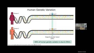
Thank you so much. Yeah, so presenting hybrid is always interesting. I will— I like to walk around usually, but I have to like control the virtual laser pointer, so I'll stand right here. We'll see how we do. I have a couple of different stories I want to tell you about, and at the end of each story I'm going to check the time because I probably won't have enough time to get through everything. But we're going to run the gamut today. I'm going to tell you about double-stranded break-reliant tools, base editing, potentially prime editing. And so we'll see how far we get in. And so the overarching goal in my lab is to better understand human genetic variation. And in particular, we are interested in point mutations. So if you compare two individuals' DNA sequences, they're going to differ at a lot of different sites. And when they differ just by the identity of a single base pair, so here person 1 has a C and person 2 has a T, this is called a single nucleotide variant or single nucleotide polymorphism. I might all refer to it as a SNV or a SNP. And so the vast majority of human genetic variation that we've identified through sequencing individuals is due to SNVs. What's more, if you look at a bunch of reasonably genetically diverse individuals and compare their sequences, you'll find that each person has a lot of genetic variants. So each person has around 4 to 5 million genetic variants in their genome if you compare to the reference genome. And so what I like to think of it as is that each one of us has a very unique combination of SNPs. And so we have projects talking about multiplexing genome editing agents to try to further our our systems where we can look at how genetic variation impacts cellular function. Today I'm just going to talk about looking at SNPs one at a time because that is how we think about things in terms of clinically characterizing the impact of human genetic variation. So thinking about what are these genetic variants doing? Are they simply contributing to diversity? So are they benign? They're just making us unique individuals. Or do they have a negative impact on our health? Are they causing disease? It's very tempting to think about it in the black and white terms or red and blue as I've shown here. Are these variants benign or pathogenic? But the reality of the situation is we've identified hundreds of millions of genetic variants through sequencing and over 99% of them don't have any type of clinical classification at all because we just We just don't understand them well enough. And the less than 1% that we have clinically characterized, it actually falls on this 5-category system ranging from pathogenic to benign. And most of these variants are in these middle 3 categories, meaning, okay, we know that they're impacting human health, but we're not 100% sure how. And so this is shown in pie chart form here. This is known as the variant interpretation problem. The fact that we're constantly generating more and more sequencing data and identifying new variants, but our ability to interpret how they're impacting human health is lagging very, very far behind. So you could see less than 1% have a clinical interpretation, and only about a third of those are actually classified as benign or pathogenic. And you can think about why, why do we care about clinically classifying these variants? Well, it leads to precision medicine. So if someone shows up to the hospital because they're suffering from a disease, the idea is we can sequence them, determine the root cause of whatever disease they're, they're suffering from, and come up with a very defined treatment plan that is catered to that individual due to the sequencing data. The way that we currently classify variants is purely computationally through sequencing data or through GWAS studies, so genome-wide association population studies. So we sequence a bunch of individuals, you know, half are suffering from a disease, the other ones aren't, and then we look at what genetic variants are popping up in each population. And most of the genetic variants that we find are actually very rare, and so we don't identify them in enough individuals to be able to classify them through purely computational methods. Additionally, any variants that are specific to non-European populations, you can see here we're just not sequencing enough individuals from ethnically diverse backgrounds to be able to start to clinically classify those variants. So the very few variants that we do understand what they're doing are specific to Europeans, which drives even larger health disparities. So what we want to do is develop experimental methods where we can take genetic variants of interest, introduce them at will to cell lines and animal models, and then study those resulting cell lines and animal models to really definitively determine how these different genetic variants are impacting cellular function and by extension human health. And we want to do this in a more— in a manner where we're looking at genetic variants from all different backgrounds equally. So this— OK, and so the first step to do this is we need to be able to introduce genetic variants at will and develop those models or generate those cellular models in a laboratory setting. And so we do this using genome editing. My first little story is going to start with traditional genome editing, as I call it, which is where we use double-stranded breaks to introduce genetic variants of interest. And so we— pretty much everybody nowadays uses CRISPR-Cas9 because it's so easy to use. The way that it works is that imagine this is our genomic DNA. This is where we want to do our genome editing. We would essentially— we take Cas9 and then design what's called a guide RNA in order to tell Cas9, bind to this exact spot here and introduce a double-stranded break, or DSB. And so the first, the 5' end of that guide RNA, so the first 20 nucleotides, is just going to base pair with our genomic DNA via Watson-Crick-Franklin base pairing properties. So it's really, really, really easy to reprogram this tool to bind to a new location in the genome. It just involves highlighting the sequence and copy-pasting into IDT. And that's why this technology has been so revolutionary. It doesn't require any protein engineering or anything. Okay, so from there, we have a double-stranded break. We've chopped the DNA in half, and there are two different DNA repair pathways that are going to compete with each other to process this double-stranded break. And so the first one, the more efficient pathway that is very ubiquitous, so it's always active throughout the cell cycle in all the cells in our body, These are called end joining pathways. And so usually they simply just re-ligate the two ends of the double-stranded break back together in order to fix it with high fidelity to give us our original sequence. Under genome editing conditions, though, Cas9 will just rebind and re-cleave. And what happens is eventually these end joining pathways make mistakes of insertions and deletions of random bases at the site of the double-stranded break. So that's what this red is showing. However, it has been shown recently that for a given double-stranded break site, these indel sequences are basically the same sequences occur over and over again for a given double-stranded break introduction site. So they are quite reproducible, but they can be kind of difficult difficult to predict. And they're certainly not for you to dictate what they are. Okay, so they're usually— these are not very helpful to us. Okay, if we're trying to do genome editing, we're just getting these, these different sequences of indels. However, what we do is we co-opt the HDR pathway, which stands for homology-directed repair. And usually this uses a sister chromatid as a template to repair a double-stranded break. But what happens is we will introduce a sort of fake sister chromatid into the cell at the time of genome editing. So this is a piece of DNA that has high homology to the site of the double-stranded break, but we've encoded in it our mutation of interest. So that's shown in blue. So HDR uses this as a sister chromatid to fix the double-stranded break, and in the process, it incorporates our mutation of interest. Okay. So as I mentioned, this these end joining pathways are much more efficient than HDR. HDR is also only active during the S and G2/M phases of the cell cycle. So typically, we'll get a couple of percent of our cells have HDR outcomes, and then the rest are all indels. And so this has been a huge challenge in the field, because obviously, we want HDR to be as efficient as possible, because we want this desired precise DNA outcome, DNA modification outcome. So this first story is a project that was done by my postdoc, Julep Bodai, in collaboration with Valentino Gantz, who's another investigator on campus. And what we did was we were looking at, again, these sequences of these indels. We were trying to do HDR-mediated genome editing. And for a particular site, we would always see the same the same, like, single base pair insertion popping up over and over again. And so we thought, hey, well, you know, since this particular indel sequence is always happening, is there a way to sort of then take these DNA sequences and then re-cleave to give us a second chance at HDR? And so we call this the double-tap method. And what happens is, We will do an initial set of genome editing experiments, and then we will sequence the outcome and characterize what these indel sequences are. And then we repeat the experiment but add secondary guide RNAs. Okay, so here we have a primary guide RNA, which matches the original genomic DNA sequence. And then there are other Cas9 guide RNA molecules in the cell that have these secondary guide RNAs.. And these match these most common indel sequence outcomes. And so within a given cell, if we get an N-joining outcome instead of an HDR outcome, while we have these other Cas9 guide RNA complexes kind of waiting in the wings to bind to that particular sequence and then cleave. And then this uses the exact same HDR template that we were using for the original double-stranded break site.. And it can give us, again, a second chance at HDR. So here's an initial example of what we did. This is a particular genomic DNA sequence. Here's our double-stranded break location. And we're trying to install a C-to-T mutation right here. So if we just use a primary guide RNA, just one guide RNA, this is a normal HDR experiment. We see, you know, 3% HDR outcome, which is pretty typical in the field. And then if we look at the sequences of all the indels that have occurred, you can see up here at the top, about 50% of our DNA outcomes all have the single A base pair inserted at the site of our double-stranded break. So we repeated the experiment, and simply just co-transfected another guide RNA expression plasmid in there that had a guide RNA that would target the single A base pair insertion. And you can see here that we actually doubled the HDR yields, which is pretty phenomenal, actually. It seems like 6% is still pretty small, but actually in the context of the field, that's quite impressive. So here's another site where instead of— in the previous one, we saw primarily just one particular indel sequence. Here we're trying to install a G to A mutation by installing a double-stranded break right here. Again, 1.8% HDR. And if we look at the sequences, there are, there are 3 different indel sequences with high-ish editing efficiencies, so around 10% for this 1 base pair deletion, and this 2 base pair deletion, and then around 3% for the single nucleotide insertion of a G. And so we tried just targeting this 1 base pair deletion, and you can see we increase HDR yields. If we throw in 2 secondary guide RNAs, so targeting both the 1 base pair deletion and the 2 base pair deletion, we get an additional boost. And then if we target all 3, we get an additional boost as well. And so looking at this over a variety of different sites, with varying sort of introduction efficiencies of those different indels. We can actually plot the efficiency of the indel that we're targeting with respect to the fold change that we see after using DoubleTap, and we kind of get a linear line. And we can actually fit a line to this. And we found in additional experiments that we can predict fold changes depending on the initial introduction efficiency of the indels that we're targeting. And it's actually shockingly accurate. Another thing we were looking at is overall indel rates. So here in green, these are at a bunch of different sites using DoubleTap. Green is the HDR yields. And so you can see that we increase HDR yields. Again, this is all just dependent on how efficient those indels are that we're targeting. And then in blue, this is the collective indel efficiency. And so this is compared to just using a primary guide RNA. And so when we use a primary and a secondary guide RNA, we see the indel, the overall indel rates actually drop at all sites. And then here, this is looking at the same sites, but now I'm just looking at overall indel percentage. And I've colored the indels according to ones that we're targeting with the secondary guide RNA and ones that we're not. And so in yellow are the particular indels that we're targeting with secondary guide RNAs. And you can see that— so these are all coming in pairs. So especially for something like this, this particular indel sequence goes from about 50% down to less than 5%. So we really are, you know, boosting HDR by targeting those particular indel sequences with these secondary guide RNAs. So everybody always asks about zygosity. And so we actually did that experiment. So we did both regular HDR with just a primary guide RNA, and then double-TAP using a secondary guide RNA. And then we clonally expanded the cells, and then sequenced to see what— if we have homozygous or heterozygous, you know, and if it's heterozygous, is it, you know, wild type on one allele or an indel on one allele? And so here we have 41 cells for each of these bar charts. And then you can see in red is the percent of cell lines that we generated that are homozygous for that mutation of interest. And that increased over threefold. And then we actually decreased the number of cell lines that were— that had the mutation of interest and an indel. And then, again, we have— and we've also increased the number of heterozygous cell lines, which are actually quite difficult to make. True heterozygous, where one allele is the mutation of interest and the other is wild type. So when you get heterozygous cell lines, it's usually one allele is the one you want and the other is an indel. You can see over here, we don't have— with just the primary guide RNA, we actually have no true heterozygous cell lines, and then we can get a couple using the double-TAP method. So one other thing I want to talk about is we can combine double-TAP with other HDR-enhancing methods to improve it even more. Over here, this is just at one of our sites, the primary guide RNA only. And then when we use double-TAP, we get about a twofold improvement. And so we combine the switch known with a technique that's known as blocking mutations. So what can be responsible for really high indel rates when you're attempting to do HDR-mediated genome editing is if your mutation of interest occurs outside that region that's base pairing with the guide RNA. So let's say in green here, this is the mutation that we're introducing. So if the protospacer region, which is what Cas9 uses to bind, is still intact, you can actually get Cas9 rebinding and recleaving, even though you had a successful HDR event. And then kind of funnel more and more of these through NHEJ. So you can get a lot of events actually that have your mutation of interest and an indel in the same piece of DNA, which is very frustrating. So what sometimes what we'll do is we'll just add an extra mutation in like the PAM region or in the protospacer region to block Cas9 from rebinding and recleaving. And so this, we don't like to do this because it's like an additional mutation that we don't really want in there. Sometimes this blocking mutation can be silent, so it's okay. But nevertheless, people have used this a lot. Because as you can see here, this is just, if you compare this, the pink numbers here to here, this is just by simply using a blocking mutation, you can actually boost HDR about 5-fold here. And then if we combine a blocking mutation with DoubleTap, you can see over here we've boosted HDR by almost tenfold, which is really great. And so here, like, again, this is just at a different site showing primary guide RNA only versus with DoubleTap. And then we tried adding these— the small molecule called Alt-R, which is this mixture that inhibits and HUJ proteins. And so comparing the pink values here to here, you get a slight boost. And we can combine it with DoubleTap to increase HDR yields even more. Here is a Cas9 HDR protein fusion that people have used. And so again, comparing the pink values here to here, we get a slight boost. And we can combine that with DoubleTap again and get an even larger increase. These types of methods for improving HDR, though, since they're messing with the endogenous DNA repair factors, we actually get really, really high cytotoxicity. And so what's nice is that we can get an even larger boost using double-TAP, and we don't have to kind of mess with cell cycle or any of these DNA repair proteins. And so, what time is it? Okay, I'll finish the story. So one other thing, we can also do large insertions. So I've shown you just single nucleotide variants. That's what we're interested in. But a lot of other labs are very interested with like tagging endogenous proteins with fluorescent markers, for example. And so we can do that and boost those yields as well with DoubleTap. So here's an example of a particular gene. Here's our start codon. We can introduce a double-stranded break. Near the beginning of the gene, and then use a larger plasmid-based donor in order to introduce GFP at the end terminus of the gene. And so in that way, we can sort of tag the ACTB protein with GFP and kind of visualize it, the localization of the protein. Okay, and so we did this with both ACTB and lamin A genes. This is with just a primary guide RNA, we saw about 2.5% HDR, again, in the realm of what we normally get. And we characterized what the most common indel sequences were and targeted those with DoubleTap and got about a 2-fold improvement. And so you can see, this is just showing the facts plotted out. And again, it's about a 2-fold improvement. Finally, so we compared it to prime editing. And so prime editing is one of these non-traditional genome editing tools that we can use to really increase the precision of genome editing. And so we wanted to install two sort of disease-causing mutations so we could sort of model them. One is an A to T mutation in the HBB gene. And then another is a 4-base pair insertion in the hexa gene. And so initially, what we want to do, you want, in order to use the DoubleTap and have it really be very helpful, is you need to find a guide RNA that facilitates, like, one particular indel sequence being introduced with really high efficiency. So at some sites, you can get just a huge mess of all these different indel sequences. And that's not going to be very helpful for using DoubleTap. But so again, we took the region where we want to introduce our mutation, and we kind of scanned 5 guide RNAs across, and then looked at the top 3 indel sequences. And so like, what is the collective introduction efficiency of these indel sequences? And so in here, what you want is you want these yellow and orange bars to be as high as possible. Because then you can be targeting a really high number of these indel sequences using double-TAP. And so after this, we chose the two sort of best guide RNAs based off of this analysis for each site, and then designed donor templates to introduce these mutations. And then we can see here that— With primary and secondary guide RNAs, we can get our HDR efficiency up to about 15%. And this is the prime editor, second and third generation constructs, which are kind of like state of the art right now. And with the prime editor, the second generation prime editor, we can beat that. And then the third generation editor, we're a little bit lower. But at this other site, the hexa site, which again is just a It has one very, very high efficiency indel sequence that we could target. We're actually nearly beating the third-generation prime editor. And so the prime editor actually has certain downsides to it. It's a really, really large construct. It's very difficult to design the guide RNAs to introduce those— to introduce different modifications. And it also has really high indel rates, actually, due to the mechanism by which it works. And so most labs prefer to just use HDR-mediated genome editing because it is so straightforward and very easy to do. Everyone has, like, the techniques and the different constructs that you need are actually very straightforward, and most labs can do that. And so that's all I'm going to talk about with the double-TAP. I'm now going to dive into base editing and what I call non-traditional genome editing tools. So this is what I just talked about, traditional genome editing. It has a lot of pros. Again, most labs do prefer to use HDR because it is so straightforward. But again, we have this major issue between HDR and NHEJ. Okay, so this is what I call genome editing precision. The precision is pretty low because we have such high indel rates. And this is through— this is because of just the way that double-stranded break repair works in our cells. And so during my postdoc work, I developed base editing, which is one of these next-generation genome editing tools. I call it a non-traditional genome editing tool. And instead of using double-stranded breaks, we use alternative DNA damage intermediates. And so these are comprised of Cas9 and a guide RNA. Okay, so we can still program it to go to a particular location in the cell just through simple DNA base pairing properties. But we've catalytically inactivated the Cas protein so that it doesn't cleave the DNA. Okay, and so we have a couple of different flavors of base editors. Some, the Cas9 is completely inactivated. And in some cases, it's a nickase. I'll show you why in a second. But then fused to Cas9 is a DNA-modifying enzyme. And so this enzyme can bind to single-stranded DNA specifically and do chemistry on the actual DNA nucleobase. Okay, so over with traditional editing, we cleave the DNA backbone. In base editing, what we're going to be doing is modifying the actual DNA nucleobase. And so with— there are two different types of base editors. One is called a cytosine base editor, or a CBE. And in this case, we have a cytosine deaminase enzyme fused to Cas9. And so Cas9 binds, opens up the DNA through that Watson-Crick base pairing action, and then if any cytosines that are within this very specific window get deaminated into uracil. And uracil has the base pairing properties of thymine. So the— this UG intermediate gets processed into a TA base pair eventually. And as I mentioned, we have dead Cas9 base editors, in which case the intermediate actually does look like this. We also have Cas9 nickase base editors where we would actually nick the top strand right here. So we would have a break in the backbone just on the top strand. And what happens is that kind of tags that top strand for repair. So the cell will replace the top strand using the uracil as an intermediate. And we actually get much increased base editing or overall point mutation introduction yields when we use nicking base editors. Okay. And so these are super new. So we have a CBE that I just mentioned, so cytosine to uracil to thymine. And we also have adenosine-based editors, or ABEs. So these take AT base pairs. The adenosine gets deaminated into an inosine, and that is read as a G. Okay, so they facilitate these two types of point mutation introductions. Even for the nickase-based editors, we have very, very, very low indel introduction efficiencies. Okay, so maybe like 1% or less usually. And our SNV introduction efficiencies are upwards of 70-80%. So the precision of these tools is really, really high. There are obvious limitations in that they can only facilitate certain types of genome edits. Okay. And because these tools are so new, we actually have— we know very little about how they work. And so that's what I want to talk to you about right now is a little subgroup of my lab where we're studying how these tools work. So we know that double-stranded break-reliant tools work through HDR and NHEJ. But how do base editors work? We weren't sure. So this is a project done by two graduate students in my lab, Cameron Burnett and Ashley Wong. And so they wanted to look at what the cell cycle dependence of base editing is. So as I mentioned before, double-stranded break reliant tools in order to do precision editing with them, so introduction of particular types of mutations using HDR requires S or G2 phase of the cell cycle. If you have non-dividing cells, such as neurons, those are kind of like always stuck in G1 or in G0 phase, you actually— it's really, really, really difficult to do precision genome editing using double-stranded breaks in those cell types because the machinery is just not expressed. So we wanted to ask— we know that double-stranded break reliant tools and HDR is only active during S and G2 phase. We don't know what is processing these base editor intermediates. But why don't we chemically synchronize cells, which we can do with these small molecules. So nocotizole can synchronize cells in G2 phase, and thymidine can synchronize cells in G1 phase. So we said, okay, let's synchronize the cells, perform base editing, and then see how that changes overall efficiencies. Of base editing and we can start to understand the mechanism. So first up, I'm going to tell you about the adenine-based editors or ABEs. And so again, these work by deaminating adenosine to inosine, and then this IT intermediate gets processed to a GC base pair. We looked at both the dead Cas9 base editors where we're not doing anything to that bottom strand, And then also the base editors that use the Cas9 nickase, where we're nicking this bottom strand right here. And so over here, this is looking at synchronization effects on the dead Cas9 ABE. And so black is asynchronous. These are 3 different sites that we're doing editing at throughout the genome. And then in purple is when we synchronized in G1. And in turquoise is when we synchronized in G2/M. And what we saw was huge decreases in editing efficiency with both synchronization conditions, so over 60%. We weren't sure if this was just due to some factor with these small molecules and potentially the cells being unhealthy. But when we repeated the experiment using the nickase-based editor, we actually saw very, very small decreases in editing efficiency there.. And so you can see here again, these are the same 3 sites, but we're just using this nickase editor instead of the dCas9 one. And so overall efficiencies are higher because we're using the nickase, so in the asynchronous cells, and then we also don't really see much of a decrease when we synchronize the cells. And so this is really interesting. It shows there's a very fundamental difference between how these dCas9 base editors function versus the nickase editors. And also, it's really exciting because it shows mechanistically that base editors are viable genome editing tools to use in non-dividing cells. And other labs have used them successfully in neurons and other cell types where double-stranded break reliant tools haven't been useful. And so with the cytosine base editor, we saw largely the exact same outcome. So again, here we have a CG base pair. We deaminate the cytosine to a uracil. And then this gets processed to a TA base pair. And we looked at both the dead Cas9 base editor, our CBE version, and the CBE with the nickase, where we're nicking this bottom strand. And again, huge decreases in editing efficiency across the board when we synchronize in G1 or G2M. So again, I'm comparing the black to the two colors. And when we used the nickase-based editor, again, we saw much, much smaller decreases in editing efficiency and very, very similar to what we saw with the adenine-based editors. So again, just recapitulating what we saw before, even with the cytosine-based editor where we're using this inosine intermediate, the dead Cas9 version versus the nickase version, very, large fundamental differences. And this is, you know, good mechanistic data kind of showing that we can use these in non-dividing cells, and we don't have to— we're not relying on G1 or G2/M or S phase. However, the dead Cas9 base editors clearly do depend on S phase. So one other thing I wanted to mention is With our cytosine-based editors, we've actually, we've engineered the construct extensively to sort of, to work against this protein in the cell called UNG. So that stands for uracil N-glycosylase. This is a DNA repair protein whose whole function is to find uracils in our genome and rip them out. So uracil is a pretty common type of DNA damage in our cells that occurs naturally. So we have an enzyme to sort of combat that. Otherwise, we'd have C-to-T base editing happening in our cells all the time without a base editor. But so what can happen is in the process of base editing, you can get your C-to-U, and then UNG will come in and rip out our uracil before it can get processed into a thymine. And then this abasic site intermediate can actually get processed into it can get scrambled, essentially. So we can get C to T, C to G, and C to A editing outcomes. And it's initiated— we've done knockout studies to show that it is caused by this UNG-mediated uracil excision. However, we can inhibit this by fusing this peptide called UGI, so uracil glycosylase inhibitor, We incorporate that into the base editor architecture in the CBE. So that was done previously. And here you can look at— this is like the fraction of edited DNA sequences that is due to a C to T in yellow. That's what we want. And then a C to G in blue or a C to A in black. And so you can see this is very— sequence dependent. So it happens we can get very high C-to-non-T editing at certain sites and not others. And if we take off this UGI component, so we just exclude it from the CBE architecture, this is what it looks like at the same 3 sites, the same experiments, except we're just not using this UGI component. So it's called the CBE delta UGI. And you see really high C-to-non-T editing efficiencies. And so we wanted to use this construct and see how the C-to-non-T editing efficiencies changed with respect to chemical synchronization. And what we saw was that when we synchronize in G1 with thymidine, we actually— you can see here the black bars here have increased significantly. So we saw huge increases in C-to-A editing when we synchronize in G1. And this is sort of normalized to overall editing efficiency. But what you can see down here, this is what I want to show, is individual, like, absolute C-to-A editing efficiencies right here, here, and here. And you can see the purple bar, so that's the G1 synchronized conditions, has a huge increase in C-to-A editing. And going back to up here, when we synchronized in G2/M, we didn't see any significant changes in the sort of product distribution compared to the asynchronous conditions. And so there's been a lot of effort of actually trying to leverage this like C-to-non-T editing efficiency and outcomes of the cytosine-based editor and then trying to to engineer new tools using the basic CBE architecture that has, like, precise C-to-G editing or C-to-A editing. And so, you know, this potentially we can identify what DNA repair proteins are involved in the C-to-A outcome, those that are clearly upregulated in G1 phase, to try to understand this basic mechanism so we can expand the genome editing toolbox. So one other thing I want to talk about is we can also have low levels of indels introduced with our cytosine base editor. And we believe that it's potentially initiated by this intermediate here. So our A basic site with our nick on the opposite strand, but we weren't really sure. And so what we did was we looked at the sequences of these indels. And we saw that they're actually very different than what we usually see with double-stranded break-reliant tools. And so here is the site of our nick. And also, if we were using wild-type Cas9, this would be the site of our double-stranded break. And what we would see with double-stranded break-reliant tools is we'd see insertions and deletions that are kind of centered around this double-stranded break site. But what we saw was that we saw these indels, so the deletions are shown in gray, we saw no insertions at all. And we saw deletions that sort of spanned in between the site of the nick, and then the site of the target cytosines that we are chemically modifying into uracils. And so that was an indication that potentially this is our indel intermediate. And we looked at changes in these different sequences with respect to synchronization. And we didn't really see any trends that stuck out. So we kind of ignore that for now. But what we did was then we sort of changed the overall architecture of our cytosine base editor and looked at how that changed the overall indel introduction efficiencies. And so this is indel introduction efficiencies with our nicking cytosine base editor that does not have the uracil glycosylase inhibitor component. And we can see we actually get like 10% indel introduction efficiencies with this, which is quite high. Nobody would ever use this editor, by the way. You'd use one of the better ones. And so if you compare it to our regular cytosine base editor, where again, we included the uracil glycosylase inhibitor component, our indels dropped over tenfold. Okay, so that's a good indication that UNG is definitely involved in installing these indels. And additionally, if instead of using the nickase base editor here, we use the deadCas9 base editor, we saw indel rates drop tenfold as well. So again, an indication that this nick down here is required for introducing these indel sequences. And that's where I'm going to stop with this story here, looking at the cell cycle dependence of base editing. I will mention that when we do these types of experiments with double-stranded break-reliant tools and look at HDR yields, we see huge decreases in editing upon G1 synchronization. We were super excited to not see that with our base editors. Back to the variant interpretation problem, I haven't lost sight of my goal, I swear. I'm going to tell you one very small story about using base editors to install point mutations into cell lines and generate models of variants of uncertain significance. Point mutations that have been identified in individuals, but we don't understand what exactly they do in terms of being benign or pathogenic. And so this is a project that's being done by Carlos Vázquez, a really talented graduate student in my lab who's one of the students who's received so many fellowships, he had to turn down some of the fellowships. And so what he's interested in is a protein called MUTYH, which is responsible for protecting our cells from oxidative DNA damage. So the most commonly oxidized spot in our genome are at guanines. So guanine normally pairs with cytosine, and under conditions of reactive oxygen species, we can get oxidation at the 8 position of guanine right here. And the reason this is bad is because when guanine has been oxidized in such a manner, it actually preferentially mispairs with adenine instead of cytosine. So it flips into this Hoogsteen position and shows a different edge of the base so that it will mispair with an adenine. And so overall, this can lead to G-to-T point mutations that accumulate in your genome. Okay, so if your oxidized guanines don't get repaired, these sort of G-to-T mutations can pop up in oncogenes, and that can result in cancer. So MUTYH is responsible for recognizing 8-oxoG when it's mispaired with adenine, and it excises the adenine and then sort of coordinates putting a cytosine back in. And then from there, this other protein, OGG1, then rips out the 8-oxoG and coordinates putting an undamaged guanine base back in. So mutations in both of these proteins are associated with early onset of cancer or predisposition to cancer. And in particular, if we look at all the point mutations that have been identified in MUTYH, The vast majority of them are considered variants of uncertain significance or VUS. The variants that are pathogenic cause colorectal cancer or MUTYH-associated polyposis or MAP. What's more is that this is specific to homozygous mutations. So if an individual has a heterozygous mutation, they actually don't develop MAP. But so if we want to start characterizing these VUSs, so if you're sort of like, if you've sequenced early on in your life and it's discovered that you have a MAP-causing mutation, you actually can just get colonoscopies early on in your life to sort of monitor the situation. And your prognosis is much better than individuals who don't know. So if you have— if you're sequenced and you have a VUS, both you and your doctor are like, I don't know what to do. So it would be nice if we could actually better understand what these VUSs are doing and characterize if they're pathogenic or benign. But again, we have to— in order to model them, we need to generate homozygous cell lines. So early on in the pandemic, Carlos and Quinn actually came up with this protocol for generating isogenic cell lines using base editors. And we compared them to using just HDR-mediated editing with double-stranded breaks. And we found that the outcome was actually significantly improved if you use base editors. And so what we do is we'll take our base editor and the guide RNA that we've designed to introduce a particular mutation of interest. And we transfect them into cells. We always have a GFP marker. We usually will sequence them in bulk to see, like, how are we doing? What's our overall editing efficiency? So we can know how many isogenic lines to clonally expand. You can see right here, we're trying to install a T-to-C mutation by modifying the A that's base paired with this T. You can just use Sanger sequencing to monitor.. And so the efficiency is high enough that we can then go repeat the experiment, take the cells to a FACS facility, and then sort one cell per well in a 96-well plate format. And so from there, depending on how good you are at sort of babying your cells and getting them to grow up, we get around 8 clones per 96-well. A lot of them die. They don't like this clonal expansion process. But from there, we found that when you're using base editing and you have— this is around sort of 30% editing efficiency in bulk— will yield about 50% cell lines that are wild type, so no edit. But then about 25% will be heterozygous and 25% will be homozygous. And so I showed you kind of like a breakdown of these isogenic cell lines and the zygosity back in the DoubleTap story when using HDR, but it is not this easy. So typically, we screen through like over 100 clones when we're using HDR, because again, we get— especially to get this heterozygous cell line. For this particular story, since we're mostly interested in the homozygous cell lines, it wasn't as big of a deal, but It's notoriously difficult to generate true heterozygous cell lines using double-stranded break reliant methods, because you always get an indel on one of your alleles. So this, we wrote up this protocol, you can, you can read it if you want. It's very, very long, but very, very detailed. And so from there, Carlos has generated both homozygous and heterozygous cell lines with about 10 different MUTYH mutations. All in-house, took a couple of months, but it's much better than having to send it out to a company who will charge you an egregious amount of money. And he took his cell lines, and he actually measured MUTYH enzymatic activity in these cells. So how well do these cells repair 8-oxoG that's mispaired with an adenine? And so the way that we do this, we can use some chemical biology methods. To take a large piece of DNA and site-specifically install an 8-oxoGA mismatch into it. We put this at a particular location in a GFP gene, such that when we transfect it into cells, if they can't repair the 8-oxoGA mismatch, the cells will not have any GFP fluorescence. And so we have an mCherry marker as a transfection control. So this is what it looks like if the cells aren't able to repair. And if they are able to repair the 8-oxoGA mismatch back to a GC base pair, we'll get both mCherry and GFP fluorescence. And so quantifying on a flow cytometry plot, we can see this is what it looks like. So just mCherry over here, this is what that looks like. And then when we get 8-oxoGA correction, we see the cells are sort of like all on this line that's like on the diagonal. Okay, so if we transfect this reporter into wild-type cells, so they have an intact wild-type MUTYH gene, you can see again, cells are all along this diagonal. So we're repairing 8-oxoG quite well. This P295L mutation is a known pathogenic mutation. In, in MYH. And with the heterozygous cell line, it actually looks similar to wild type. So we still see a lot of the cells are being— are along this diagonal. So our 8-oxoG is being repaired. But then when we have our homozygous cell line, you see there's actually a huge shift. And we see most of our cells are in this quadrant here with only mCherry fluorescence. And even the cells that do have some GFP fluorescence, it's actually a much lower signal than the wild type. Cell lines. And so with that, I actually do have a little bit of time. But so with that, we've actually— we've generated a bunch of variants of uncertain significance, and we've been able to clinically characterize them based off of MYH enzymatic activity. We need to do more studies looking at, you know, cell growth and different cancer-relevant assays. This is just looking again on the protein level. But we're really excited that we've even been able to generate these cell lines in the first place, because prior to using base editing, it would have been really, really difficult using just HDR, even with the double-tap method. And so I always— I like to, when I have time, I like to end with just talking a little bit about some of the outreach that we've done in my lab. I think it's really important to reach out to the local communities.. And so we, we started small and we've gotten a little bigger. Right now I actually have 3 of my students are at Sage Creek High School in Carlsbad doing an outreach activity that I'll show you on the next slide. But so we, we started out small. This is the local BYEWISE community, so stands for Better Education for Women in Science and Engineering. It's mostly middle school and high school students. Who are interested in science. And so we brought them here to UCSD campus and gave them tours of a couple of different labs. And we taught them about base editing through this like fluorescent protein assay. And so here you can see this is GFP. It looks green. This is bacteria with the GFP gene in it. This is the fluorophore of GFP. So the 3 amino acids that are responsible for its green color. And so this middle one has to be a tyrosine for it to be green. And if you mutate that to a histidine, it is now blue fluorescent protein. And we actually can interconvert between GFP and BFP using cytosine and adenine base editors, which is pretty cool. So we just taught them about it and then kind of showed them some plates with different fluorescent proteins in them and stuff. We said, oh yeah, we swear you can interconvert them. But then what we did was we came up with this lab activity. And again, my students are doing it at a high school right now. We don't know if it's worked yet. This is the first time we've been able to actually do it in person because of the pandemic. But yesterday we kind of taught them about genome editing and base editing, and we gave them a little worksheet where they had to like write out guide RNAs. It was really fun. And then we gave them them today that we gave them E. coli cells that have GFP-itis, where we've completely inactivated GFP. So we've mutated that fluorophore, the first amino acid in the fluorophore, we mutated it to isoleucine, so that it's completely non-fluorescent. And then we give them a choice of 3 plasmids that have 3 different guide RNAs, and they have to choose the correct one that will take the adenosine base editor to the spot in the genome where this dead GFP is and then correct it to wild type. And so we would get adenine base editing, so A to G. And so in our hands, it looks like this. So this is what the GFP IDA cells look like. If they pick the wrong guide RNA, it looks like this. And then if they pick the right one, it looks like this. But, you know, we've never done it in a high school classroom before. So I will update these slides as soon as we get the results. Results tomorrow. But my students are having a great time. They said it went really well. So we'll see if it works. And so with that, I just want to thank my awesome lab. This is our first picture that we were able to take after the pandemic. So no masks on. This is us on Twitter. Come find us. Thanks to my funding. And thank you guys for showing up in person really exciting to give, to give a talk in person again. So that—
I'll take any questions. Thank you so much, Alexis. That was really awesome, especially your high school thing at the end. Do we have any questions from the audience before we see if we have any from our Zoom people? Well, you guys are thinking, I'll sneak one in.
Okay.
I was curious why your inactivated Cas9 versus your nickase had such a big difference in the cell cycle dependency.
Yeah. So I think that, yeah, when we're not nicking that top strand, it's like more passive. We're just sort of waiting for the cell to replicate over that modified base. And so that would make sense since we synchronize in G1, and then in G2, huge decreases. And we don't have any chemical synchronization agent to like keep it in S phase. But I think like by process of elimination, I think that means that it's highly dependent on S phase. And so we need DNA synthesis across in the absence of that nick. So when we install the nick, it's like more of an active process, we're telling the cell like, hey, there's an issue here, replace this top strand, and then it uses we're sort of forcing it to use that modified base as a template without having DNA synthesis to happen.
That makes sense. Okay, do we have any Zoom questions? We have no Zoom questions.
Okay. All right. Well, Alexis, thank you so much.
So easy. Crystal clear. That's really cool stuff.
Thank you so much. All right. Well, thank you for having me.
More from Joshua







Scientist Stories: Hans Waterham, The value of phenotypic assays in peroxisomal disorders




Scientist Stories: Joel Zlotogora, Genetic carrier screening programs for reproductive purposes