Scientist Stories: Bin Zhang, Genome Organization through Phase Separation
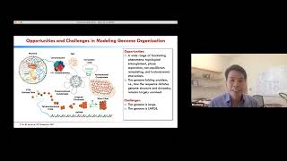
In this seminar, Bin Zhang outlines his laboratory’s computational approach to understanding the three‑dimensional organization of the human genome and its relationship to membraneless nuclear bodies. He begins by emphasizing the massive scale of the genome (≈6 billion base pairs, 2 m when stretched) and the hierarchical packaging that enables gene regulation, from nucleosomes to chromatin loops. To tackle the size problem, the lab builds a coarse‑grained bead‑on‑a‑string model where each bead represents 1 Mbp, confined within a spherical nucleus. An energy function governing bead‑bead and bead‑protein interactions is iteratively parameterized using Hi‑C contact maps via an information‑theoretic approach, producing ensembles of structures that reproduce Hi‑C data, chromosome territories, euchromatin/heterochromatin segregation, and radial chromosome positions—none of which were directly trained. The validated genome model is then extended with explicit coarse‑grained protein particles that mimic nucleolar and speckle components. Dynamical simulations of phase separation generate multiple, kinetically arrested droplets, matching experimental observations of nucleoli number, coarsening kinetics (size exponent ≈0.1) and fusion kinetics (neck‑radius exponent ≈0.5). Free‑energy calculations reveal an entropic barrier arising from protein‑chromatin tethering, explaining why droplets do not always fuse into a single droplet. The framework is applied to tumor versus normal cells: Hi‑C‑derived interaction changes shift heterochromatin away from the nuclear periphery and increase nucleolar number, predictions that are confirmed by electron microscopy. Finally, an integrated nucleus model adds speckles and lamina, proposing a co‑phase‑separation mechanism that simultaneously yields precise, reproducible distances between specific genomic loci and nuclear bodies while allowing cell‑to‑cell heterogeneity. The talk concludes with a brief Q&A on model details such as interaction strength, protein concentration, and simulation stopping criteria.
Chapters
for putting this together in this wonderful meeting and giving me the opportunity to present. So my lab focuses on computational modelling of genome organisation. And the reason we are interested in the genome is because it contains the complete set of DNA, and it's supposed to instruct the development and operation of an entire organism. And because of its importance, the genome has often been poetically called the blueprint of life. Now, since its discovery as the genetic material, significant effort has been devoted to study the DNA. And an obvious question is, how does the information transfer occur? How do you go from base pair to a walking, talking person? A lot of progress has been made in that regard. And one of the important findings is that the genome structure is actually quite complicated. So one cannot simply view the genome as just a very long double-stranded helix, at least not for human. So instead, the genome is actually packaged in a very exquisite way to both fit inside of the nucleus and to enable gene regulation. So as shown here, the human genome actually consists of more than 6 billion base pairs. That's about 2 meters long when fully extended. And this set of molecules is not confined into a nucleus, but only about 10 micrometers. So there are just 5 orders of magnitude difference in terms of the length scale. And this And there's a landscape difference that necessitates genome packaging. And as shown here, the genome is kind of packed in a— you know, genome packaging occurs over multiple length scales in kind of a hierarchical manner. And remarkably, cells have also evolved ways to essentially take advantage of this packaging kind of every step along the way. And for example, the formation of the nucleosome essentially precludes other proteins from accessing the DNA. So here the DNA is shown in gold here. In this sort of closed conformation, there's a tighter bond to the histone proteins. So then essentially, by regulating this population of the closed and open conformation, you can sort of regulate DNA accessibility and in turn gene expression. So, and, you know, sort of a similar argument can be made for a strand of nucleosome. Here you have open and closed chromatin, which can again sort of regulate DNA accessibility and gene expression. And now at a larger scale, over millions of base pairs, the formation of so-called chromatin loops, they can, again, bring DNA segments that are kind of far apart in sequence now into spatial proximity. And this sort of 3D context further allows cells to encode complex gene regulation. So in some sense, I like to think of the genome really kind of like an enzyme instead of a long linear polymer. It really has to fold in 3D to form active sites, okay, like to catalyze chemical reaction. And in this case, the reaction is, of course, gene transcription. Now, in addition to its biological significance, the reason that we are particularly interested in the genome is also because it's actually a very fun and complex problem. And there's a lot of sort of fascinating phenomena that's occurring. And better yet, in a matter regarding sort of how the genome sort of is established in 3D, The underlying mechanism remains unknown. So there's a lot of opportunities for discovery. Now, of course, opportunity always comes with challenges. And the first challenge is that the genome is very large, right? As I mentioned, 6 billion base pairs, that's a lot of atoms. And just the sheer size of the system actually creates a lot of issues. We as computational chemists over the years have become very good at modeling sort of small-scale systems. But for this kind of larger-scale system, there's still a lot of difficulty. And the second challenge is that, you know, the genome is really, really large. I couldn't emphasize that enough, because, you know, 2 meters long, that's a really macroscopic scale molecule. And so because of this complexity of genome organisation, we try to study the genome at different scales with different approaches. On the one hand, we try to study the whole genome organisation There we use an integrative approach that combines modeling with experimental data. And on the other hand, we also try to study the genome at a smaller scale with the atomistic details. And today I will focus on our whole genome modeling effort. So there, you know, we are particularly interested in the principle of genome organization, the mechanism that establishes the genome structure in 3D. So as you might expect, the genome is very complex, and so is its mechanism, right? So today we're going to focus on one particular aspect that has gained a lot of momentum recently. Now, before I dive into detail, I want to remind you that in the nucleus, in addition to DNA, as shown here, there are also these membraneless structures called nuclear bodies. They include nucleolus and the speckles. And so there is evidence that both the genome and nuclear body, they use liquid-liquid phase separation for their organisation and for their, for their formation. And for example, EM image of the eukaryotic genome had a long review that there are two types of chromatin that occupy spatially distinct regions, there is euchromatin in bright and heterochromatin. In dark colours. And this organisation is kind of reminiscent or consistent with a picture of block of polymer phase separation, as shown here. Now, the liquid property of nuclear bodies include their round morphology and their fusion upon contact. So, this is basically a time trace of two contacting nucleoli that eventually fuse into a larger droplet. And this is kind of a typical phenomenon of liquid droplets. Now, you know, phase separation is a very intuitive idea, and it has sort of— it's consistent with a wide variety of experimental observations. These are just two examples. However, I want to emphasize that, you know, the phase separation also cannot explain many other experiments. In particular, recently, sort of several groups have found that certain genomic loci actually have very well-defined distance to nuclear speckles. And, you know, this kind of very precise distance is actually inconsistent with a liquid picture. You know, if you— if we think of the nucleus as just a, you know, a liquid, then by definition it's supposed to be very dynamic and homogeneous, which it really can— I cannot encode, you know, very precise distances. So another puzzling observation is the apparent coexistence of multiple droplets. So here the cells are stained with antibody to basically highlight the number of nucleoli. And this is a quantification of the image shown on the left. So here you clearly see multiple nucleoli. Now, why is this interesting? It is interesting because it seems to be inconsistent with this classical nucleation theory, as you might know. And so this theory tells you that the stability or the free energy of a droplet can depend on two terms. One term comes from the surface tension, sigma, here, and is proportional to the surface area. And there's another term that, uh, that arises from the interaction among particles and is proportional to the volume. And with this expression, you can show that the free energy, uh, will always decrease when the droplets fuse, merge with each other. So then basically the thermodynamic equilibrium literally just consists of one single droplet, as shown in this cartoon here. So then the obvious question is, what drives the inconsistency between experiment and theory? And what is maybe so special about the nucleating environment that stabilises the apparent multiple droplet state over the entire cell cycle? So these are the kind of questions that we would like to understand better with computational modelling. But to study the genome organisation and phase separation in the nucleus, We first need to build a model for the genome. But as I mentioned in the beginning, modeling the genome can actually be quite challenging just because of its sheer size. To basically make any meaningful progress, we essentially have to throw away our beloved atomistic detail and work with coarse-grained models. This is basically a cartoon for our 3D model. We have each one of the 46 chromosomes. We represented them as a bead on a string, and the chromosomes are confined in a sphere. To mimic the nuclear envelope. So in one resolution that we work with, each chromosome is represented— sorry, each bead in the chromosome represents 1 million base pairs. And now with this coarse-grained representation, you can define an energy function, basically kind of quantify the stability of different genome structures. So the design of the energy function itself is actually a very interesting topic and basically sort of Through trial and error, you can sort of actually learn a lot about the mechanism of genome organisation. But in particular, we show that a simple block of polymerase actually cannot explain many features of the human genome organisation, but I won't go into too much detail. But now once you have the energy and function, right, once you establish the functional form, you still need to sort of parameterise the interaction within the particles. Because those interactions really quantify the energy and the final structure that you're going to get. Now, to be frank, coarse-graining is actually a very popular technique in the theoretical chemistry community. However, we cannot just directly borrow existing techniques and try to parameterize the genome model. And the reason is that, as I mentioned, each one of our beads actually contains many atoms, and just the sheer size and the the complexity of the chemical composition of each bead basically renders this traditional so-called bottom-up approach inapplicable. In particular, we cannot afford to just run quantum mechanical calculation or atom simulation to try to parameterize the interactions and parameterize the genome model. Basically, to parameterize the model, we really have to think outside the box. In the end, We established— we call this an information theoretical approach. And what this allows us to do is basically extract parameters from experimental data. So the experiment that we use is called Hi-C. And it's actually very similar to NMR, but for the genome. So what it does is basically it detects contact between the DNA segments in the nucleus with cross-linking, as sort of illustrated here. And the result is typically shown in the contact map, and each data point in this contact map basically tells you the probability for a pair of DNA segments to be in contact in the nucleus. Now here in this color scale, the probability decreases from yellow to red to white. Okay, and this diagram basically illustrates how our sort of methodology works. We start with an energy function And with this energy function, we can then basically simulate an ensemble of genome structures as shown here. And from this ensemble of structures, we can then compute an encymical contact map that we can look at the difference between the simulation and experiment. And we can use that difference to, to basically update the parameter to improve the energy function. And we can do this iteratively so that in the end, we arrive at a converted energy function and a converted genome structure that reproduces experimental data. So I mean, I won't go into detail, but we do have a little nice little argument to say that, you know, the model we have is supposed to be optimal given experimental data. So after we obtain an ensemble of structures, we carried out extensive validations to ensure their biological relevance. So it's probably not too surprising that our model was able to reproduce Hi-C data given that they were actually provided for model training. So here, the experiment is shown in bottom, and the simulation is shown at the top. And these are basically just a zoom-in of a different part of the contact map. And you can appreciate that our model was able to reproduce the contact across several orders of magnitude. And we do have quantitative metrics in addition to this diagram to ensure that the simulation indeed agrees with the experiment very well. And importantly, in addition to the high C, we also compared with a set of qualitative and quantitative image results. In particular, the chromosomes are known to form territories. And you can readily see that from our simulation structure as well. Here, each chromosome is assigned with a unique color. And this is basically a blow-up of the simulation structure. And you can see that each chromosome is more or less sort of collapsed. There's very little intermingling among them. And now if we instead color the genome with different chromatin types, you can see that there is a clear phase separation between euchromatin, which is shown in red, and heterochromatin shown in blue. The heterochromatin also localizes at the periphery, which is consistent with the EM image that I showed previously. Finally, when we look at the radial position of individual chromosomes, you can see that the simulation value actually correlated very well with the experimental results. Again, I want to emphasize that none of these results were actually provided for model training. The fact that we're able to reproduce them provides confidence in both our modeling strategy and in the biological relevance of the structures. So now that we have a structure, you might wonder, you know, what can we do with that, right? What additional value do they add to the genome? And so I will say that one advantage of having the structure is that they're actually very useful for building intuitions. In particular, here in a collaboration with Brad Bernstein, we looked at the the genome organization in normal and tumor cells. So here, if you just look at the Hi-C data, you sort of see some differences. But for example, the contact in tumor is a little bit weaker, but it's not clear to conceptualize, you know, what exactly does that mean in terms of the 3D structure. But I hope we can appreciate this larger-scale change, you know, from our simulations. In particular, unlike what you see in tumors, Sorry, unlike what you see in normal cells, in tumor, the heterochromatin, they actually now move away from the envelope and sort of now kind of aggregate into the nuclear interior. So we were very excited about this prediction, and we wrote the manuscript. And as you might expect, we encountered reviewer number 3, who were not very impressed, and they wanted to see more and more validation for the simulation predictions. And fortunately, we have excellent collaborators, and they performed electron microscopy on the on these tumor and normal samples. And lo and behold, they found exactly the same as our simulation prediction. In particular, here the heterochromatin are again showing dark color. You can see that in normal cell, they are— they look like at the nuclear periphery. But in tumors, and you see that they indeed now sort of dissociate from the nuclear envelope and move to the interior. So that was kind of a eureka moment for me that, you know, the simulation agreed very well with the experiment. All right. And now that we have a reliable model for the genome, we can actually also now begin to think about the phase separation in the nucleus. To do that, we introduced the additional coarse-grained particles. And this particle, they basically try to mimic the nuclear proteins, which are sort of the key component of the nuclear body. And we basically make sure that the concentration, the size, and the interaction with the chromatin of this nuclear— of this corresponding particle sort of match with those of the nuclear proteins. And so this particle, they sort of have a track to interacting with each other to promote phase separation. They also can sort of interact with the chromatin favourably, really interact with the specific region of chromatin favourably. So basically, after introducing this corresponding particle, we can essentially perform a dynamical simulation of phase separation in a nucleus. So here the protein particles, they are showing in orange, and the chromosomes are showing in gray. And I'm just going to play one movie that sort of is a typical simulation trajectory. And as you can see that as the simulation evolves, yeah, we started from a kind of a random distribution of the protein particles. As time evolves, you see that the particles, they will aggregate into small clusters, and small clusters will coalesce into bigger clusters. And, and interestingly, in the end, the simulation actually settled into two droplets, not one but two droplets. And that's interesting because that's again kind of inconsistent with the expectation of the classical nucleation theory, but it is consistent with the biological observations. And we can also monitor the number of clusters as a function of time. You can see that, you know, basically you have an initial burst of clusters at the beginning, but then gradually coarsened, but it eventually settled down to droplets. And we always performed additional simulations and just to see whether this is statistically significant. And as you can see here, in all but one simulation, we actually always arrive at multiple droplets in the end of the simulation. So now this actually becomes quite interesting. Now, before we get too excited, we also performed additional comparisons with the dynamical measurements from experiment to sort of see whether the dynamics of our simulation makes sense or not. So, in particular, here we looked at the study from the Brangwynne group. So, they actually monitored the coarsening kinetics of droplets directly in the cell nucleus. It's actually a very cool experiment. And what they did is they monitored the size of— the average size of the droplets as a function of time. You can see that this side increase, a power law behavior of the side as a function of time, and the exponent is around 0.1. This exponent itself is peculiar because it deviated from the canonical value, which is around 1/3, but our simulation was actually able to reproduce that very nicely. In this other experiment from the Wesker lab, So they— here they directly monitor the fusion kinetics of two droplets, and they sort of measure the neck radius of the fusion as a function of time. And you can see that this radius increase sort of, again, sort of follows a power law, and the exponent is around 1/2, and that agrees with our simulation as well. And as a final check, we also went back to our tumors and we, you know, we decided, we determined, we basically performed a dynamic simulation of phase separation in tumors. And here, everything was kept the same regarding the protein particles, the concentration, the interaction with the chromatin, everything was kept the same. The only thing that we changed was the interaction between chromosomes. So here now, the interaction was derived from tumor Hi-C instead of the normal Hi-C in our previous simulations. And interestingly, that change actually resulted in more nucleoli in tumors compared to normal samples, as you can see here. And that's interesting because tumors are actually known to have more nucleoli. So the fact that we were able to reproduce that trend, at least qualitatively, is quite exciting. So again, all of this qualitative and quantitative comparison, again, sort of supports the biological relevance of our simulation. So now we can maybe go back to look at the simulation details and maybe learn something about the system. In particular, we want to understand why are we seeing multiple droplets in our simulation, right? And as a chemist, I like to sort of think of this from a reaction perspective. So we introduce a reaction coordinate, and this reaction coordinate basically measure the distance between the two droplets. So at a larger distance, the two droplets are sort of separated from each other, and at a small distance, they will fuse. And we then computed a free energy profile along this reaction coordinate. So what you see is that, interestingly, we saw a free energy barrier, and this barrier appears when the two droplets sort of begin to touch each other. This is, again, sort of inconsistent with maybe the which you might expect from the classical theory of phase separation. And then we sort of try to understand, you know, why do we see a barrier here? And we perform a free energy decomposition. We found that the barrier mostly arises from an entropic origin. And then we scratched our head a little bit and we still thought, you know, and we thought that there could actually be a very intuitive explanation for this entropic barrier. And the reason is that the protein droplets, they are essentially tethered to the surrounding chromatin network because of favourable protein-chromatin interaction. So then as the droplets try to sort of fuse and then move closer to each other, they will basically, you know, pull on the chromatin network, they will basically put more constraint on the chromatin network. And the chromatin network is made of polymers, I guess, in a naive approximation, they are kind of like rubbers. And when you pull on the rubbers, they're going to be a sort of countering force. And this countering force is known to have an entropic origin, and that will give rise to this barrier. So this is kind of reminding me of this cable crossover that I kind of enjoyed doing before. But this is more like overcoming gravity, not really entropy, but you get the idea. So our study on this nucleolite kind of supported that the chromatin network could actually impact the formation of nuclear bodies. But in the meantime, you can expect that the nuclear body, they will also impact the chromatin network as well. And that point actually really become very clear in our latest study where we try to now build a more complete picture for the nucleus. So here, in addition to the genome and nucleoli, we also included the speckle and nuclear lamina. Those are sort of key components of the nucleus. So in this model, in addition to reproducing the genome organisation, we are now also able to reproduce the contact between the genome and the various nuclear landmarks. So this is supposed to provide a more accurate picture for the nucleus. But interestingly, now this model actually also helps us to understand how would the precise distance that I mentioned in the beginning would arise in the nucleus. And the idea is that we think that the genome and nuclear body, they sort of conform through a self-assembly process through basically a co-phase separation mechanism. And sort of shown here in this mechanism, the nuclear body, they will basically nucleate around the chromatin segments as sort of a— because of favourable interactions. And this nucleation essentially ensures they're sort of a close contact within individual cells. And because this nucleation also happens in all cells, so this will essentially ensure that the certain genomic region will always have very well-defined distance with the nuclear speckles. And this explains the precise distance that had been shown in previous experiments. But in the meantime, because phase separation is nonspecific, in different cells, you also— basically, the nucleation could use a different set of chromatin segments. So there is a heterogeneity in the chromatin segments that are nucleating the nuclear body in different cells. And this heterogeneous context could sort of give rise to the heterogeneous chromatin some positioning in individual nucleus as well. And so there's basically a simple coface separation mechanism that can actually reconcile this, you know, both of these precise and heterogeneous features of genome organisation, which may sort of seem a little bit contradictory, you know, at first sight. Okay, just— yeah, so yeah, I showed you one mechanism about genome organisation, but we are trying to sort of use that to try to predict genome organisation and to see the impact on genome function. Function. And I'm running out of time. So I want to thank my lab member who did the work. I was fortunate to work with an excellent group of people and thank my funding. And thank you for your attention. Okay.
Very, very exciting talk. So we see a lot of questions. Let me just read a few of them. You already answered my question on this polymer constraint thing. Robin asked you By starting with the Hi-C map, are you not introducing by hand the incomplete phase separation that you are wanting to explain?
So with, with the Hi-C, yes, we do in sort of, uh, uh, we do. Yes, I mean, Hi-C in fact more or less tells you this phase separation. That is correct. I mean, yeah, so Hi-C encodes that, but, but Hi-C does not really tell you much about the the nuclear body phase separation, I guess. I don't know which part of the phase separation Robin was referring to. But you know, it— yes, I mean, the fact that we were able to reproduce this phase separation, that is totally from Hi-C data.
I was referring particularly to the chromosome territories, because the Hi-C map in a way includes all the physics which leads to the chromosome territories.
That's fair. Yeah, you are absolutely correct in that. So yeah, that's why you can sort of see that they're the very kind of strong contact in each block that actually comes from the individual chromosomes. But I would say this part is, you know, it's definitely not apparent from Hi-C that the fact that we were able to reproduce the radial position of chromosomes. So I mean, it is in some sense still encoded in Hi-C, but I would say it's not obvious that, you know, a model just based on Hi-C would be able to reproduce that in terms of the radial position of chromosomes. Thank you. Thank you.
So yeah, there are some questions on the details of your simulation. One thing, Pinaki asked you, do the number of droplets in simulation depend on the interaction strength between chromatin beads and the protein beads?
Yes, that's a great question. Absolutely. So we actually show that— I didn't show that result here. We actually have that in our manuscript. So basically, as we increase the strength of the interaction, we actually see more droplets. And the idea is that basically the droplets that form through nucleation around, around the chromatin, then they will sort of basically fuse. And at a stronger strength, it will make the nucleation easier. So initially, you have more droplets in the beginning. And because the coarsening is sort of kinetically arrested, if you start, if you nucleate more, you're going to end up with a more proper list. Yeah, that's a great question.
And also probably also the protein concentration, right?
The protein concentration, yeah, that's true as well. Yeah, yeah, we sort of just try to sort of more or less fix the quantum concentration based on the biological values.
But yeah, correct.
Yeah.
Rafael asked you, how do you determine when to stop the simulation in the phase separation study? How does number of droplets depend on simulation time? So I want to make sure it's, you know, whether it's metastable or not, right?
That's an excellent point. I mean, I guess you can see that, uh, I mean, we can sort of more or less map our simulation time scale to sort of experimental time scale. You know, we sort of run about several hours. Um, but you know, if you look at the free energy, I mean, you are correct that the single droplet state is still the somewhat dynamic minima. So that if you run that infinitely long, you would probably in the end still end up in this, you know, single droplet state. That's why, you know, we call this a kinetic arrest. But, you know, in our reasonable simulation time scales, you know, we can map to biological data on the order of maybe several hours a day. I mean, this basically gets arrested in this multiple droplet state. We do not see sort of further coarsening anymore. Yeah.
So let me see, probably last question is Jingting asks you, are the droplets associated with the heterochromatin and euchromatin domain respectively, or other distinct connected domains?
Sorry, could you say that again?
Are the droplets associated with the heterochromatin and euchromatin domains respectively, or other distinct connected domains?
So what's the nature of the droplets?
They connect to the— associated with the heterochromatin, euchromatin, or there are some other features?
I see. So this is a droplet. So these are— we're trying to model the nucleolus. So the nucleolus, they are sort of— they are functioning for this ribosomal RNA. So they actually sort of associate with a very specific set of chromatin, in particular those sort of segments for transcribing the ribosomal RNA. But they also have sort of a maybe more, more than those, those regions I called nucleolus-associated domains. So yeah, so I mean, those are typically heterochromatin, you are correct. Yeah, but it's not just for heterochromatin, they are also important for transcription as well.
More from Joshua







Scientist Stories: Hans Waterham, The value of phenotypic assays in peroxisomal disorders




Scientist Stories: Joel Zlotogora, Genetic carrier screening programs for reproductive purposes