Scientist Stories: Blake Wiedenheft, Microbial Immune Systems
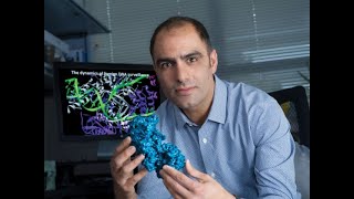
Blake Wiedenheft describes how CRISPR systems recognize and cut foreign DNA, then pivoted his lab during COVID to track variants in wastewater and develop a diagnostic that detects RNA directly from clinical samples using the Csm complex's natural ability to generate fluorescent signals.
Chapters
Hi everyone, um, happy New Year 2023! And, uh, new year, new location. In this case only because Building 10 had some flooding, but, um, still, you know, it's kind of convenient and nice to come home and just walk downstairs and see everyone. So real pleasure to see you all and today to host Blake Wiedenhoeft for the first NHGRI seminar series for 2023. And it's a real pleasure to have Blake visiting today, both as a scientist and really as a mentor, sort of teaching us about a lot of the conversations that I hope we'll have with Blake are really about the intersection of doing amazing science with amazing people. But even more than that, Blake is, is, you know, thinking about how to encourage scientific curiosity and grow that amongst the early learners, doing a Montana wild virus hunt for high school students and even younger students going into the classroom. And taking a lot of pride in his own scientific efforts, but as much really even just in the achievements of the people who work with him and that sense of scientific community that we're building. And so when I asked Blake what he wanted me to highlight on his illustrious CV, he did mention that I should talk about the undergrads, the grad students, and the postdocs who have won awards from Rhodes Scholarships to K99s. And we were fortunate to have Andrew come and give one of the NIH talks earlier this year, who's now gone on from— to win a K99. So Blake has really a wonderful pedigree himself, but has done it with his own sense of integration and style and has— was originally a bachelor's and PhD at the Montana State University. And he describes it that he was fortunate to be recruited back there as a junior faculty. But I think that they feel that they were fortunate to have recruited him. He started working in CRISPR and the work that he's going to talk about today really dates back a decade, even from when he was an HHMI LSRF fellow at Berkeley with Jennifer Doudna. And over the last 10 years has just been really doing amazing, illustrious work in this field with CRISPRs and editing and looking at microbial communities that's been recognized with a number of awards, including the PCASS Award from President Obama and the NIGMS Director's Early Career Scientific Award. So with that, I am happy to bring Blake up. Great. Thank you. So Blake, welcome to NHGRI and NIH.
Thanks a lot. Thanks, William. Thanks for the really kind introduction. Very much appreciated. And thanks, Amanda, for the nomination and anybody else who was involved in the selection committee. It's really an honor to be here. It's a privilege. And it's really one of the most enjoyable aspects of this job, in my opinion, to be able to travel around the globe and chat with some of the most interesting, curious people that you would otherwise never come across. And I really enjoy it. So thanks for taking time out of your schedules to listen to some of the work that we've been doing, but share some of your own as well. Today, I'm going to tell you about some of the work that we've been doing on mechanisms of CRISPR-mediated immunity and some of the applications that go beyond editing. And as Julie mentioned, one of the individuals that was spearheading a lot of this work is Andrew Santiago-Frango, whose initial work was supported by the Life Sciences Research Foundation. Julie mentioned that I was one of these fellows as well, so it was exceptionally rewarding to have one of my own trainees then end up with this fellowship that was sponsored by the Simon Foundation and also the Burroughs Wellcome Fund. And Andrew recently got a K99 from the NIH, and he was working with a graduate student Wilhelm Riechelt's— oh, one of the things I meant to have in motion here was they've been working on the structure of this CRISPR, this Cas integrase, and a complex that delivers foreign DNA to a specific location in the genome with single nucleotide resolution. And I think understanding how that works specifically has pretty big implications for some biotech applications, but just from a fundamental understanding of how these machines deliver foreign DNA in a very reproducible way. And then once I tell you about the structure that explains how this complex delivers foreign DNA to the bacterial genome with single nucleotide resolution, I'm going to shift gears a little bit and take you back in time 2 years to tell you about how our work on the pandemic collided with our interest in CRISPR in an unexpected way. All right, but before I do any of that, I'm going to take a few seconds here to just quickly give an introduction to CRISPR-mediated immune systems. And I know that most of you could probably come up here and give this slide yourself. So I promise I'll keep my intro to 60 seconds or less so that I don't lose you. But I just want to remind you that CRISPRs are found in about 40% of sequenced bacterial genomes and almost 100% of archaeal genomes, but 0% of eukaryotic genomes, including eukaryotic DNA that was bacterial-derived, like mitochondrial, chloroplast, class DNA. And I think that the skewed distribution, particularly in the prokaryotic domain where there's 40% and 100% CRISPRs, is not well appreciated. Why is that? We don't know extremely well, I think, why there's such a distribution. But what we do know is that wherever there are CRISPRs, they seem to be involved in adaptive immunity. And these immune systems work in 3 stages. And then the first stage— and I'm going to spend a fair bit of time talking about this this afternoon is this DNA is recognized as antigenic, and that in itself is quite a feat. How does this DNA get distinguished from the host DNA? And then a short snippet of that DNA is site-specifically integrated at one end of the CRISPR in a way that maintains this polarized— or there's this polarized evolution that maintains a chronological record of all previously encountered foreign nucleic acids. But the CRISPR is just molecular memory. In order to elicit an immune response, the CRISPR has to be transcribed, and this long pre-CRISPR transcript is subsequently diced or processed into a library of short CRISPR-derived RNAs that each have a unique sequence that was derived from and is then by definition complementary to this previously encountered foreign nucleic acid. Each of these RNA-guided surveillance complexes then patrol the intracellular environment and look for target nucleic acids. But they don't do this by looking for a complementary target, as you might think. At least they don't do that initially, because most viruses that infect bacteria that we know of at least are double-stranded DNA. So since the target sequence is buried in this double-stranded DNA duplex, it would be probably energetically expensive and slow to unwind all the nucleic acid in the cell to look for a complementary target. So that's not what they do first. Instead, they scan for a motif called a PAM, or a protospacer adjacent motif. This sequence of colors is called the spacer, and the spacer is flanked by repeats. And the origin of the spacer is the protospacer, and it's an identical sequence that's now in the phage, but that sequence isn't flanked by repeats. Instead, it's flanked by this protospacer adjacent motif that's 2 to 5 base pairs long. And when this complex recognizes the PAM, it distorts the DNA in a way that facilitates RNA-guided strand invasion. And once those two criteria are met, both PAM recognition followed by directional unwinding of the DNA and complementary base pairing, that activates the nuclease, which of course, is a discovery that enabled targeted genome engineering. All right, but if these systems are so efficient at eliminating invading DNA, Then why do viruses persist? And as many of you know, viruses that infect bacteria are among the most diverse and abundant biological agents on the planet. There's an estimated 10 to the 31 viruses on Earth. That's roughly 1 trillion viruses for every grain of sand. And they cause a lot of infections. Many of these infections are lethal. And in fact, phage infection is responsible for turning over 20% to 30% of the entire biomass in the open ocean every day. So if these immune systems are so good at eliminating foreign DNA, and yet foreign DNA remains this biologically diverse and highly abundant element on our planet, then how do we reconcile this contradiction? And of course, one way that viruses escape these systems are through mutations in the PAM. The other possible way is mutations in the protospacer. And each of these instances, these viruses then go undetected by the RNA-guided surveillance complex. But possibly a more interesting and proactive method for escaping CRISPR systems is virally encoded anti-CRISPRs. These are small proteins, usually 10 kB, maybe 20 kB, that are immediately early expressed upon entry into the host. And these small proteins then bind oftentimes bind to these large ribonucleoprotein complexes in a way that really blocks the function, either blocks DNA binding or blocks activation of the nucleus, one of these critical functions. And because they do that, they've been really critical tools for teaching us about how CRISPRs work. Anti-CRISPRs serve as these molecular beacons that point experimentalists to the parts of the machine that are most critical for their function. All right, but this kind of conflict, of course, a consequence of this kind of conflict drives diversification, and that's exactly what you see in CRISPR systems. CRISPR systems have evolved independently at least two different times in the so-called Class I and Class II kinds of CRISPR systems. Everybody's familiar with Class II systems. They've been popularized, of course, by the Cas9 RNA-guided endonuclease, which is critical for the Class II Type II immune systems that are RNA-guided DNA nucleases. But my lab, or the work that we've been doing, has been primarily focused on the Class I Type I systems, which are biologically far more abundant, but less popular, given that they're not the primary tool for targeted genome engineering. But these complexes consist of multi-subunit assemblies that recruit, oftentimes recruit a transacting nuclease for destroying the DNA. And I'm going to spend most of today talking about the Type I F system from Pseudomonas aeruginosa, pneumonosa, although this Type I-F system is found in many different types of bacteria. Then in the end of the talk, I'm going to transition to these Type III systems. You can think of Type III CRISPR systems as sort of the Swiss Army knife. They include a polymerase within the CRISPR complex. They cleave both DNA and RNA. And I'm going to explain some of the sophistication of these systems and how we've repurposed them for a diagnostic, and why I think these particular Type III diagnostics are have a distinguishing feature that makes them maybe perhaps more valuable or useful for diagnostics than some of the other alternatives. All right, so let's zoom in on these Type I-F systems. And as I said, I'm going to talk primarily about the Type I-F system that happens to be in the Pseudomonas aeruginosa genome, pa14. If you look upstream of the CRISPR, there's an AT-rich leader sequence that's known as the leader, and it contains a a transcriptional promoter, results in transcription of the CRISPR, which includes a repeat, a spacer, repeat, and so on. And downstream of the CRISPR, in this particular case, there's 6 Cas genes. We know that Cas1 and Cas2 are among the most highly conserved Cas genes of all the different systems, and that they're involved in adaptation. That means putting foreign DNA into the CRISPR locus. We know that Cas1 forms a stable homodimer, and that Cas2, the other component of this adaptation complex, complex also forms a stable homodimer. But strangely, in this system, and unique to the system, there's a unique fusion that's conserved in all Type I S systems where the Cas2 adaptation protein is fused to this large nuclease that's involved in target destruction. It includes both an HD nuclease and a Superfamily II helicase. And the Cas2 protein, like Cas1s, also forms a stable homodimer. And these two homodimers assemble into a heterohexameric integration complex that's responsible for inserting foreign DNA here. But since this complex contains this unique fusion of Cas2 fused to this Cas3 nuclease that destroys invading DNA, the question has been, how does this complex achieve these two different goals, both adaptation and interference? So I'm going to talk about the interference complex here. What we know about this is that the CRISPR again is transcribed in the repeats within this CRISPR locus are palindromic, and they form these stable hairpin structures. These stable hairpins are recognized both sequence and structure by an enzyme called Cas6 that binds to the stable stem loop and cleaves at the 3' end, resulting in a mature CRISPR RNA species that includes 8 nucleotides from the repeat on the 5' end, the entire phage-derived sequence, and then this 3' stable stem loop structure. And the Cas6 protein remains stably associated with this hairpin with picomolar binding affinities. We think that this is a subcomplex that the rest of the complex assembles around. And this complex includes 6 subunits of the Cas7 protein that oligomerize along the entire length of the CRISPR RNA. And we've in fact shown that if you lengthen, artificially lengthen the CRISPR RNA by 6 nucleotides, you can add 1 more subunit, 12 nucleotides, 2 more subunits, 18, and so on and so forth. So it's really an RNA template-driven assembly of this complex that's capped at the head by Cas6. And on the other end of the complex, it's capped by the tail that's composed of a Cas8 and Cas5 protein that form a stable heterodimer that forms the tail of this complex. And it has a distinctive vice-like feature here that grips onto DNA. And I'll show you how that works here. But I think you could kind get a sense for that just from the complex. So the way that we think this thing works is the phage attaches to the surface, of course, and it injects its nucleic acid, which in most cases is double-stranded DNA, and that these complexes initially look for a PAM sequence, as I described. They don't unwind all of the double-stranded DNA to look for a complementary target. Instead, they probably slide, hop, and scan along the DNA, until they encounter a PAM, in which case the vice closes around the PAM and it drives a conformational change in this complex as it unwinds the double-stranded DNA duplex. This is a really critical aspect of this complex that I'll talk more about here in the upcoming slides, but it recruits the transacting nuclease. Here we're zooming in on the PAM and the mechanism for PAM recognition. This vice closes specifically around a PAM, making nucleobase-specific interactions in both the major and minor groove. But also when it does that, it drives this amino acid side chain, which serves as a molecular wedge that splits the DNA. One strand of the DNA base pairs to the RNA guide, and the other strand gets displaced entirely and is what's targeted by the transacting nuclease. So I'll show you here a couple of snapshots of this structure that I think demonstrate the function of how the complex recruits this nucleus. This is the RNA-guided surveillance complex without binding to any target. Um, Suram's lab actually determined a structure of this complex where the complex was bound to an artificial DNA target that's duplex here in the beginning and contains a PAM, but then it was non-complementary and only complementary to the RNA guide, and then it has this flap of DNA that was hanging off the end. And what they learned was that this complex, the vice closes on the double-stranded DNA around the PAM, as I described before, and then hybridization drives an elongation of this complex. And a consequence of that elongation is that it creates a big gap up here at the head. And when they published the structure, we thought we were scooped, but we continued to build our model mostly because we were stubborn, not because we really had any particular insight. And it was far too late that we realized that we had used a different DNA substrate, and this made a big structural consequence that had functional implications. So we used a completely complementary double-stranded DNA substrate. And much like what they saw, we saw that the vice closed around the PAM. But as this complex unwinds a double-stranded DNA, this displaced strand is critical for driving a conformational change in this helical bundle that you see here, it rotates almost 180 degrees and translates up to fill this gap that they saw in their structure. And here's And here's why that's important. You can see that this target DNA in yellow is solvent exposed right here. But what happens when this helical bundle rotates 180 degrees and moves up into the slot is that this feature right here and this feature, these features lock this helical bundle over the top of that foreign DNA, locking this complex onto a target in a way that it basically has no off rate. Once it finds a target, it's on and it stays on. And the other thing that we learned about this structure that we might have been able to surmise, but really came from a structural prediction, was that this is the surface that recruits the nuclease for target degradation. So in this complex, that this surface right here is buried. In this complex, the same is true, that opposite surface is buried. It's only when this thing undergoes 180-degree rotation and rotates up into this position that this entire face of this helical bundle is now exposed. And what we learned after determining the structure is that when we submitted these proteins to a structural homology search, basically a BLAST, but at the structural level, we found that this helical bundle had a structural homolog that was encoded in a phage genome. And this is a virally encoded anti-CRISPR. And there was a structure of this virally encoded anti-CRISPR bound to the Cas3 nuclease. And by simply taking that structure and doing a structural superposition of the structure of this protein onto the structure that we just determined, we determined precisely how this transacting nuclease gets recruited to this complex that has captured a piece of foreign DNA. And I'll show you that just a little bit more here in this movie, where you can see that— what I'm going to show you is that this target-bound complex recruits this transacting nuclease. Initially, there's this major clash here between the Cas1s and the backbone of this complex. So that complex has to disassemble. And when it does, the Cas3 nuclease fits perfectly on top of this DNA-bound complex in a way where the shape and charge of the nuclease fits with the complex that's recruiting it. All right, so what I've shown you so far is that CRISPRs are transcribed, they're processed into these mature RNAs, and these mature RNAs serve as a template for the ordered assembly of all these Cas proteins into these complicated RNA-guided DNA surveillance complexes. And that this DNA surveillance complex undergoes a pretty radical conformational change upon binding. And this exposure of this surface here is a surface that then recruits the transacting nuclease for target destruction. But when Andrew came to my lab, he wanted to understand what's the role of this complex in capturing foreign DNA and inserting it precisely into the CRISPR. How does that work? And initially, we thought that his work might be just a bunch of single molecule experiments that we have kind of roughed out, really trying to understand the kinetics of the assembly of these machines and the delivery of foreign DNA to this position. But he noticed he had an observation that really changed the course of his work. And I'll try to highlight that for you here. But before I do that, I should say that we weren't the only people working on these kinds of things. My previous advisor, had continued to pioneer some of these kinds of questions. And Jennifer's lab had made a couple of really important discoveries. And I'm going to highlight them here and then explain how this sent us in a slightly different direction. So I already told you that Cas1 is a critical and conserved protein of all CRISPR systems. It forms a homodimer. It forms two homodimers on opposite sides of a Cas2 homodimer, creating this dog bone-shaped-like complex that's responsible for capturing and delivering DNA to this position. But what they discovered was there's another protein involved in this process that wasn't previously known. It's a protein called IHF, and IHF plays a kind of an ironic role in this position. Some of you will know that IHF was discovered— this stands for integration host factor— was discovered maybe back in the 1950s by Esser and Joshua Lederberg when they were studying lambda phage and showed that integration of lambda phage into the E. coli genome requires a host factor called integration host factor for inserting the lambda phage genome into the E. coli chromosome where it replicates as a prophage. That was a really fundamental aspect of biology, I suppose, and played important roles in science history in terms of understanding how viruses replicate and hide out in terms of retroviruses and so on. But nevertheless, the reason I described it as an ironic role here is because here IHF is playing a critical role in integrating phage DNA, just like it did during development of the, the lysogen. But here it's just a small fragment of DNA, 32 bases of DNA that's inserted specifically at the leader end of the CRISPR. And the way that it does that is that IHF is a DNA kinking protein that bends the DNA and presents an upstream sequence motif to the CRISPR complex, or the CRISPR integrase, which is now bound to a piece of foreign DNA. And this complex gets recruited to the toe of this horseshoe, this DNA horseshoe that's created by IHF, and that creates critical interactions between the upstream DNA and the Cas1 protein and Cas1 and IHF. So that's all shown here in these structures and the biochemistry that they use to complement this. I won't go through the details, but I think you can see the upstream sequence gets bent by almost 180 degrees by IHF. And as a consequence of that, there's this upstream sequence motif that gets presented to the integrase. And if you make mutations in the DNA, or in the amino acids responsible for these interactions, you perturb new sequence acquisition. The CRISPR can't evolve if you make mutations in those critical positions. So again, when Andrew came to my lab, one of the things he wanted to do was to study the kinetics of this entire process. But while he was out at Berkeley working in Carlos Bustamante's lab on one of these single molecule experiments for, for doing these sorts watching each molecule get recruited to a CRISPR, he wrote me an email. It was more elegant than this, but the gist of it was the distance between the first repeat and the IHF binding site is 8 bases, 8 base pairs more than what's seen in E. coli. And initially, I was kind of concerned about Andrew. I thought, wow, that seems like a fairly detailed, nuanced aspect of this system. Is that really the most important question that we could chase? And And maybe it's already obvious to you why that's really critical, although not a huge number of bases. But as a consequence of inserting 8 base pairs there, not only do you break this interaction because you translate IHF away from the Cas1 protein along the helical axis of the DNA, but DNA also has phase to it. So as a consequence of inserting 8 base pairs there, you introduce a 260-degree rotation of the upstream DNA. So both of these critical interactions that have been described in the structure couldn't possibly be true for this complex. And if you just model it, you know, very sort of back-of-the-envelope style structural modeling here, not only is this interaction broken, but the interaction that was up here is now rotated to the other side of the complex and clashes with the other molecule of the the Cas1 homodimer. So it seemed like that was completely impossible structural explanation for how new sequences would be inserted here. But one of the questions we had was that, back to what I was concerned about in the beginning, is this just some sort of mutational idiosyncrasy between these two genomes, or is this more of a common trend than what might be apparent from this simple pairwise comparison? So instead of looking at two genomes, we looked at 20,000, and we compared 15,000 CRISPR leaders. And what we're doing here is taking the first repeat of a CRISPR and then looking upstream and asking, do we see that IHF binding site? And if you look in E. coli genomes, there's a strong conservation of an IHF binding site. But what was a little bit surprising already is that the understanding at the time was that this was paradigm for all Type 1e immune systems. They would require this IHF binding site. But lo and behold, two-thirds of the 1E immune systems don't have an IHF binding site. Instead, they have a highly conserved motif that's probably involved in new sequence acquisition, but nobody knew about it and it still hasn't been functionally characterized. Moreover, if you look upstream of this novel leader, what we call the LAM or leader adjacent motif, there's another sequence motif even further upstream, also completely uncharacterized. So I guess the point that I'm trying to make at a bigger level is I think if you ask anybody off the street, they know that Cas genes are pretty diverse. They'll probably even tell you that CRISPR repeat sequences are diverse. But I think what we're finding is that leader sequences are extremely diverse and probably more biologically function and has been important than what has been previously appreciated. All right, so if we come back to the to the E. coli model. Here's the IHF binding site. Here's the upstream sequence motif that's shown to be critical in the structure and the corresponding biology. But now let's go back to the question that we started with when we started analyzing all these genomes. Is this difference in 8 nucleotides, is that something that's found in more than just these 2 different genomes? And in fact, if you look in all the Type I F systems, you see that there's this distance this difference in distance between the 1E systems and the 1F systems. So at least it's not idiosyncratic just between these two genomes. It's something that seems to be highly conserved. And in fact, if you look up further upstream, there's not just one IHF binding site, but there's two. And this IHF binding site is flanked by two upstream sequence motifs that happen to be inverted repeats. So the question is, what is this, what are these conserved sequence motifs doing upstream of the CRISPR that haven't thought to be previously involved in any aspect of CRISPR biology. And we just took an artistic approach to this again at the beginning and just started doing some modeling asking, what would this complex look if we introduced two 180-degree kinks? And how would that impact this inverted repeat sequence that's upstream? You can sort of think of this as an enhancer in some ways. This inverted repeat sequence, we we speculated might be involved in interacting with a homodimeric DNA binding protein that would coordinate these two different sequences. So we thought there might be a host factor involved. But in fact, when we reconstituted this system in vitro, we could do in vitro integration reactions, delivering foreign fragments of foreign DNA to a specific site without any other host factor. So it suggested that there's no missing factor here. What we have is necessary to do all the integration reaction. So what we thought was happening is that this 1-2-3 complex is snatching a piece of foreign DNA and delivering it to this leader-repeat junction, and doing it in a way that splits the repeat such that you maintain the repeat spacer repeat architecture. If you just insert new spacers, then you end up with an array of spacers, but you lose the repeat spacer repeat architecture that's absolutely critical for every aspect of this system. And previous work had been done to show that this model was generally correct, that the first repeat served as a template for making the, the next repeat that would be created, meaning if you make a mutation in the first repeat, then that mutation would be carried on for subsequent repeats that are added. And then this situation is resolved by DNA repair enzymes that would fill in these these remaining CRISPR sequences, or sorry, repeat sequences on either side of the spacer. All right, so what's happening in the leader sequence that might participate in this reaction? So as I told you previously, we found these IHF binding sites in the leader sequence and these upstream sequence motifs. So Andrew set out to test what might be happening between leader the CRISPR, this 400-kilodalton integrase complex, and these IHF binding proteins. And he did that by purifying the Cas2/3 protein, the Cas1 protein, the IHF heterodimer, all the DNA sequences involved, and then purified this complex using SEC. And then we put this sample on a new cryo-electron microscope at Montana State University. And I need to give a shout out to Martin Lawrence, a colleague of mine in biochemistry, who is the PI on an NSF MRI grant that enabled us to get this microscope. And without his hard work, we would have never been able to collect the preliminary data necessary to get time to come to New York to collect data at the National Center for Cryo-EM Access and Training. And with Ed Eng at NCAT and some of the people from his team, we collected or recorded 11,000 movies and selected over a million different particles and then classified those particles into 2D class averages, some of which you could see considerable structural detail, which was pretty encouraging, and others looked more comet-like. Or contaminant-like. So through this iterative process of pruning, 2D classification and 3D classification, we eliminated most of those particles and resolved in a stack that included 200 particles that all contained 2D class averages that captured the complex in many different orientations, and then use that complex to determine the 3-dimensional structure. And I'll show you that now. What you're looking at here is the repeat sequence, the first repeat of the CRISPR, and then looking upstream. And as I told you before, upstream of the CRISPR, there's an IHF binding site and an inverted repeat motif and an IHF binding site, and then the other inverted repeat. And in this movie, you'll see that these IHF proteins bind to the DNA and kink the DNA in a way that positions this complex onto this position here for target integration. All right, so just to be clear, we don't know that this is exactly the order of events that happens, but this is how we imagine this process occurring. So this IHF binding site gets kinked, creating these two DNA pillars that present these inverted repeat motifs for symmetrical interactions on either side of a Cas2 homodimer. These proteins recognize these inverted repeat motifs through non-sequence-specific interactions. And I think that that's pretty interesting. Every protein in this complex recognizes DNA, all of which are through non-sequence-specific interactions. The repeat itself is going to continue through this pore right here, although we don't have the electron density to see it. And there's an active site in the Cas this one molecule over here. And what we're just showing here are some dimensions in the DNA that explain why the complex is driven to the integration reaction rather than disintegration. The reaction can go in both directions. So you can— the other thing I kind of wanted to highlight here is this upstream sequence motif is recognized by Cas2, and it introduces a DNA bend. And that same kind of thing happens on the other side of the complex. The other Cas2 subunit recognizes the other inverted repeat, also introduces the DNA bend. Collectively, the IHF proteins and these Cas2 are bending the DNA by almost 360 degrees sum total. So it's creating this quite contorted structure, and you can see here that the foreign DNA is trapped on one face of this integration complex under this DNA bridge that's created by this IHF protein. So this major groove of this foreign DNA is pinned between the major groove of this upstream genomic DNA on this side. And the same is true on the other side, suggesting that the integrase has to capture foreign DNA first before it docks onto that complex because it's exceedingly unlikely, I think, that this foreign DNA could be threaded under this DNA bridge after the integration complex is formed. Alright, so what I just showed you in that structure, I think, explains how this complex captures foreign DNA, facilitates this transesterification reaction, where it trades a phosphodiester bond from the foreign DNA to one strand of the repeat sequence, and then the other strand of the foreign DNA then makes a bond with the other end of the repeat sequence, effectively tying a non-covalent knot around the Cas2 dimer in the middle that has to be resolved in a— through a mechanism that we don't understand that creates a scenario where you have the foreign DNA that's duplex flanked by a single-stranded repeat on either side, and that single-stranded repeat then gets filled in by the DNA repair machinery. The last part of this model we don't understand. That's a little bit speculative. All right, I think I've covered some of the main points of what I think we've learned from this integration complex. So I'm going to shift gears a little bit here and take you back in time 2 years, which maybe nobody else, maybe nobody wants to go back in time 2 years. But I think there was a real reality at the time that changed the course of my research program in a way that I couldn't really anticipate. And it ended up colliding with CRISPR in a way that was unintentional, but I think is interesting and valuable. And I hope you share that. But I'm going to tell you a little bit about our foray into SARS coronavirus and how it ultimately led back to CRISPRs. So when the pandemic hit, I was on an airplane actually back from Hawaii, and as soon as I got off the airplane, one of the people in the lab had said, hey, there's this pandemic that is, it's probably going to be a big deal, and our vice president for research at MSU had sent an email that articulated this same concern and asked us, what are we gonna do to protect our students and citizens? What does MSU have to offer in this response? And at first I really thought probably, at least for me, not too much. I felt intimidated by the question. But once one of the individuals in the lab said that SARS-CoV-1 was detected in wastewater, I literally got in the car and drove down to the wastewater water treatment plant because, as Julie mentioned, we had a biosafety protocol for sampling wastewater for viruses that infect bacteria. So although the language in my biosafety protocol probably wasn't exclusive, well, it wasn't at all to do with SARS-CoV-2, I think we were within the constraints of the protocol to go there and sample for SARS-CoV-2. And what we learned from this temporal analysis was that at a time when tests were extremely, uh, uh, in short supply, we could use one test and measure changes of spread of SARS-CoV-2 in the population, in the community. So we measured the concentration of SARS-CoV-2 in the wastewater multiple times a week, and we showed using epidemiological data from the county that we could predict the surge in the community by 4 to 5 days in advance by testing wastewater. And I think that that makes sense in retrospect because people that aren't symptomatic are still shedding the virus and there's 100% compliance. Everybody's contributing and people who aren't symptomatic but still shedding the virus were contributing to the viral load. I think that was a relatively simple but really important contribution. In fact, it ends up being one of my most highly cited papers in even the last 2 years. But at the same time, in addition to sampling the wastewater, we were also helping the local hospital set up a SARS-CoV-2 diagnostic testing center. And as part of the deal for helping them get set up, we also started collecting those samples and bringing them to our biosafety level 3. And we were sequencing the SARS-CoV-2 genomes on a small scale compared to what a lot of other people were doing. Ultimately, we ended ended up sequencing thousands of SARS-CoV-2 genomes. But even so, not a huge contribution, a small contribution from a big state. But what we were surprised to find is that the first 7 genome sequences that we sequenced in Bozeman all contained a deletion mutation in ORF7a. And we thought, wow, what are the odds that in Bozeman, Montana, we would stumble across a variant that's gonna end up being, you know, a global problem. And so we were initially kind of concerned about that. And so we asked the first question, which was, is anybody else seeing this mutation and just not reported it? Because most of the attention was focused on the spike. And it turns out that that's true. Once we downloaded, at the time it was probably a million genomes from GISAID, and did a phylogenetic analysis, in fact, we found other people were also sequencing SARS-CoV-2 genomes around the globe that also had deletion mutations in ORF7a. So we wanted to ask, what's the phenotype of this deletion mutation? So we isolated the virus and competed against the Wuhan wild type and showed that it has a replication defect. And the reason that it has a replication defect is because ORF7a is an immune suppressor. It suppresses the interferon response. So if you delete ORF7a, you get more of an interferon response, and that knocks down the virus. If you overexpress the less ORF7a, then you get more immunosuppression. All right, but we were doing all this work at a time I told you we were at the hospital trying to set up the diagnostic center. And so one of the things that as a consequence of being for the first time, you know, we're at an ag school, not a med school. So having this connection to clinicians and being just a little bit closer to patients, we were intimately aware of the backlog of samples that were waiting to be tested by quantitative PCR and the time delays that were adding to part of the problem with the spread of SARS coronavirus. And we wondered, would there be a role for CRISPRs in some sort of aspect that could accelerate diagnostics? But as I just told you, most CRISPR systems are RNA-guided DNA targeting systems. I just spent the last 20 minutes telling you about these Type I CRISPR systems, these large protein complexes that unwind double-stranded DNA and destroy it. And that's true for a lot of the Class II systems too, these single, uh, PET protein effectors that bind to nucleic acids and target double-stranded DNA. But there's both Class II and Class I effectors that target RNA, not DNA. They're rare, but they're around. And some of them are called type VI systems. These are classified as Cas13s. But the ones that we were focused on were these type III systems. And I think that for diagnostics, these offer some really differentiating features that might be particularly valuable for detecting any RNA in a fairly programmable way. So I'm going to spend a little bit of time talking about the type III systems. Different than the Type I systems, these Type III systems, they target RNA, and here's how they work in nature. This complex binds to a target, a complementary RNA, typically from a DNA virus that makes a transcript, or in the rare case that there is an RNA phage, some of them can target the RNA phage genome as well. And when they do that, this complementary binding drives a conformational change that activates a polymerase domain that was originally identified by Kira Makarova and Eugene Coonan here at the NIH, probably a decade before anybody proved, showed that biochemically they actually polymerize. And they polymerize something that's very specific. They take 4 riboATPs and convert it into this cyclic molecule. It's not always CA4, sometimes it's CA6, CA3, a library really, an entire lexicon encoded by nucleic acids that then signal a very complicated downstream immune response. In the simplest case, the cyclic nucleotide binds to an otherwise dormant nuclease and activates that nuclease, which then starts to cleave RNA in a largely indiscriminate fashion. And as a consequence of that, it pushes the cell into dormancy. The other outcome, of course, is that if it stays in this activated state, then the nucleus stays activated and the cell commits suicide, and that's an abortive phenotype. But built into these complexes is this timing mechanism that shuts this machine off. So the Apo complex binds to a complementary RNA that generates these cyclic nucleotides that activate these downstream effectors. In simplest case, the downstream effectors are nucleus but they can also be proteases. They can also be porins. They can also be transcriptional regulators. They're extremely diverse and really interesting, and they respond to a variety of different cyclic nucleotide activators. And I think we're starting to see a theme here where bacteria are producing these nucleotide-based signaling molecules from many different kinds of immune responses, C-gases and these kinds of things that trigger these really complicated immune stream systems downstream. So nevertheless, in some instances, these complexes then cleave this RNA in this timed fashion, and then they resort back to this unactivated complex that's no longer polymerizing ATP. But what we did was, in the interest of making a diagnostic, which I would argue that's exactly what CRISPR systems naturally are, they're viral diagnostics, they're just bacterial diagnostics that are sensing viral infections in bacteria. We thought we'd just exploit that natural function to do— to have programmed this complex to recognize a virus that it would never would in nature. And we eliminated the nuclease activity that's normally part of this complex, so that it only binds, activates, and then stays perpetually activated, creating this signal amplification effect that's unique to something that has of polymerase activity. All right, so this complex, this, this work was originally done by Andrew Santiago, Frangos again, and I'll show you how another postdoc has picked this up and kind of taken it to the next level. But initially what we showed was that this complex could bind to SARS-CoV-2 RNA, produce these 3 different products, the cyclic nucleotide that I already told you, and just like any other polymerase, it pyrophosphate, and protons. We could detect each of these different signals. The cyclic nucleotide is detected by activating these CARF-containing nucleases that are activated by the cyclic nucleotide, and they cleave a tether that links a fluorophore to a quencher, and this liberated fluorophore, of course, is a signal that we can detect using a fluorimeter. The pyrophosphates we can detect by calcium and a change in pH, which is the increase in protons we can detect using a pH-sensitive dye in much the same way that like a LAMP-based assay works. But the problem with these assays is that they weren't sensitive enough to be clinically relevant. So we did what a lot of other CRISPR-based technologies were doing at the time. We took the clinical sample, we extracted the nucleic acid, we did RT to convert the viral RNA to DNA. And at the same time, we inserted T7 promoter sites so we could turn this amplified DNA back into RNA, which would then be recognized by the CRISPR Type 3 CRISPR complex called CSM, which would then make the cyclic nucleotide, which would then activate the nuclease, which would then liberate the fluorophores. Even exhausting to say, right? And I think as a consequence of that, it was probably obviously not really going to have too much of a market impact. But I think what we did show was that there's no cross-reactivity to common pathogens. The sensitivity was modest but still clinically relevant, 200 copies per microliter, which is around a CT value of about 30. All right. But then Ani came along and she thought, well, maybe we can do better. And her ambition was to eliminate all the intermediate steps and just go straight from a clinical sample directly to a fluorescent signal, eliminating even this RNA extraction step, which is a step that many people just ignore both in terms of costs and time to extract the nucleic acid prior to performing a diagnostic assay. And the way that she did this I think was pretty clever. She decorated magnetic beads with these Type III CSM complexes and added those directly to a patient sample. In this case, you can use a large volume sample where you add the complex to a large volume that contains a low concentration of a target RNA of interest. It doesn't have to be SARS-CoV-2. Whatever RNA that you're interested in, if it's in a complex mixture of RNA, DNA, metabolites, whatever else you might find up somebody else's, somebody's nose, you can extract and isolate that rare RNA to a very small volume by just now adding a magnet, pelleting these target-bound complexes to a small volume, decanting whatever's not bound there, and then with a single liquid handling step, add ATP, a new improved nuclease— we're making improvements all the time— and an RNA reporter. And I think as a consequence of this, what we showed is that we can reduce sampling handling reduce sample handling and time to result. There's no RNA extraction that requires, you know, a high complexity lab or organic solvents or any of that kind of thing. And there's no requirement for preamplification. So there's none of the artifacts that also come with amplifying nucleic acids, or, and it's not sort of susceptible to many of the polymerase inhibitors that you find in a lot of different samples. All right, so I guess with that, I just want to thank some of the people in the lab and acknowledge their efforts, in particular Andrew Santiago-Frango's, Anna, Artem, and Will, and many of the funding sources. In particular, I guess it's apropos to really say thank you to the NIH, who's consistently funded my research program for the last decade, most recently through support with an R35, as well as continuing to support some of the trainees in the group, including Andrew, who has a K99 from General Medicine, and Artem, who has a K99 through AID. I mentioned that we wouldn't have been able to do the microscopy without support from the NSF and the microscope that we purchased with support from both NSF and Murdoch. We do some work on algae and biofuel engineering through the Department of Energy, and I have conflicts of interest with Virus Detection Systems and Surgion. And with that, I thank you for your attention and take any questions.
Well, thanks very much. And Sean, you can lead the way by going to one of the microscopes— the microscopes— microphones. And we'll also welcome questions from the online group. So start us off.
Yeah. Very cool.
Thanks.
I'm interested in these changes of binding sites in that leader sequence, and also the arms race between phage and bacteria. And I was wondering if there's a correlation between the categories and the kinds of anti-CRISPR proteins that are generated within those classes, and if this is one of their escape mechanisms for for preventing inhibition of the CRISPRs? Yeah, it's a really good question. In fact, we have the same kinds of questions. I guess what I can tell you about that is yes to the first part of your question, that they do seem to be categorized according to the different types. And you could even see that in the phylogenetic analysis that I showed. They categorize pretty well, but not— nowhere near perfect. And in fact, what we've been doing recently is developing new software that complements CRISPR detection software that's out there now. It seems obvious that you detect CRISPRs. CRISPRs have, you know, we've been doing this for so long now, you'd think you'd be able to at least find them in genomes. But in fact, there's lots of confusion and it's an imperfect process when you try to do this in a high-throughput fashion. In fact, there was a recent paper that reported over 13,000 CRISPRs in the human genome. So I'd be curious on your thoughts on that. But nevertheless, I think I say that just to highlight the fact that an unsupervised algorithm like this that's just looking for these patterns of repeats and there's lots of variability between repeats, even within an array, becomes extremely hard. So one of the things that we've been doing is categorizing CRISPR leaders according to subtype and according to their 16S-based taxonomic classification, and then looking for new sequence motifs that are more indicative or predictive of CRISPRs, and then using those algorithms to complement existing CRISPR detection software to look for new CRISPR systems. So that doesn't answer your question about conflict, but I think there's a lot of value in looking at these leaders. And I'll just say that to date, I don't think there's any virally encoded anti-CRISPRs that stop acquisition in particular, at least that have been discovered. Yeah, so with the, with the output, the readout for your your assay. In diagnostic assay. Yeah, being this cyclic nucleotide. Does that prevent a barrier to multiplexing? Do you see a path forward for multiplexing these either using different multiple systems or something? Yeah, I think that we're about as far as you got in the last 5 minutes. We've been wondering about multiplexing as well. I think you can use different systems to generate different cyclic nucleotides. Which could have different readouts. But we haven't made any real meaningful progress in that area. I think the multiplexing that we've talked about that would be a little bit more practical is to try to separate one complex that's bound to one target into one geographic location on a chip, for example, and another in a different location, but then there's complications with that too. Lots of people are interested in that. If you have any good ideas, please let me know. Thanks.
I'm just wondering, where do you see the role of academic labs in this viral diagnostic space or just maybe even diagnostic space? Because it seems to me that there's the large assays that could be run in diagnostic companies. Quest or something. Then there's something that could be done point of contact at a doctor's office, and then there's something that could be done at home, and maybe there's other things. Do you think that— that are you looking at an academic lab to be, um, work—
like working towards one of those three spaces, or do you think that these kinds of diagnostics could be across that in terms of what you're, you know, where you're seeing Well, I mean, I think that the platform itself is generally applicable and really kind of agnostic to whether it's a centralized diagnostic or a point of care or whether it's being used in ag or in the healthcare setting. Something I learned about Montana recently is that we happen to test all the seed potatoes for the entire Northwest. So one of the major things in potatoes is a virus called PVY. And just the lab across the hall, or really it's across the street, that I didn't even know about is one of the major testers for PBY in seed potatoes. So we're right now seeing if our technology can outperform their technology in seed potatoes, which I haven't seen their system yet, but I'm eager to go over there. I understand it's some sort of high-throughput mashed potato kind of generator thing. But I guess, I mean, more to the philosophical part of your question, like, what role does an academic lab have to play in this? And I guess my approach to this has been, can we learn fundamental aspects? Like, what are the kinetics of these interactions? What's the on and off rates of nucleic acid binding? What are the fundamental aspects that might have biological insights, but also biotechnical applications and the real kind of applied thing, really bringing it to market. That's the role for industry. But I think by understanding some of those fundamentals, we'll have some insights in both biology and biotechnology that are valuable. That's at least our ambition. I mean, I don't want any of my students working on making nuanced changes to a concoction that might lead to slightly better marketability of some product, but rather sort of what cyclic nucleotides are made. If it's not just one, then what are the other ones that are made in different stoichiometries? What are they signaling to? Or is this just noise that's made by these polymerases? Those kinds of questions.
Well, that's a great answer. That is what we're all after, is this basic understanding. And so thank you for bringing it to that. With that, I'd like to close the seminar. Thank you all for coming today. It's really nice to see everyone, and thanks again, Blake, for coming and giving us such a great talk.
Yeah, thanks, Sandra.
More from Joshua







Scientist Stories: Hans Waterham, The value of phenotypic assays in peroxisomal disorders




Scientist Stories: Joel Zlotogora, Genetic carrier screening programs for reproductive purposes