Scientist Stories: Bo Huang, Mapping the Inner World of Cells
In this seminar, Professor Bo Huang (UCSF) presents his laboratory’s strategy for constructing a comprehensive, dynamic map of the cell by combining scalable endogenous protein tagging with advanced live‑cell imaging. He outlines the motivation to move beyond static illustrations toward real, quantitative movies of subcellular organization. The talk details the development of compact split‑fluorescent protein tags (GFP11, AmnionGreen1‑10/11, sfCherry) that enable high‑efficiency CRISPR‑mediated knock‑ins across thousands of loci, overcoming the size and background limitations of full‑length GFP. Huang describes the pipeline—design of 200‑nt donor oligos, RNP electroporation, FACS sorting—and reports a 30‑48 % integration success rate for 48 targets in three weeks, with higher detection using the brighter AmnionGreen series. He showcases applications: (1) imaging phase‑separated oncogenic granules formed by the EML4‑ALK fusion, revealing enrichment of specific adaptors (GRAP2, SRC) and the effect of ALK inhibition; (2) the OpenCell project, which has generated >1,500 endogenously tagged human cell lines, paired with live‑cell confocal snapshots, proteomics, and a public web portal. The talk also covers expansion of the tagging toolbox (split HaloTag, SpyTag/AlphaTag, photo‑activatable FP, biotin tags) for purification, cryo‑EM, and interaction scaffolding. On the imaging side, Huang introduces a single‑objective light‑sheet microscope that captures long‑duration 3‑D movies with minimal photobleaching, and mentions computational pipelines for resolution enhancement. He contrasts the high‑throughput, polyclonal OpenCell approach with the precision‑focused Allen Institute strategy, emphasizing community‑ready protocols that enable any lab to “fish” their own tagged lines. The session ends with a Q&A on multicolor imaging, sequential knock‑ins, and future phase‑separation modeling, followed by a preview of an upcoming talk on spatial omics.
Chapters
Hi everyone, today we are very excited to have Professor Bo Huang from UCSF, and he's going to talk about the subcellular imaging world using mostly live imaging. But briefly and strongly, Dr. Huang is currently a professor at UCSF in the Department of Pharmaceutical Chemistry and Department of Chemistry and Biophysics, and previously he was at Harvard with Dr. Charlie Swang doing STORM, the super-resolution microscopy techniques. And you may have read his fantastic papers in Nature Science discussing how we can do 3-dimensional STORM. And then prior to that, he was a PhD student at Stanford with Dr. Richard Zehr doing chemical analysis of single cells. And he earlier on then received his degrees from China. And he has received many prestigious awards, including Charles South Scholar Award, NIH Director's New Award, Packard Fellowship Award, and many others—Zuckerberg Biohub Investigator Award. And basically, you can keep on going, keep listing and adding more awards into his. And without further ado, I think I'll turn to Beau. And then we'll hear about his work. Thank you.
Thank you, and thanks for such a nice and almost embarrassing introduction. And it's really my great honor to be able to get connected to the spatial omics community and have discussion with all of you here, because this is the field that I I always admire that you can really systematically understand, have a truly systematic understanding of a system, not just from the spatial part but from the content part. And for us, my lab has been, well, from the title, my lab has been focused on one thing, is the microscope, and because our interest of Understanding how things are going on inside a cell in a tiny volume, how things are organized, how things are running around doing their own jobs. With a microscope, our hope is to see what's inside a cell. And this is just like really, really bad. Okay, forgive me for my bad art taste. And this is my hand drawing. Of a map of the cell with different things in the cell. And other things, there are like in terms of the research projects going on on that, one thing is we really want to have a comprehensive map of the cell. So this is the Sub-Zero Atlas project going on in the lab that we're working on the labeling, imaging, and analysis aspects. We have spent a lot of effort developing super-resolution-like WASP techniques and apply it to understanding how protein complexes are built up in the cell. And very recently we got very interested into understanding how protein phase separation or biological condensates setting up compartments for cell signaling. In this particular case, activates cancer cell signalling from oncogenic kinase mutations. And very recently, we've joined the Nucleome Consortium as trying to understand how the nucleus is organised and different factors contributing to the nuclear organisation, including the phase condensation behaviour. And for this talk, and given the interest of this group, I want to focus on our effort of the Subcellular Protein Atlas project. Basically, well, now we can just like get through the Zoom window and jump off the computer screen and go to get to the other side of the Earth. But in the good old days when we fly to a new city, what we need is a map for us to get around. We want a map to have the landmarks. We want the map to know where we are or where to go if we want to look for something. And with the advanced imaging techniques from the satellite, what we need is a satellite image and marks on specific locations so that we know we can start to paint together map. So this is a map of San Francisco City from NASA, an image I took just across the street from UCSF at the Chase Center, which is the, in the basketball stadium. Unfortunately, no one can get in there to watch the games anymore. Yeah, this is my eldest son looking at, pointing. This is also where the UCSF Mission Bay campus is.
Okay.
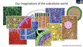
To get the world map for the Earth and to get the map for the cell, there's a lot of similarity. To get the map of cell, there's also a lot of challenge because things are so small and there's just too many things in there. So far, a lot of understanding of how things are arranged and organized in the cell has been relying on our imagination. So these are really, really, really pretty illustrations from David Goodsell, and it is always my dream to turn those beautiful pictures into real photos and real data. To map the cell, there are 3 steps. You have the cell. The first step is we need to put a label on the molecule of interest. Speaking of force microscopy, because we can identify exactly what our signal is coming from because that's what I've labeled. There can be two general approaches. For fixed sample, you can just use specific binders like antibodies or FISH probes to bring in an organic dye. For live sample, the most common approach is to use a a genetically encodable fluorophore like GFP. Then with the labeled cell, you put it on the microscope, take the image, and send it to a computer. Of course, it's not going to be a small laptop like what's shown here, but more nowadays with big computers to get all the data together that will allow us to paint a map. This is essentially what we are thinking of. In particular, we want a map that's not just static. We want a map that's a movie so that we know not just where things are, but how they are moving, what they are doing. So for this purpose, we're going to use the genetic engoblement for, for like frozen proteins. And the idea is We're going to create a library of cell lines, and within these cell lines, each cell in this library, each cell line is going to carry a tagged protein, a protein tagged with GFP. And more importantly, we don't want to just like overexpress those tagged proteins because over the years we have learned that the behavior of many proteins are dependent on their expression level, and sometimes even a tiny amount of expression can mess up with the localization or function. This thought has been inspired by the work from Aaron O'Shea and Jonathan Weisband more than 10 years ago. In that work, they created the EAST library. In this, it's a collection of over 2,000 yeast strains. Each of the strains has one protein in the others tagged by GFP. That is an immensely powerful resource for, well, quantifying the abundance, the expression of proteins, the subcellular localization of proteins, and more importantly, for screening for, say, in the process of stress or aging of yeast, in how the cell responding to it. Basically, the thought is that in fluorescence microscopy is a very, very powerful analysis tool because you label the protein of interest. Whenever you see the signal, you know where the signal is coming from because that's the protein you have labeled. But that's also the biggest limitation of fluorescence microscopy. Because you have to know what to look at before you label it and image it. Now with this library, actually with the yeast library, it has turned fluorescent microscopy into a discovery tool that we can say just pick the process of interest and unbiasedly analyze the library to identify the players very much in the same way as genetics screening, CRISPR screening, or proteomics type of screening can provide us. OK, for yeast, it has been made possible because of the easy genetics of yeast. It has very strong homologous combination. And what has been done before is to take the GFP gene with a homology arm, transform the yeast, yeast will take it up with homologous recombination, and that generates the library. For mammalian cells, it has been a lot more difficult until recently with the advancement of the CRISPR-Cas9-based gene editing method that with the double-strand break made by the Cas9 protein and the homology-directed repair process, we can incorporate the donor DNA much more efficiently. So there has been a lot of successful examples that people can tag protein endogenously with the GFP using CRISPR-Cas9-based genetic engineering. But there's still challenges. The challenge comes from the fact that GFP is a pretty big gene. It's over 1,700 base pairs long, and that needs also a fairly long homology arm to make the process efficient. Now, if you have just one target or just a handful of targets, it's pretty trivial to clone the donor DNA and try the conditions and make it work. But if our goal is to scale this up to hundreds or even thousands or tens of thousands, That becomes overly expensive to deal with. And this is what we have set out to solve, the challenge we try to address. And we're thinking, okay, the problem is because GSP is big. Can we have a much, much smaller tag that is also fluorescent? And this is the question that Daiichi Kamiyama, a scientist in the lab who is now running his own lab at the University of Georgia Athens. And Sayaka Sekine is a postdoc in the lab who's now about to start her own lab at the University of Tohoku in Japan. And well, GFP is nice because it's entirely genetically encoded, that you only need to put in the protein sequence, it by itself, it will generate the chromophore. That's hard to achieve by any of the other proteins, not to mention short peptides. Now, if we cannot replace GFP itself, we can still have a way around. That is utilizing the split fluorescent protein system that Jeffrey Waldenapf has developed back in 2005. In this case, we don't need to put the entire GFP sequence. Into the target. Instead, we only need to put a small fragment of it and express the rest of the GFP just freely in the cytoplasm. Neither of those two fragments are frozen by itself. Particularly the bigger fragment requires a residue on the small fragment for the chromophore maturation. But these two fragments have very high affinity between them. So once they are, as long as they are in the same subcellular compartment, they're going to bind and reconstitute a fully functional force and protein. And it's different from other systems where people use split force and proteins to detect protein-protein interactions. In this system, they just bind by themselves without assistance of other protein-protein interactions. So that's the case of putting GFP11 on the lamin and expressing GFP1-10 in the cell. You can nicely see the signal from the nuclear envelope. As compared to the case of knocking in full-length GFP, GFP11 is only 16 amino acids with a linker with a short homology arm that's enabled by this small donor. We can encode everything in a 200-nucleotide oligo that we can directly buy from nucleic acid synthesis like IDT. That makes the whole process extremely efficient. You don't need multiple steps of cloning to generate that donor DNA. To demonstrate the knocking at a scale of the ventricular system, doing knocking at a scale, we've teamed with Jonathan Weissman Lab, in particular with the postdoc Manu Naneti, Jonathan's lab, who's now a group leader at the Chan Zuckerberg Biohub. And Manu has been working out, has worked out the pipeline of using in vitro transcribed guide RNA combined with purified Cas9 RNP and electroporation together into the cells that's expressing GFP 1-10 already. Then for cells that has correct integration, it will generate the fluorescent signal, which allow us to isolate those cells with the integration with flow, flow-activated cell sorting. And this is the case for the knocking on the NAMM gene. We got like 36% of cells that has an inclusion, And sort out them, we see nicely the nuclear immunodeck signal. Now this pipeline, what the experiment takes one day and wait for a couple of days for cell recover and we can sort the cell. Very efficient. And I've asked Daiji and the Serkan and the Manu to, well, to pick their target and try to demonstrate for a small library. They picked about 48 targets and took 3 weeks to get to this stage. So 3 weeks, like 1 week to pick the target and design a sequence, 1 week to wait for IDT to send us the articles, and 1 week later we have 30 cell lines. So 30 out of 48 successful. And that is in fact an even higher success rate compared to the yeast GFP library effort. Now the ones that are not successful, the majority of them are the ones that are expressing at a level that's below the FAST detection limit because we can plate the unsorted cells, and we can see the fraction of cells containing the correct signal. To allow us to cover more of the proteins, we want a better probe. So this is the work started by Siyufeng, a very talented graduate student in the lab who's now a postdoc from Genentech. MITech. And she single-handedly built up the entire directed evolution pipeline for protein engineering. This is one of the proteins that she has engineered, the split AmnionGreen2 system. We can use the AmnionGreen2-11 fragment to label endogenous proteins. And you can see here are some of the images in there. The nice thing for the AmnionGreen2 system is that it has much higher signal-to-background ratio compared to the fluorescent protein, compared to the SpeedGFP system. I mean, first, AmnionGreen is a brighter fluorescent protein than GFP. On the other hand, well, even better is that we noticed that GFP 1:10 this fragment somehow still contains a little bit, a very, very weak autofluorescence background. So look at this, this FACS histogram. And on the right is the log of the fluorescence from individual cells. And this is GFP 1-10 by itself expressed with strong promoter. You can see the signal is about substantially higher signal compared to the autofluorescence background in wild-type cells. Even with a weak promoter, the PGK promoter for the GFP 1-10 fragment, it still did an appreciable background. Whereas in mNuGreen 1-10, even expressed at a higher promoter, there's no detectable background signal compared to the wild-type. Cell. When we are tagging a protein with low expression, like this is compared with the GFP11 versus AmnionGreen11 versus AmnionGreen1-10, you can see that the integrated cell is much better sorted out with the improved system. Now it's not that the AmnionGreen system does not have disadvantages. GFP 1 to 10 has much higher affinity to the GFP 11 fragment compared to the affinity between the AmnionGreen 11 and AmnionGreen 2 1 to 10. So that's the competition efficiency higher. But overall, when you— we see AmnionGreen is just unanimously better than GFP— GFP system for the subtargets that we have tested. And very recently, we have the third generation of AmnionGreen3 that has improved computation efficiency compared to the AmnionGreen2 system. So with the AmnionGreen system with ability to build small libraries, I'm going to very quickly go through one example that we have applied this method for the screening of proteins in the pathway. So this is when we are studying the oncogenic mutations involved in the receptor tyrosine kinase-RAS-Raf-MAP kinase signaling pathway, a collaboration between our lab and the Trevor Beverley lab at UCSF. And classically, the receptor tyrosine pathway, I would say, is very, very well studied in controlling cell proliferation. And the textbook model is that when we have a ligand, it starts to dimerize on the plasma membrane, and that will cause the kinase to— in other phosphorylated kinases will recruit adaptor proteins and the guanine exchange factors to activate the Ras protein by exchanging GDP for GTP, and the activated Ras protein on the plasma membrane will activate Raf and then MEK and ERK translocating into the nucleus and activate the proliferation signal in the cell. So it's very important in the development of an organism and And you also understand when proliferation is out of control, you're going to have a problem. So it's no wonder that many of those proteins in this pathway is heavily represented in mutations that's causing cancer. One of the mutations that in one of those oncogenic mutations that we're studying is the EMO4-ALK fusion mutation. ALK is another receptor tyrosine kinase, and EMO4 is a microtubule-binding protein. In this fusion mutation, it's the kinase domain of ALK fused to a fragment of the EMO4 protein. Now this fusion protein has completely lost its transmembrane domain, so it's no longer associated with the plasma membrane, but still it's very efficient in activating the RAS signaling pathway. And from earlier work in the Bevon lab, they have found that the EMO4-ALK fusion protein in the patient-derived cell line exists in those clusters in the cytoplasm. And it's been puzzling because in a textbook model, everything from receptor tyrosine kinase to Ras happens on the plasma membrane. Ras is post-translationally modified to go to the plasma membrane. And how are these sarcasmic granules somehow connected to the Ras activation? So this is the work by Jian, a very talented postdoc in the lab who's now running her own lab at University of Florida. And Dana Neal is an MD-PhD student in Trevor's lab who's now a resident in Harvard Hospital. And Asmin Topol is a postdoc in Trevor's lab who's now a senior fellow at UCSF. To make a very long story short, we've shown that those clusters we see on the microscope are phase-separated protein granules coming from the multivalent interactions mediated by Umo4out protein and the Grap2 adaptor protein binding. And to illustrate how those granules could activate signaling, we need to know the players in those granules. In doing proteomics on those phase-separated protein granules, in fact, has been extremely difficult. And even till now, we haven't had satisfactory data yet. So taking advantage of our ability to endogenously tag protein at scale, we're taking an imaging-based approach. Basically, using our AmnionGreen2/11 knock-in, we tagged everything in the RAS signaling pathway and imaged them in the cells expressing the EMO4-ALK protein and see whether they are enriched in those protein granules. So showing here are 3 examples of classic scaffolding proteins, GRAP2, and adaptor proteins GAP1 and SAS1, by themselves they are more or less diffusively cytoplasmic, but when we have immuno-4L granules in there, they are clearly enriched in those granules. And those enrichment can be reversed by crizaninib, which is an ALK kinase inhibitor. So it's specific. We show that the RAS activator Src is enriched in there. The RAS Gaps, which deactivate RAS, is not. And skipping all the other controls, just come to the conclusion of a model which showed that Imo4Alc, because of the bulk polymerization, ability of MO4 that gets initial activation of ALK through its phosphorylation. And then with the additional interaction or multivalent interaction brought in by a GRAP2 binding to phosphorylated ALK, the MO4-ALK starts to form granules, and those granules enriched in adaptors and Ras activator Src1 at a concentration that is so high that we can even activate RAS, that it is in the cytoplasm, noting that there's actually a sizable fraction of RAS that is in the cytoplasm in addition to the membrane portion. So this project by itself has a lot of other aspects in terms of how, say, those fusion mutations, they separate into granules, and what it means for cancer signaling and, well, potential treatment targets for this type of cancer. And for this talk, I just show— it's just a case that's an example that's showing how we can use even a small-scale library of of endogenous tagged proteins for the purpose of discovery, for the purpose of identifying players in a certain pathway. It would definitely be great that we can scale this up to the entire human proteome. So this is the OpenCell project at BioHub that's led by Manu Narayanan. So Manoah has taken this— well, taking the SPIT/Phospho14 tagging system, and he has been scaling up, established a highly scalable CRISPR tagging pipeline to generate so far over 1,500 cell lines and Hepto-93T cell lines that has endogenous tagged protein. And he's performed live imaging, both snapshot and confocal microscope, as well as in collaboration with Matthias Mannab proteomics analysis on all these cell lines. This is a web page that's kind of still under internal test at this time. And I hope that the data is going to be released to the public in a few months after the internal test. Just a screenshot of the images in this library and the collection of different proteins that has been tagged. I will say, okay, quoting the words from another postdoc in the lab, this is a cell biologist. Gold mine, that just by looking at those images, there's a lot we can see and a lot of surprises, in fact, that proteins are either doing things that they're not expected to be doing or having a hugely unexpected cell-to-cell variability. So for each of those proteins, Manu has had the imaging data. And the proteomics analysis of the interaction partners showing here SMC1 is one of the cohesin complexes. And with the pull-down using endogenous protein, using tagged endogenous protein, it's much, much cleaner compared to the case of any overexpression case. So that said, I hope this database will come online in a few months, and we're going to learn a lot from this database. For us, we just keep on developing the technique. One thing that we really want is a second color so that we can see protein-protein collocations and interactions. And this started with the SFCherry, the SpeedSFCherry system that was and designed and tested by a rotation student, by Marcy Ryan, in the lab. And Siyu, together with Shuting Zhou, is a very talented undergrad student, visiting undergrad student in the lab from Tsinghua University, and has used direct modulation to improve it so that we have the SF Cherry 2 and SF Cherry 3 system now. And we've demonstrated in it also has a tag for many different substrate proteins overexpressed, and in this case, endogenously tagged. You can notice that whenever you move to endogenously tagged target, the signal is just much, much weaker compared to even casual overexpression of the protein. Now with the second color, we could do double knocking. And this is looking at two ER proteins. Sec61B has been widely used as an ER marker, so that will— because it's an ER translocon complex, is not a structural protein, or is supposed to be everywhere in the ER. But surprisingly, in those images you can see that it is not enriched in those like dead-end ER tubules that is marked by the ER tubule protein R6IP1. Around a couple of months later, we learned from Pietro Di Cominio's lab that they happen to be studying this pair, well, the R6IP1, and noticed the same thing of R6IP1 being enriched in those newly formed ER tubules, and their hypothesis is that this being related to the ER genesis of that. And for us, it's just another example, is just by having the ability of looking at, of imaging, unbiasedly, endogenous TAT proteins, we can always, we can easily have surprising discoveries. Now for us, We've been thinking, well, can we generalize this method even more? The whole concept is that we have the spFosM protein that's been divided into two fragments, one small, one big. When we try to tag our target protein with the big For some protein, we don't need to put everything in. We just need a small fragment on the protein of interest, and then it will recruit the bigger functional fragment to reconstitute the fully functional label. There are many other intracellular peptide binders. SpyTag SpyCatch is a famous one, and more recent one is AlphaTag. NB-Alpha is a nanobody-based one. We could use this as a generic fragment neighboring concept. And to show a number of cases of examples, for example, when the lab has been doing super microscopy for a while, so it would be really— we really want to have a photo-activatable, uh, fluorescent protein that can be tagged in a similar way. In fact, very amazingly, just by simply merging, okay, merging the point mutations to convert mCherry into photoactivatable mCherry and a point mutation to convert mCherry into superfolder Cherry, we obtained a photoactivatable superfolder Cherry. Well, this is nice that we could do the tagging very much in the same way, just replace the 1-10 fragment. But the challenge is that PM Cherry is non-fluorescent before activation, so we cannot do the sorting in the same way as when we're tagging a protein with simple SF Cherry. But because this is, this is the binary system, we can switch the functional fragment we could express SF-cherry and sort the target. And then when we're imaging, we express with PASF-cherry so that in this population, as illustrated for the Class III labeling, we can get STORM images of endogenously tagged Class III. Very similarly, Veronica Pacinos, another postdoc— well, another grad student in the lab who's now finished, already finished her postdoc and been working at a startup, has been looking at tagging the proteins with biotags so that we can use the high affinity the spike catcher for enabling after-fixation, for efficient labeling after-fixation. In this case, we could use the, the tandem-infused FP11 tag as the selection marker and then express the FP11 to sort it so we can get a Good, well, we can get the knocking cell population, and we could either go with trineon transfection so that the sorted tag would go away. So you only need— we only have a short peptide tag on the target protein. Or in the case of SpyTag, in fact, it can tolerate having the GFP on it and still bind very efficiently. So that's CRISPR3C again. It's just now fixed and stained with SpyCatcher for us to take a STORM image. We've validated this system. This is definitely higher tagging efficiency compared to even the highest affinity epitope tags epitope tag antibody combinations. More recently, we've expanded this idea of having tandem tags, one as a selection marker and the other one as the purification handle, and collaborate with the Yifan Chen's lab of using this way of doing single particle cryo-EM structural imaging of endogenously expressed proteins instead of having to overexpress and reconstitute the protein complex. Now we can directly purify the complex from endogenously tagged proteins. Sriraj, a student, engineering student who just graduated, and Dave, another postdoc, NAP has expanded, further expanded this idea of tandem tagging. In this case, we can have just two tags and two affinity binders in there, and then these two binders can scaffold another type of protein communication on top of them, on top of those tandem tags. This is the desire for us to enable tagging proteins with the split HaloTag. And for some people who might be familiar with HaloTag, because it's enzymatic tag that can, that can covalently bind to the fluorophore, and because it's the covalent covalently bind the ligand, the ligand can carry an organic fluorophore that is providing much higher photostability and a signal compared to the fluorescent protein. So it has been very, very widely used in live imaging, particularly single molecule tracking in living cells. The issue we wanted to have a split version of the HaloTag for this type of fragment labeling, but it has been difficult to obtain an asymmetric split of a HaloTag while still having high affinity between the two fragments. In fact, the ones that's available right now is a symmetric split, that both fragments are quite big, so you cannot do the simple small fragment insertion and big fragment recruitment. But instead, we can use our tag binder system. In this case, we use the SpyTag SpyCatcher and AlphaTag and the alpha to do this. So this is the knock-in of GFP11 SpyTag. And expression of GFP 1-10 and spike catcher, and we see the endogenously enabled NAMming on the nuclear envelope. We could replace the GFP with mBiα. It's actually an even better system. Now this becomes a really highly modular system. Because we can just like put a tag in and put the binder, whichever binder that's needed for a particular application. And for example, we can have two tags, two binders on each of the tags. And in this case, we can use HaloTag to detect interaction between two endogenously tagged proteins. And we did a very simple demonstration again on NAMming, but in this case with double knocking of NAMming of SpyTag and AlphaTag. Because NAMming dimerizes, we can see this, see the complementation signal on the nuclear amino. We could even just do a knocking and then try to transfect the cell. With the binary and split halo system. So that really becomes a modular, highly modular system to analyze interaction of endogenous and tagged protein. And finally, we can introduce this into the endogenous— well, into not just cells but in whole organisms. And this actually has been another motivation for us to get onto the speech system. That's when Daiji in the lab has been studying the development of drosophila motor neurons. And he's been focusing on, well, the spatial and temporal signal for the ACC motor neuron to develop dendrites. At the very bottom of its axonal projection. So this is very precise in time and very precise in space. When trying to figure out exactly what's sending the spatial signal, he noticed that the position is exactly the same location where the ACC motor neuron is crossing the MP1 neuron. Like this is the confocal image of the two neuronal projections. Then, well, from a genetic analysis, he identified that the signaling molecule between these two neurons could be— would be DSGAM, the protein DSGAM, or Down syndrome cell adhesion molecule. DSCAM is a very, very famous cell surface molecule for Drosophila because it's involved in neuron recognizing itself. It's famous because through alternative splicing, a single gene of DSCAM produces 40,000 random splicing results in 40,000 isoforms. And each neuron is going to have a stochastic subset of those isoforms. And that is one of the— well, that is the mechanism for drosophila neurons to use the sgam to recognize, say, if it's running into another neuronal projection, whether it's coming from itself or not. Now, that's the challenge. DSCAM is on all neurons, and this is the space that is filled with all kinds of neurons. So if you do an immunostaining of DSCAM, it's messy. But if you express the transgene like with the neuron-specific promoter, you cannot recapture that splicing pattern. You really want to have a way to tag endogenous proteins with cell specificity. So to solve this, what we did, what Daiji did, is to knock in the GFP11 in the endogenous DSCAM locus and express GFP1-10 with a neuronal-specific promoter. So only in the ACC neuron now we got specific labeling of the endogenous DSCAM. And here we have specific labeling. It's very clear that DSCAM is right at the base of those, of those dendritic projections and at the interface between ACC and MP1 neuron. So this is an application for a whole organism. Just in a quick summary, this is with those fragment tags, we are literally having a universal adapter for imaging, for purification, for— we're thinking of ways of using this for perturbation like degradation and I've showed you that this can be a handle to scaffold protein-protein interactions and controlling multipolarization and basically very, very versatile functions that we can add to the target protein. OK, so in the last few minutes, I'm going to switch gear and very quickly talking about some of our efforts on the imaging side. And as I mentioned, whenever we switch to the endogenous tagged protein, the signal is much, much weaker, and we're suffering a lot from photobleaching during live imaging. And yes, the open cell images are mostly just like confocal snapshots instead of like long movies. There are definitely some movies though. To take movies of those proteins, and one way to solve the fluorophore problem while still being able to acquire 3D snapshot and 3D stats is to switch to light sheet microscopy, because in this case we only expose the focal plane for which we are imaging. But in typical light sheet microscopes, having two objectives sticking to the sample is not really compatible with the multiple plates. And it's basically not compatible with the regular dishes that cell biologists are used to use. So Bing Yang in the lab has had this clever, really, really clever idea of adapting the single-objective nice sheet configuration that's started by Chris Dansby and then advanced by Xiang Zhang and Elizabeth Human Labs to high-resolution subspectral imaging. The concept is that we can use the same objective to generate a tilted light sheet and capture the image, and then use remote focusing to untilt that image in focal plane to allow the camera to capture the in-focus tilted image. And the trick, the really clever trick that Bingyang came up with, is to use a pair of air and water objectives so that we can untilt the focal plane without loss of signal and loss of resolution. And so this is a moving of, um, I don't know why it's not playing, but this is the movie of NAMI knocking cells, a 3D movie that we can take over a long time. And Xinye, a visiting PhD student, has built this on an inverted microscope body, and it's basically operating like a confocal microscope, spinning disk microscope, but has a resolution that's kind of approaching that of a lattice light sheet microscope. And there are some more recent advances of the untwisting pair, they call this SNALDI, by Andy York at Calico, to make everything more operable. One example, in this case we are imaging Unfortunately, it's not playing. The movies of 16 cell lines, 16 endogenous TAD cell lines that we can acquire in parallel on the same multi-well plate over the duration of 9 hours. It's a very cool movie if it plays. And now with the effort from Rachel and Rashmi and two postdocs in the lab, we're working with Malu to imaging through the cell in the cell line OpenCellLibrary. And Emma and Michael are two grad students and postdocs in the lab who's working on machine learning to better improve the— well, to improve the optical resolution as well as to build up the analysis pipeline. Just finally, I hope this image can play. I don't know why. I have to say this is absolutely a gorgeous image that unfortunately doesn't play. That's where we are imaging the dynamics of nucleolus through the cell division at a— with an— it's a 3-colour 3D movie with 10-second—
Maybe you can exit the presentation mode, that might allow you to play. Sounds like this is super cool movie. We really want to have a look.
I'm going to—
Yeah, I will quickly start.
It is a super cool movie. Somehow it's not playing, but— Your screen, okay, screen share, this is easy. I'm just going with movie play. Okay. So, yeah, look at the, this nuke cell is gonna be, it's gonna divide. And under anaphase, the pink part, the granule core, just dissolves. But the green one is associated with the ribosomal DNA, still there. And then you can see the chromosome is going to pull apart in mitosis. And after mitosis, nucleolus is going to rebuild. But the granule component, you can see, is pulled into the nucleus. But there are also like these clusters everywhere in the cell. So this is the phase condensation. But those clusters away from the core of the nucleus, the nucleoli core start to grow, but other clusters start to evaporate and recondense on those cores. And yeah, to us this is a super cool movie because all three proteins that were tagged are endogenous attack proteins, and we can literally measure their abundance, their concentration, and kinetics. We are working with a number of physicists building up a quantitative model of this whole process of of nucleoli, say, decondensation and recondensation process and hoping to understand the mechanism of how the phase separation is there to help nucleoli getting together and organize the cell. So that is the movie. I can see it's 120 minutes now, 2 hours in the movie. Yeah, it's a very large dataset that you can see there. You can see the screen share again, right? Somehow it just took in place. Yeah, so that's the end, and that ends my talk. And I'd like to thank the, well, people in the lab who's been doing the real work. And this is the format of the group meetings nowadays. There's a long list of collaborators as well as, well, all these funding agencies that make things happen and everyone for listening. Yeah, thank you very much.
So, um, I think, Ahmed, you, um, okay, you, uh, if you want to help kind of write the questions in the chat box.
I can read if you like them. There's a question in the chat box from Kimberly. It seems like it would be useful to have cells labeled with just one color and neon, but add 3-4 endogenous proteins, nucleus, mito, actin, etc., to sort of highlight the relative locations of various structures. Have you considered doing that?
Yes, we are. Okay, first there has been— well, BioHub has done a lot of really cool analysis on subterranean landmarks. So far all their images has simply just using DNA stain as the reference mark. And for this Nuclear imaging, we are— well, because there are 3 components in there, and we pretty much exhaust the 3 most imaging-friendly color channels on the microscope. And this is our initial study that we are looking at the relative kinetics of those 3 different compartments. And from that, we're definitely going to start to make other multicolor cell lines to visualize, say, things relative to one another for the different compartments. Another thing that we've been looking at, and in fact the postdoc has just showed some really cool movies in the group meeting a couple of hours earlier than this, so I haven't had a chance to move those movies into into this presentation. We have been looking at the overall morphology and particularly many of these organelle markers during the cell cycle, particularly the entry and exit of the mitosis. And there's some really, really cool and surprising details that we can see from those movies.
And as people are asking, I have a quick question about what if you also combine multispectral imaging, like 20 channels live movies into your platform, would that work?
So you're talking about spectral imaging to image many, many channels? Absolutely, yes. The challenge is to get that many different colors on live labeling. I know the work done by Jennifer Nicole Schwartz was using a range of different fluorescent proteins with different spectrum. But in that case, just getting the cell transfected by— well, getting a stable cell line expressing 6 different plasmids is not trivial. Task. Well, maybe we can, we can make the cell surviving 6 rounds of knock-ins.
Actually, can you do like sequential type approach as well, like 1, 2, 3, and add more and more to the same cell line?
Yes, we can. Yes, we can do sequential approach. We can do things simultaneously or sequentially. In fact, speaking of that, the best situation for that type of knock-ins potentially in iPS cells or ES cells because their chromatin is much better guarded, unlike those like HEK cells that are hugely transformed. So as long as in that case you're careful, each round to pick the cells that are happy, most likely they can stay happy after multiple rounds of editing. And Manuel has done a lot of knocking on iPS cells already. They tend to be very friendly to— well, they're not friendly to cell culture, but they are really friendly and robust to CRISPR knocking. That would be exciting.
So I have actually a number of questions. And the first one was actually related to what Ahmed asked. So how to do multiplexing? I think it sounds like you already have a project ongoing. But I'm thinking probably Very naive question. So, can you kind of design something like a reconfigurable GFP? You can control which light lights up, and you can quench, and the rate kind of light up. That might give you multiple rounds of imaging, but without doing harsh chemical processing.
Well, that would be my dream. OK. But so far, so far, it's, yeah, it's really limited. The only thing I can think of is photoactivatable fluorescent proteins that you can image and bleach and then activate while bleaching, activate a second protein that is in the same color channel. But the issue for live imaging is you need— we need to care about temporal resolution, and we need to— we need something that, that can be repeatedly switched. That's the, that's the difficulty.
Yeah, yeah, great. I think for, uh, live cell time-lapse imaging, I think it's, uh, very, very challenging to, to do so many different colors. Um, so I think last week we had the Zhu from from Allen Institute, and a couple of weeks ago, we had Emma Lundberg from Sweden. Seems like what they do very complementary to your data. Yeah, I just want to kind of hear whether or not you're thinking so if you can not just cross-validate it, but really kind of augment your data by combining either their assay or their data sets.
Well, first, Manu, who's in charge of the Open Cell project at Bauhaus, has been having discussions with the Allen Institute from the very beginning, even before they started. We've been having discussions with them even before they made the first knock-in. And Emma Nauenberg has spent a couple of years sabbatical at BioHav in the past several years. At Stanford. Yeah. Everyone's well connected and well aware of what everyone else has been working on. The Human Protein Atlas by Emma Nuremberg is focusing on antibody staining. So that is a different scenario compared to live imaging. And a more interesting comparison is the work by Allen Institute and the OpenCell. And I think this is two different philosophy, and both are valid and both are important. Allen Institute has been focusing on precision. That means picking a handful of morphology markers and the most robust cell line, the iPS cell line, and each one of them generating the knocking with that whole genome sequencing, careful validation of everything. And that is one approach, whereas the OpenCell Project is much more emphasizing throughput. Because if you want to, say, do that for 30 cell lines, it's okay. If you want to do that for 1,500 cell lines, that's just way too much resource that needed to do all these clonal selection and validation. We, ours are heterozygous knocking or polyclonal. Cell lines, and we haven't done sequence validation for, for them. But the idea in this case is that we're going to go and opt for throughput so that we can make initial discovery and narrow down our target. And after narrowing down the target, we can then start with more careful, more careful validation. It's just two different approaches, and I think suitable for different type of investigations. The other thing that— the other difference is obviously Allen Institute is sharing all their cell lines, whereas for the OpenCell Project, it's kind of impractical to maintain 1,500 million cell lines and set up shipment for 1,500 cell lines. It's just like too much resource needed for that. So our goal is, in this case, is to, well, establish a robust enough protocol and test it, guide RNA donor DNA sequence, so that people who want to tag protein endogenously can do it themselves with ease, very importantly in their own favorite cell line. Because even iPS cell is not the best, it's a very important a very versatile cell line, but it's not necessarily the best cell line for all cell biology investigations. And different people may have different cell lines that they prefer to work in. In this case, it's our desire to show people how to fish instead of just giving them the fish. Yeah. And I also can quote a comment from Veronica when I asked her to clone a couple of PKA-GFP fusion plasmids, and she told me that it's actually easier and faster to generate a PKA knockout line than to clone a GFP fusion classmate in her case. And that's, that's where I would like everyone to get to.
Exciting. And we're getting late, but I've got one more final question. If anybody has, of course, feel free to unmute yourself and ask. The question is about the phase separation game that you alluded to. It's actually a pretty exciting field. I've been recently reading about the quantitative models for phase separation, and I think your dataset and your approach could be perfect for this. And which part of phase separation would you be more illuminating light? Is it more the cell division? I mean, there are also membranes, organelles, and others. Would you be interested in going towards those directions too?
Right now, our main focus is two things. One is the EMAL4-ALK system and other oncogenic kinase fusions. It's actually a very complicated system. For example, they're not liquid droplets. They have a high heterogeneity among them. But it seems to be a really generic feature. For many of the oncogenic kinase fusion mutations. That's one focus. The nucleolar system I mentioned previously, a postdoc in the lab actually has done an analysis of the entire human protein sequence for proteins that can kind of having a tendency of phase separation. A lot of them are DNA-binding proteins. And this— so for the 40 Nucleome Project, we're collaborating with Gita Narayanan and Xiaoping Xue and Vijay Ramani at UCSF, trying to see how these— well, you can call it transcription factor— in these DNA-binding proteins play in the phase separation, their phase separation. Play a role in, yeah, in organizing the nucleus and controlling gene expression. So that's our current, that's basically our current focus. There are a lot of unanswered questions. In particular, sometimes phase separation can be very clear in vitro or by overexpression, but at endogenous level, it's just like hard to tell. And that's our focus.
Yeah, that's exactly, I think, what people are thinking of the major challenge in phase separation studies. So we can see beautiful results in vitro, but in the cells, oftentimes that's quite a different story.
Yeah, so any other questions? If not, I think let's thank Bo again. I think it's slapping time, the Interhumanities Digital.
Yeah, we're going to do a real one.
Thank you so much.
And before we— Thank you.
Thank you so much. And before we also conclude, I'd like to also highlight that next week we will be hosting Dennis Shapiro from Harvard Medical School. And he's working in the lab of Aviv Regev and the Sorgel Lab. And he's going to talk about Google Maps for tissue biology Mapping the tumor microenvironment with spatial omics technologies. And with this, I'd like to thank everyone for their participation and hope to see you next week.
Yeah, have a good weekend. Bye.
More from Joshua







Scientist Stories: Hans Waterham, The value of phenotypic assays in peroxisomal disorders




Scientist Stories: Joel Zlotogora, Genetic carrier screening programs for reproductive purposes