Scientist Stories: Carolyn Bertozzi, Bioorthogonal Chemistry & Glycobiology
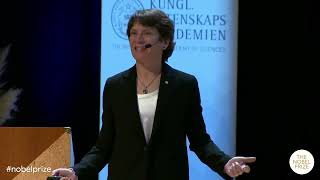
In this talk, Nobel laureate Carolyn Bertozzi recounts the conception and evolution of bioorthogonal chemistry, a set of chemical reactions that can occur inside living systems without interfering with native biology. She begins by describing the unmet need to image cell‑surface glycans, especially sialic acids, and how this motivated the design of a two‑step metabolic labeling strategy. The first breakthrough was the Staudinger ligation, a modified Staudinger reaction that forms a stable amide linkage in aqueous environments, demonstrated in cultured cells and later in mice. Recognizing the slow kinetics of the ligation, her lab pursued faster reactions, leading to copper‑free click chemistry based on strained cyclooctynes. By exploiting ring strain and fluorine substitution, they created a series of cyclooctyne reagents (e.g., DIFO, DIPhO) with rate constants up to 4,000‑fold faster than the original ligation. These reagents enabled the first live‑animal imaging of glycans in zebrafish embryos, revealing dynamic glycan distribution during development. The talk then surveys the rapid expansion of the bioorthogonal toolbox (azide‑alkyne cycloadditions, tetrazine‑strained alkene reactions) and the broad application of these chemistries to label proteins, nucleic acids, lipids, and other biomolecules. Bertozzi highlights translational advances, including antibody‑drug conjugates, vaccine conjugates, and clinical trials using bioorthogonal chemistry for targeted drug delivery. She concludes by emphasizing the crucial role of curiosity‑driven basic science and the contributions of many students and collaborators that turned a modest idea into a versatile platform impacting both fundamental biology and medicine.
Chapters
Carolyn Bertozzi was born 1966 in Boston, Massachusetts, USA. She was awarded a PhD from the University of California at Berkeley in 1993 and is now active as the Ann T. and Robert M. Bass Professor at Stanford University, where she is also an investigator at the Howard Hughes Medical Institute. So by that, Please welcome me in— welcome— please join me in welcoming Dr. Bertozzi up on stage. Thank you. This is the honor of a lifetime, to say the least, and it's my great pleasure to tell you about the work that we did to develop bio-orthogonal chemistry and to apply this new chemistry to imaging of cell surface glycans and many other applications related to that. Let me start by just introducing one of the basic tenets of the field of chemical biology, which is my field. And one of our tenets is that there are many unmet needs in the biological sciences and in medicine that drive the development of chemical technologies. And bioorthogonal chemistry is very much one of those technologies that was inspired by an unmet need in biology. And at a very high level, bioorthogonal chemistry, was developed by my coworkers and myself when I started my career at UC Berkeley. And the fundamental goal was to invent a new type of chemistry that could be performed in what I think of as the most complex reaction vessel you could think of, which is the living organism. And this flies in the face of the traditional ways in which we think about chemical reactions. When we do reactions in our chemistry laboratories, we're typically using this kind of a reaction vessel. It's a round-bottom flask where the chemist can control every single parameter of that reaction. By contrast, if your goal is to do chemistry in a living system, as was our goal, You're no longer in the reaction flask, and you no longer have control over all of the parameters. Instead, you might be doing your chemistry in a live cell, in a cell culture dish, for example. And there are a lot of interesting problems you could solve by doing chemistry in the live cell. For example, to attach imaging probes to biological molecules that you want to study in their native habitats. And that's a very complex environment in which to think about chemistry. But even more complicated is the chemistry you might want to do in living organisms, like a laboratory mouse, or, as I'll show later in this talk, the zebrafish, which is a model for development of vertebrates. And probably the highest level of complexity where one might want to do a chemical reaction is the human. And in fact, as you'll see, bioorthogonal chemistry is actually now being tested in human cancer patients for the purposes of drug delivery. So for all of these kinds of reaction vessels, we knew that we needed a different brand of chemistry. And so we coined this term bioorthogonal to describe the qualities that you would need to do chemistry in these living systems. So our definition is that bio-orthogonal reactions are reactions among components that neither interact with nor interfere with a biological system. And one way to think about that is that there is an enormous reactivity space that's described by biology. I like to think of biological reactivity space as a galaxy of chemicals within a much larger universe of chemicals. And it's a large galaxy, but there's a boundary around it. And somewhere outside of that biological reactivity space is the bio-orthogonal reactivity space, a different galaxy with different chemicals, functional groups that you would not find in a biological system, but that will react with each other regardless of being surrounded by all of those biological functionalities. So that's the type of chemistry we'll be talking about today. Now, as I mentioned, chemical biologists often develop new technologies in response to an unmet need in biology. And in my lab, the unmet need that we became interested in was to study changes in cell surface glycosylation. That's another word for the complex carbohydrates that decorate the surface of all of our cells. And even back in the 1990s, there were decades of literature showing that when cells undergo a transformation of their state, there are changes in the patterns of cell surface glycans. And sometimes those changes in cell surface glycosylation are associated with disease. For example, it was already known back then that when healthy cells transform into a cancerous state in the context of a tumor, there are cell surface glycosylation changes that often include an overproduction of a particular sugar that's known as sialic acid. And it's one of the building blocks in the complex carbohydrates on the surface of your cells. And we had a fairly straightforward objective, which was to develop a technology that would allow us to profile the abundance of sialic acids on healthy cells compared to cancer cells, and to develop a strategy for molecular imaging of changes in sialic acid in order to study cancer. But at that time, there was no technology available to image cell surface sugars, including cell surface sialic acids. So that was the unmet need that we were focused on solving. How do you image a sugar? Well, you have to do it in a different way than you would image other biomolecules like proteins or nucleic acids. For those other molecules, there were some good technologies available at the time, but for sugars, there was not. And during my postdoctoral years— so this takes me now back to around 1991 or 1992— I was thinking about this problem, the need to image the cell surface sugars, and I happened to attend a conference in Southampton, England, and this was a serendipitous moment for me because I heard a lecture by the late Professor Werner Reuter, a very well-known German biochemist at Free University Berlin. And his lab had been studying the biosynthetic pathway that produces sialic acids and delivers them to the cell surface. And what he had observed, and he presented this at this conference, is that one could feed cells a chemically altered version of a metabolic precursor of sialic acid that's called N-acetylglutamine. Menosamine. And normally, N-acetylmenosamine has an acetyl substituent here at this pink position, where this R group would be a methyl group. And what he found is that one could make conservative changes to this R group, just extend the side chain out by a few carbon atoms, and those subtly altered N-acetylmenosamine derivatives would be subject to enzymatic metabolism inside the cell, which led to the production of an altered sialic acid residue, which was then incorporated into the complex carbohydrates on the surface of those cells. So you feed the cell a chemically altered sugar, and they produce a chemically altered sialic acid. And when I saw that talk, a thought came to mind, which was that maybe we could put a chemical group that's reactive at that position, and once that chemically reactive group appears on the surface of the cell, we could chemically attach an imaging probe. So that was one of the research proposals that I crafted and submitted when I applied for a faculty position at UC Berkeley. And here is a cartoon that's almost literally right from my job application from 1994, where I proposed a two-step process for imaging cell surface glycans. The first step would be to feed the cell or the organism, or maybe the human patient someday, that chemically altered simple precursor sugar. And that sugar would be metabolized by the cell, converted to the cell surface product, where this chemical group X— and I was agnostic about X at that time because I wasn't quite sure what X would be— but some chemical group X would come along for the ride. Now it's presented on the cell surface, And we would do a chemical reaction with an imaging probe that had a complementary functional group Y. And the hope was that X and Y would react with each other, form a bond, and that would attach the probe to the sugar, and we would now be able to image the sugar. But there was one problem with this cartoon, which is that we would need a chemical reaction between two functional groups that could proceed in a living system, in cells, in animals, the natural environment of these cell surface sugars, which means that the chemical reaction would have to be bio-orthogonal. And this was the problem that was the genesis of bio-orthogonal chemistry. So the reaction that we started with, and which ultimately was the first bio-orthogonal chemical reaction, was a derivative of a very well-known chemistry from the German literature of the previous century. This is the classic reaction of triphenylphosphine with what turned out to be a very important functional group. You'll hear a lot more about this in the other talks this morning, which is the azide. And phosphines and azides will react with each other to form an adduct called an azaylid. This was a chemistry first reported by Hermann Staudinger over 100 years ago, 1919. And in fact, Professor Staudinger won the Nobel Prize in Chemistry about 40 years after this initial report. Now, phosphines and azides do not exist in biological systems, And they react with each other very selectively without any interference by the biological system. So on its face, this reaction, as I've shown it, looks like it could be a bioorthogonal chemistry. But there was one liability with this classic Staudinger reaction, which is that the azylid product is not stable in water. And if you're doing reactions in living systems, Water is your solvent. There's no choice. So we had to solve this problem of the hydrolytic sensitivity of the azahylate. And one of my graduate students, Eliana Saxon, who joined my lab around 1998, came up with the solution to the problem. She made a simple modification of triphenylphosphine, introducing this methyl ester. So now when the phosphine reacts with the azide to form this azaylid, the nitrogen atom, which is nucleophilic, can attack the ester in an intramolecular reaction, very rapidly forming this cyclic product. So now when water ultimately hydrolyzes this phosphorus-nitrogen bond, the amide bond that had formed in that intramolecular reaction keeps this product together as a ligation product. So this variation on the classic Staudinger reaction we called the Staudinger ligation, and this was the first example of a bioorthogonal reaction. And here is Eliana. I found this old photograph the day that she got her first big result, where she showed the Staudinger ligation could be performed on cultured cells in a dish with no harm to those cells. And she came into my office and she showed me the data. And I remember thinking, I think this is a really big result. But this was very early in the days of my young lab. So once we had this chemistry worked out, we then demonstrated that we could introduce the azide into a variety of different cell surface sugars, all of which are interesting in their own ways. So for example, I mentioned that imaging sialic acid was one of our initial goals. And so we showed that we could introduce the azide on the side chain of N-acetylminosamine. And when fed to cells and metabolized inside the cell, the corresponding azido-sialic acid would appear on the cell surface. Primed for reaction by the Staudinger ligation. And likewise, we figured out how to put an azide into a sugar called fucose, which is also an important sugar for various biological systems. And in addition, we showed that we could put azides into N-acetyl galactosamine, or GalNAc, using this azidoacetyl variant of the parent sugar. And each of these sugars we chose because there were some interesting biological stories behind them. Another big moment in the lab came when two of my former graduate students, Jen Prescher and Danielle Duby, demonstrated that the Staudinger ligation could be used to attach probes to sugars in living mice. This was another big moment in the lab because it was the first demonstration that a bioorthogonal reaction could be so well tolerated by a living system that you could do the chemistry in the mouse with no toxic side effects. And in fact, Jen and Danielle, I'm so privileged to say, are with us in the audience today. And here is an image from this paper they published showing that they could inject into mice azidoacetylmanosamine. It would get metabolized by the cells in the body of the animal, and the azidosialic acid would appear on the surface of those cells. Then in a second step, they would introduce a phosphine probe set up for the Staudinger ligation, and in the body of the animal, the phosphine would find the azide, undergo the reaction, and formed this product, which we could then detect in a variety of ways, including with fluorescent-tagged monoclonal antibodies. Another major achievement by these fearless students who were willing to turn the mouse into a reaction flask. But we knew that there was more work we had to do in reaction development. And the reason was that the Staudinger ligation for all of its amazing qualities, was a rather slow reaction at biological temperatures. It's a bimolecular reaction, which means that it follows bimolecular reaction kinetics. And so hopefully the students in the audience will recognize that for such a reaction, the rate is equal to an intrinsic rate constant times the concentration of the two reactive partners. And in a biological system, like a living animal, we learned some things that are important. For example, we learned that the concentrations of these reactants were probably going to be very low. The animal is metabolizing these reagents as soon as we put them in the body. This is not a reaction flask in your fume hood. And there's only so much of a molecule you can introduce into an animal because of issues like solubility in aqueous systems or nonspecific toxicities for molecules that really don't belong in the animal's body. So given the limits on the concentrations of these two parameters, the intrinsic rate constant of the reaction is very important if you want the reaction to proceed at a reasonable rate. And for the Staudinger ligation, which had a second-order rate constant around 10��³ per molar per second, we discovered that this was a little too slow. This reaction took hours to proceed, sometimes a day. And there's a lot of biology that takes place on a much shorter time scale than a whole day. So we were interested in alternative bio-orthogonal reactions that would leverage the azide as a reactive partner. Now, it turns out that azides can also undergo what we call 1,3-dipolar cycloadditions with alkynes. This is another chemistry that was well established in the previous century, and Professor Rolf Wieskin from University of Munich was one of the major practitioners of this chemistry, who did a lot of mechanistic work to describe how this reaction proceeds. And like the classic Staudinger reaction, this chemistry seemed to us as being almost bio-orthogonal because like the azide, alkynes are also quite rare in biological systems. We don't have them in our bodies, as far as we know, and yet these two will react with each other to form a product called the triazole. Now, the reason I say this is almost, but not quite, bio-orthogonal is because this reaction is very slow at room temperature, much slower even than the Staudinger chemistry, which is why when people do these reactions in the fume hood in the chemistry lab, they generally have to reflux the reaction at high temperatures, like 100°C. So at biological temperatures, we thought of this Whisky chemistry as just being too slow for our needs. Now in 2002, the world was changed, and that's because my co-laureates, Professor Barry Sharpless and Professor Morten Meldahl, discovered a mechanism for accelerating the rate of the azide-alkyne cycloaddition. And this is what is now almost synonymous with the term click chemistry. And as you will hear from their glorious lectures after mine, the addition of a copper catalyst changes everything. And suddenly this reaction is incredibly fast. It's over in minutes. And it is now by far one of the most important reactions in all of chemical biology and very much worth recognition by a Nobel Prize. Could this solve our problem of kinetics? And the answer was not quite. The problem was the copper catalyst was just too toxic for cells and live animals. But it was kind of on the right path, and it got us thinking about other mechanisms by which one might accelerate the rate of the cycloaddition, but without the need for a toxic metal catalyst. Now, like many of us, I teach sophomore organic chemistry, introductory organic chemistry. And when we teach this class, we always have a lecture on the concept of ring strain. And here are the two textbooks that I used to use to teach this class when I was at Berkeley, both written by Berkeley faculty, Peter Vollhardt, Clayton Heathcock, and Andy Streitwieser. And in these books, you will see a description of what happens when you make ring systems that have uncomfortable bond angles. These strained ring systems can cause functional groups to have very different reactivities. So, for example, when you put a double bond into a 3-membered ring in this form or in this form, a lot of strain energy gets pent up in that ring. And if you can react those pi systems and release the strain, that can accelerate the rate of a reaction dramatically. So I talked with my students about whether there was precedent for an azide alkyne cycloaddition with a strained ring system. And Nick Agard, who is sitting here in the audience today, went to the library, which used to exist, and he found this amazing precedent. Back in 1961, Georg Wittig, another Nobel laureate in chemistry, reported on the reaction of cyclooctyne, the smallest of the stable cycloalkynes, which has 19 kilocalories per mole of strain energy. And when Wittig and Krebs, his coworker, combined cyclooctyne with phenyl azide, you can see from this statement in German, the reaction proceeded like an explosion to form the corresponding triazole. So here was an azide alkyne cycloaddition with no catalyst that basically proceeds at room temperature without the need to heat it up. So based on that precedent, Nick and then Jeremy Baskin, who is now a professor at Cornell, and he's sitting here in the audience today as well, as well as several other coworkers, went to task to try to develop cyclooctyne-based probes for what we called a copper-free click chemistry that we could use in living systems. And to make a long story short, they spent several years building cyclooctynes with different kinds of modifications, and what we learned is that the cycloaddition of these cyclooctynes with azides can be tuned in terms of reaction kinetics over several orders of magnitude. So we had a lot of control levers on the rates of these cycloadditions. Just to give a few examples, in the upper left is a very simple cyclooctyne, which would react with a model azide with a second-order rate constant that was kind of on par with the Staudinger ligation. So that was our starting point, but we knew that we needed to enhance the rate by a few orders of magnitude. And so Jeremy Baskin had a big breakthrough in this project when he showed that addition of two fluorine atoms next to the triple bond would increase the reactivity almost 100-fold. And this reagent, which we gave the acronym DIPhO for a difluorocyclooctyne, became one of the first important reagents for imaging sugars in live systems, as I'll show you next. But just to complete the slide a little bit, we could enhance the rate yet another order of magnitude by fusing benzene rings on either side of the cyclooctyne, which increases the ring strain even a little bit more. And then the ring could be shrunk down to a 7-membered ring as long as one of the atoms in the ring was a larger atom like sulfur, and one could get yet another boost in the reactivity, where now we have a reagent that's 4,000 times more reactive than our parent compound. So this was a really fun period of time in the lab because we felt like we were returning to our roots as physical organic chemists, doing the kind of structure-reactivity correlation studies that were popular 100 years earlier. But that had fallen out of fashion in the new millennium. And yet here we are invoking some of these classic concepts to tune a reaction for biological systems. With these new reagents in hand, Jeremy Baskin and another coworker, Scott Laughlin, took on the challenge of imaging sugars in live animals. And the animal that they focused on was the zebrafish. Which is a very popular model organism to study vertebrate biology, with the benefit that zebrafish are translucent. So they're a wonderful organism for fluorescence imaging studies. You can see all of their organs under a microscope as they are alive. And so Jeremy and Scott did experiments where they would take early embryos of zebrafish developing in a dish They would feed these embryos azido sugars, which would get metabolized, and once those azido sugars appeared on the surfaces of the cells of the animal, they would do the copper-free click chemistry reaction in the live animal with a DIFO probe, introduce the fluorescent dye, and now we could image the sugars for the first time in a living animal. It was a very dramatic moment in the lab. And they did some experiments where they would pulse these embryos at different time points with different azido sugars and map out sugars appearing in different cell types. So for example, in one experiment they would do one round of labeling and then any unreacted azides still present in the fish, they would quench with a reagent that is a phosphine reagent in a classic Staudinger reduction. Then they could pulse the embryo with more azido sugars that would appear at different time points and label them with different colored dyes. And we could create fish that basically had all the colors of the rainbow, where the different colors represented different types of cell surface sugars that appeared during development at different time points. Just to give you a sense of what these images look like, up top is the head of a roughly 5-day-old zebrafish, which has had different sugars labeled at different time points with different colors. You can see how they're distributed. And below that is basically a, a round of cell division from an earlier embryo. So here's the intact early embryo. Each arrow is pointing towards cells that are undergoing mitosis. And if you expand the image and take a close-up look at cell division, what you can see is that a single parent cell— I'll wait for it to loop— duplicates its chromosomes, segregates them as the cells divide, and you can see the cell dividing through the portal of the DNA, which there are good tools for imaging, and we used one of those tools here. You could see the cell division through the portal of the proteins, which were also easy to image back then, and the proteins that we're imaging here are located on the membrane of the cell, and now it's dividing into two daughter cells. But for the first time, we could also see cell division through the portal of the cell surface glycans. And that was another first-in-class achievement by these incredibly fearless group of students. And in this image, we were quite struck to see how the glycans build up and concentrate right at the junction as the cell is completing a round of mitosis and contributing to the segregation of these daughter cells. In a way that was previously unknown. So after this point of time, the field really took off because it turns out that once you show that you can use chemistry to image sugars in a biological setting where the animals are alive, lots of people came out of the woodwork and asked us if they could use our reagents to image all kinds of other kinds of molecules that were also difficult to study. And so here is a pie chart on the cell showing roughly the breakdown of all the different types of biological molecules. And over the course of 4 or 5 years— this brings us up to about 2010— there were groups that showed that they could use the azide as what we call now a chemical reporter for virtually every type of molecule on the pie chart. They could put azides into amino acid side chains, and label proteins and use bioorthogonal chemistry to image those proteins in living systems. They could put azides into nucleosides and label the oligonucleotides and use bioorthogonal chemistry to image those molecules. They could put azides into lipids and track those lipids around in living systems using bioorthogonal chemistry. So the applications of this chemistry in the research setting expanded quite rapidly into lots of different areas of biology. Another big development around that time was expansion of the toolkit of reactions that are available for bioorthogonal chemistry. And so we started our work with the azide, and the cyclooctane partner became a really good combination for chemistry in living systems. But later, other groups discovered that Tetrazines can be bioorthogonal reagents reacting with strained alkenes like this cyclooctene or this cyclopropene. And now that toolkit continues to grow, and I would put the challenge out to students in the audience here: if you come up with a new idea for a new bioorthogonal chemistry, this could have tremendous impact because while we now have a handful at our disposal, There's always a need for more options in this space. It's a great challenge for someone who's starting out their career in organic chemistry. Also, the applications expanded. So 20 years ago, we developed these chemistries as tools for basic research in the laboratory setting. But over the last 10 years or so, there's been considerable activity in the translation of this chemistry for new medicines and new diagnostic strategies for human disease. And so I thought I would mention that there are now a number of biotechnology companies that use bioorthogonal chemistry in a variety of settings. A very popular application is for making antibody drug conjugates, and these are important medicines for the treatment of cancer. As well, there are companies that are using bioorthogonal chemistry to make new vaccine conjugates, and some of these are in right now a Phase 2 clinical trial. And then I have a company, as a disclosure, which uses bioorthogonal chemistry to make targeted degraders of extracellular molecules. And I'm very excited to be an advisor of a company called Shaski, which is performing bioorthogonal chemistry in the body of cancer patients as a mechanism to target chemotherapies very selectively to the tumor tissue and to keep those chemotherapies away from healthy tissues. So it's a very exciting time for me to see how the chemistry came from a very modest idea in the research lab aimed at imaging sugar molecules into a very generally applicable tool which people are using to make new types of medicines. Finally, I want to make one point which I think is important, because despite all of these exciting applications, I still think the most important lesson from the story of bioorthogonal chemistry has to do with the importance of curiosity-driven basic science. And those of us who are scientists are familiar with what people often describe as a tension between basic science and applied science. And funding agencies go through various phases of interest in applied science versus basic science. But bioorthogonal chemistry tells the story of how important curiosity-driven basic science can be in order to realize these applications. Because I'm pretty certain that the chemists on whose shoulders we built bioorthogonal chemistry, and by that I mean Hermann Staudinger, Rolf Wieskin, Georg Wittig, when these chemists reported on the curious reactivity of azides, phosphines, and alkynes, they would never have foreseen that someday I would stand here and show you fluorescent zebrafish. They couldn't have known that. They made observations about exotic molecules, and they published them purely based on curiosity. And here we are, 100 years later, making use of these chemistries in ways that we hope will benefit human health. So in order to get to fluorescent zebrafish and imaging of sugars, to imaging of tumors in human cancer patients, and to making new medicines, you need to have that foundation established by the basic science. So with that, let me thank the bodies of people that basically made this moment possible. First and foremost, the Royal Swedish Academy of Sciences and the Chemistry Nobel Committee. Thank you for changing my life. The Nobel Foundation for the amazing festivities of this week, which are just ramping up. My institution of origin, UC Berkeley, which we often call Cal. This is where all the work was done that I presented today. So I owe a great debt of gratitude to the students and to the faculty at Cal that gave me my big break and that did all this beautiful work. And my current institution, Stanford, is very supportive and is helping me try to translate some of these reactions into human medicines. And then finally, the people who financially supported this work from the very beginning, which is the National Institutes of Health and the Howard Hughes Medical Institute. The most important slide of all is this one, because none of this happens without the intrepid students and postdocs that occupy the academic laboratory. And this is my current group at Stanford. This is how we had group meetings during COVID We had to rent a drone to get this picture. And after the Nobel Prize announcement, my students organized a Zoom party. And these are all the faces that we could get on Zoom on short notice from all over the world, and some of whom I'm presenting this work to today. Thank you very much for the work that you did leading to this moment, sitting there in the second row. My family is here. Thank you very much for all the support you have given me since my moment of birth. And thank you very much to this audience for coming today. Thank you.
More from Joshua







Scientist Stories: Hans Waterham, The value of phenotypic assays in peroxisomal disorders




Scientist Stories: Joel Zlotogora, Genetic carrier screening programs for reproductive purposes