Scientist Stories: Chen Dong, T-cell Regulation in Immunity & Diseases
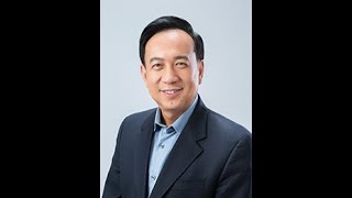
Chen Dong's lab found that fever-level heat (39.5°C) makes Th17 cells more inflammatory by triggering a protein modification of SMARCA4, which ramps up inflammatory genes. In a second study, intestinal cells produce IL-17D, which signals through CD93 on immune cells to protect against colitis and colon cancer.
Chapters
At 9:00 AM PST, it's now, and I have the pleasure to welcome everyone to another Global ImmunoTalk. We are back after a brief break, re-energized, and most importantly, with an amazing list of Global Immuno speakers that will be presenting in the next few months. Just as a reminder, Ajita Stockinger will be presenting next week. So I hope you can all join us. So I will now stop sharing the screen because I would like to introduce to you our fabulous speaker today, Dr. Chen Dong. So Chen is originally from Wuhan, China. And is currently a professor at the Institute for Immunology at the Tsinghua University and also more recently at the Jiangai Jiatong University School of Medicine. So Chen has served as a professor of immunology and the director of the Center for Inflammation and Cancer at the University of Texas MD Anderson Cancer Center before he moved to China and also was He was for 4 years, from 2016 to 2020, the Dean of the Tsinghua University School of Medicine. Chen's research, as many of you may know, focuses on understanding the molecular mechanisms whereby immune and inflammatory responses are normally regulated and to apply this knowledge to understanding and treatment of infection autoimmunity, and allergy disorders, as well as cancer. And we can say without any hesitation that Chen and his team has made tremendous contributions in immune regulation, particularly in the field of T cell differentiation from both his laboratories at US and China. As an example of Chen's impactful contribution to this field, We can highlight his work that led to the discoveries of Th17 cells, antifollicular helper cell subsets within the immune system and the elucidation of these subsets' biological and pathological functions. So Chen has over 200 publications in top-tier journals and was rated highly cited researchers for 7 years from 2014 to 2020. The honors he has received include in 2009, the American Association of Immunologists BD Bioscience Investigator Award, and in 2019, the International Cytokine and Interferon Society BioLegend William Paul Award. He's a member of the Chinese Academy of Sciences and a fellow of the American Association for the Advancement of Science and the Chinese Academy of Medicine. He's currently an editor for Immunity and a scientific advisory, uh, board member for MADD, an editor-in-chief for Frontiers in Immunology, and an associate editor for China Sciences Life Sciences. On a more personal note, I have great memories when I had the pleasure to meet Chen at the Gordon Conference. He and Carola Vinuesa invited me to attend, it was particularly antifollicular helper cells, and I could experience there Chen's amicable and generous personality. So Chen, it's a great, great honor to have you as a Global Immuno speaker today, and we are really thankful and and appreciative of all the efforts you put in preparing this talk. And we cannot wait to hear it. Chen's talk is entitled T Cell Regulation in Immunity and Diseases. So Chen, before we let you share the screen, your slides, as you know, we have a question that we would like to ask you. So we get to know you better and know your perspective, particularly here on how do scientists work together in the COVID-19 pandemic. What are your thoughts about that?
First, Nina, thank you so much for inviting me to this Global ImmunoTalks. Is a wonderland for immunologists to get reconnected again. I think the question you asked is a very important one. So we first need to stay connected because we all miss each other. We shouldn't forget about each other. We saw names in journals but we need to see each other in faces, virtually or in reality. And that's first thing, I think we need to stay connected. And the second thing is we need to work together because we all experience the globalization of sciences, but now everything is interrupted by COVID. I think we need to think of ways to resume our scientific collaboration, scientific exchange, and I think that's important. And I'm sure that scientists still need to work together. In not only dealing with the infection but also many challenges around the world. So I think we need to reverse the deglobalization of sciences and stay connected. Thank you.
Yes, thank you. Thank you so much for sharing those thoughts, and I couldn't agree more how, you know, science is a global endeavor, and yeah, borders are non-existent in science, and that's a beautiful aspect of it. So thank you so much for sharing that. Okay, Chen, we are all ready for your talk, so if you want to share your screen.
Yeah, perfect.
And presenter mode. Excellent, perfect. Thank you so much.
Okay, thank you. Uh, good morning everyone, wherever you are. Uh, in China it's, uh, midnight, so it's morning again. So I, I, I'm sure every— most people are still in the morning, so, and except European people. So, uh, my name is Chen Dong. It's my honor to present in this Global Immunotox I found this wonderland, and I'm sure I will tell others to join and to listen. And as I said, stay connected. And so today I will share with you two stories we recently published on IL-17-producing T cells and also related lymphocytes. So Th17 cells were added to the map of CD4 helper T cells in 2005, more than 15 years ago. We and others proposed that these represent the third lineage of helper T cell differentiation. These cells are named because they secrete IL-17, IL-17F,. Also, they express a discrete array of cell surface receptors. Also, the differentiation of Th17 cells occurs independent of that for Th1 or Th2 cells. We know that IL-6, together with TGF-beta, would induce early Th17 cell differentiation. And we reported IL-1 also detected early to sustain and to mediate Th17 cell differentiation. Downstream of IL-6, of course, STAT3 is necessary, and also there are two, at least two Th17-specific transcription factors. The major one is RORγT, discovered by Dan Quire and Dan Littleman to be necessary and sufficient for Th17 cell differentiation. The other one was discovered in our lab, ROR-alpha, which plays redundant and also coordinative roles as ROR-gamma during Th17 cell differentiation. So initially, when we proposed Th17 cells as a distinct lineage of helper T cell differentiation, we also proposed that these cells may have different biological functions compared to those by Th1 and Th2 cells. So in summary, Th17 cells are very important for our host defense to infection. We now have patients which, who have deficiency either in producing Th17 cells or could not respond to IL-17. These patients are sensitive to various infection, particularly by extracellular bacteria and also by fungi. Th17 cells are frequently found at the mucosal surface. Not only are they important for mucosal immunity to infection, but also they help to maintain the mucosal barrier by stimulating epithelium cells to repair and also form tight junction. Most importantly in the field is the relevance or the function of Th17 cells in chronic autoimmune diseases. Now targeting Th17 cells has been already therapeutically approved for the treatment of psoriasis and other autoimmune diseases in humans, proving that indeed these cells very important in human diseases. But so the work in the field recently has been focused on the pathogenic regulation of pathogenic function of Th17 cells. As I mentioned, Th17 cells can be found in at the resting state in the mucosal tissues, intestine for example, but they don't elicit strong inflammatory reactions in those tissues. On the other hand, in disease or disease models like EAE, Th17 cells can migrate into central nervous system and to induce chemokine production and induce tissue inflammation. So it's not clear why Th17 cells sometimes are quiescent and sometimes are even tolerant to the environment, but why in autoimmune diseases they play very important roles in sustaining or initiating tissue inflammation. So several years ago, we began to investigate this issue by looking at environmental factor that we all are familiar with, that's fever. Because I'm sure many sitting in the audience have all already been enjoying being tested for the body temperature. Upon infection, our body temperature would increase, and this would limit pathogen propagation and also has been linked with regulation of innate immunity, etc. But whether or not fever can directly regulate T cell responses was not very clear. So several years ago, the former postdoc, now associate investigator, research associate professor Xiaohou Wang in the lab, decided to investigate whether or not high temperature could influence T cell differentiation. So Xiaohou incubated T cells at 37 degrees or 39.5 degrees to drive these cells, naive CD4 T cells, into Th1, Th2, Th17 cell differentiation under two different conditions, or Treg, TGA-beta-induced Treg differentiation. So as you can see that under Th1 or Th2, differentiation condition, there was no difference between 37 or 39 degrees incubation, or Treg differentiation did not show any difference either. But under two different Th17 cell differentiation, Xiaohou observed that IL-17 staining by antibody was nearly double in cells cultured at 39.5 degrees than those cultured at 37 degrees. Then Xiaowu performed RT-PCR to further analyze a bunch of genes associated with Th17 cell differentiation or function. Surprisingly, our CEO, our alpha expression, was not affected by the increased temperature. But cytokine secretion, in particular IL-17A and F, IL-22 was all increased, and IL-10 was downregulated. Pathogenic factors, including IL-23 receptor, IL-1 receptors, all show increased expression. So therefore, fever appears to selectively affect Th17 cell differentiation in vitro, and without affecting transcription factor expression, lineage-specific transcription factors, but it has an impact on cytokine and other cell surface receptors, cytokine receptor expression. So this result was reproduced later on by a graduate student in the lab, Lu Li, and she also reproduced this many times. And Lu then further compared by RNA-seq the transcriptome of Th17 cells differentiated under 37 degrees or 39.5 degrees. She found that that as Xiaohou found with RT-PCR analysis, the mRNA expression for IL-17A, IL-17F, and also other chemokine genes was all increased as a result of culturing at 39.5 degrees. Interesting, if we use this transcriptome analysis, to compare with Th17 cells cultured in the presence of IL-1, IL-23, and IL-6, the so-called pathogenic in vitro Th17 differentiation condition. Lu found that those cultured at 39.5 degrees show more similar gene expression patterns as Th17 cells culture under pathogenic condition. Furthermore, Lu compared the in vitro culture Th17 cells as those isolated ex vivo. She found that in EAE disease, Th17 cells migrated into CNS show more similarity in the transcriptome as those cultured in vitro under 39.5 degrees, but those harvested from laminal propria of intestine did not show significant similarity in gene transcriptome analysis. So therefore, based on the RNA-seq analysis, Th17 cells induced at a high temperature appear to be similar to pathogenic Th17 cells cultured either in vitro or harvested ex vivo. So this suggested that Th17 cells induced at a high temperature may be more pro-inflammatory. To prove this, or to examine this, Lu did acute pneumonia lung inflammation model where she cultured OT2 cells at 37 or 39 degrees in vitro. And after differentiation, she transferred these OT2 cells in vivo followed by intranasal administration of oval protein. So then that will result in acute lung inflammation mostly dominated by neutrophils. Lu found that OT2 Th17 cells cultured under 39.5 degrees show increased ability to induce neutrophil infiltration into your bath or into the lung tissue. So indeed, these cells are truly more pro-inflammatory in vivo. So then the question became how T cells sense high temperature and induce more IL-17 and other pro-inflammatory mediators in the expression. So in the literature, it has been shown that the simulation is protein— total protein simulation is increased as a result of increased temperature. So Lu confirmed this by using Th17 cells cultured under 37 or 39.5 degrees. As you can see, probed by anti-SUMO2, TH17 cells cultured at 39.5 degrees indeed show more total SUMO2 conjugation at proteins, and particular SMAR4 SUMO target also shows increased sumoylation after IP. So then we wanted to confirm the roles of, the functional roles of sumoylation. She took advantage of UBC9 conditional knockout mice. UBC9 is the only E2 enzyme for sumoylation. So when you knock out UBC9, you abrogate all the sumoylation in cells. So then Luke cultured T cells, naive T cells, to differentiate into TH17 cells in vitro. You can see that although Y-type cells show increased IL-17 expression after culturing at 39.5 degrees, UBC9 knockout T cells did not show any difference. And interestingly, even at 37 degrees, UBC9 knockout T cells already show somewhat reduced IL-17 expression, suggesting stimulation is important for Th17 cell differentiation. So then stimulation is induced or potentiated after culturing T cells at 39.5 degrees. What would be the factor that is stimulated and also transmitted this signal to induce more IL-17 expression. So I already told you that SMAR4 is stimulated and its stimulation was increased after T-cell, after T-cell culturing at 39.5 degrees. Indeed, SMAR4 is the target. We first analyzed STAT3 activities, and they didn't see any difference. And in the TGF-beta pathway, SMAR4 is an interesting molecule because at 37 degrees, it's selectively required for Treg differentiation but not for Th17 cell differentiation. So this was already reported by our lab in 2008.. And Lu reproduced these results, as you can see, at 37 degrees, culturing T cells using different cytokines to potentiate Th17 cell differentiation. Lu found there was no difference between wild type and SMAR4 knockout T cells. Surprisingly, at 39.5 degrees, Although Y-type cells show increased IL-17 secretion, but SMARF4 did not show this increase, and it showed the consistent level of IL-17 expression as they were cultured at 37 degrees. So therefore, SMARF4 may be simulated, and also is required for the increase of IL-17 expression at 39.5 degrees. So then we wanted to examine what residues may be necessary for summation, or maybe the target of summation. This was already reported in the literature. There are two lysine— there's one lysine, one arginine residues that could be sumulated. And Lu decided to mutate it, these two residues, to prevent sumulation. So then she took a Y-type of SMAR4 knockout T cells, transduced with retrovirus containing either empty vector or wild-type SMAR4 or mutant SMAR4 that could not be simulated. So she found that at 37, there was no difference, but at 39.5 degrees, wild-type SMAR4 could rescue the temperature sensitivity in IL-17 expression, although empty vector showed this difference. Interesting, SMAR4 mutant did not show any ability to rescue the temperature sensitivity in SMARF4 knockout T cells. So therefore, SMARF4 summation may be important for cytokine expression. So, but why is it important? It has been already reported that SMARF4 summation regulates its nuclear localization. Localization. So Lu found that when Th17 cells were cultured at 39.5 degrees, this high temperature drives almost all of the SMARF4 into the nucleus. But when we use a sumo mutant SMARF4, they lost this ability to respond to 39.5 degrees to translocate into the nucleus. So this has put fever to SMARF4 simulation and SMARF4 nuclear localization together. And I should mention that these nuclear— the function of SMARF4 was sensitive to TGF-beta, because when we block TGF-beta using antibody, it abrogates the ability of SMAR4 to respond to high temperature. So then, is SMAR4 stimulation or SMAR4 required for Th17 cell development in vivo? So we took advantage that SMARF4 was only required in vitro for cytokine expression following 39.5-degree culture. So, lucrose SMARF4 flox mice were C4-cre to delete SMARF4 only specifically in T cells. So, in these knockout mice, when you induce EAE, no found that they are greater resistant to EAE disease and also show reduced IL-17 positive cells in the CNS. And interestingly, in the literature, there has been reported that aspirin can reduce EAE score. Lu found that aspirin could also reduce IL-17 positive cells in the CNS of EAE mice. But this effect was not observed in the SMAR4 conditional knockout, suggesting that fever that is inhibited by aspirin can be linked with SMAR4 in this EAE disease. And we believe that there are many effects of fever in autoimmunity, but it appears that SMAR4 expression in T cells and also its ability to potentiate cytokine expression is important for autoimmunity, at least in the EAE model. And then what is the function or functional roles of SMAR4. To address this, Lou did ChIP-seq with SMAR4 antibody using Th17 cells cultured at 39.5 degrees or 37 degrees. So then Lou also combined, integrated the analysis with genes that were upregulated in Th17 cells cultured at 39.5 degrees. So then when we combine all these three datasets, we could come up with 60 genes that were only bound by SMARF4 under high temperature whose expression was increased after culturing Th17 cells at 39.5 degrees. So within this list, the IL-17A, IL-17F, IL-21, these are the genes that are already told you to be affected by high temperature. So it appears that a small number could directly regulate cytokine expression in response to high temperature. Interestingly, TGF-beta receptor 1, the upstream molecule, cell surface receptor of SMAR4, was also bound by SMAR4 and also increased by SMAR4 increase at high temperature. So this suggests that SMARF4 not only augments the downstream gene expression but also may feed forward to enhance its own signaling by increasing TGF-beta receptor expression. So therefore, this work, we believe, that has linked One environmental factor, fever, commonly seen in infection and also in autoimmune diseases, through SMARF4 to regulate the pathogenicity of Th17 cells by enhancing the cytokine expression and also the receptor IL-23 and IL-1 expression. In T cells to further reinforcing the pro-inflammatory function of Th17 cells, which has implications in autoimmune diseases. Okay, so that's my first part. So the second part, let me touch on the regulation of ILC-3. So these are— ILC-3 belongs to the ILC lineage. And IC3 is known to express our gamma just like Th17 cells.
Chen, I interrupt you one sec. There is a little bit of noise, but I think it's the connection. Maybe if you turn off, you just remove the background, the virtual background, or turn off the camera, that the connection will become a little bit better. I mean, we can hear you fine. It's just that maybe you can make it a little be better.
Okay, is it better?
Maybe I can speak more loudly. Yeah, yeah, we can, we can, we can try that. Uh, there was a little—
maybe I'll use the headset. Maybe I can use headset.
Okay, uh, just a second. Hello?
Yes.
Hello? Yes. Okay, we can hear you there. Let's try. Yeah, yeah, it's better.
There's less noise.
Yeah. Okay. All right, you can do presentation mode. Yeah, okay, for the PowerPoint. Yeah, uh, so it's— yeah, perfect, excellent.
Thank you so much, sorry for the interruption. Ah, it's all right, thank you. Uh, so IL-6, IL-3, uh, they are known to secrete not only IL-17 but more importantly IL-22, which will stimulate the epithelial cells to produce antimicrobial peptides, and also is important for the wound healing and repair of the mucosal surface. So the reason we came to this cell type is because we have a long-standing interest in IL-17 family cytokines and the receptors. So in this family, I should say that— describe that these cytokines serve at the interface of lymphocytes and epithelium. For example, IL-17 and IL-17F, I already told you, are produced by lymphocytes. They bind to cell surface receptors expressed by epithelial cells to induce inflammation and also gene expression. On the other hand, IL-17B, C, and E are known to be expressed by epithelium cells. Not only they act to signal within the epithelium, but also they signal and regulate particular type of lymphocytes. For example, IL-17E, also known as IL-25. Its receptor is selectively expressed by type 2 lymphocytes. On the other hand, IL-17C binds to IL-17RE, which is highly expressed by type 3 lymphocytes, including gamma delta 17 and Th17 cells. So these cytokines have emerged as critical players in mucosal diseases, and also they are targeted by various antibody drugs, and that have been already approved in the clinic or being tested in the clinic. So in this family, there's often cytokine called IL-17D. This cytokine, of course, shares homology with the other I17 cytokine family, but its receptor and function was not clear, which is the subject I will discuss right now. So a former postdoc in the lab, Younghee Lee, coming from Seoul, South Korea, South Korea, she decided to work on IL-17D, and she took IL-17D knockout mice and treated these mice with DSS. And so for 5 days, after 5 days, she stopped and changed to normal water as drinking water. You can see that wild-type mice, after 5 days, they started to regain weight, and the epithelium starts to be repaired, because DSS will kill the epithelium cells. On the other hand, IL-17d knockout mice show continuous weight loss, and they— in the intestine, these mice show worse inflammation and also impaired wound repair. So then, Heian left the lab. This project was taken over by another postdoc, Jilin Huang, in the lab. Jilin not only reproduced the DSS phenotypes, but also she examined the expression of IL-17D in different tissues. You can see that I17D mRNA not only was expressed in spleen, but also highly expressed in lung and intestine tissues. So then, Jilin further examined the expression of I17D in epithelial cells and also in CD45-positive leukocytes in the intestine. She found that IL-17D was mostly expressed by CD45-negative epithelium cells. And interestingly, in the expression of IL-17D mRNA, there was no difference between SPF or germ-free mice, suggesting that this expression was not regulated by microbiota. So to prove that IL-17D expression by epithelial cells may be most important for its function, Jilin did a bone marrow transfer experiment where she took Y-type or knockout IL-17D knockout bone marrow cells and transferred into irradiated Y-type or IL-17D knockout mice, and then she further induced DSS colitis, acute colitis, by feeding DSS. So as summarized here, using colon cancer as an example of inflammation in the intestine, you can appreciate that no matter what the donor cells, or as long as the recipient mice were knocked out, they all show the worsened DSS colitis disease. So this proved that a non-hemopoietic source of IL-17D may be important for its function to regulate wound repair following DSS treatment. So then at that point, we don't know the target of all the receptors for IL-17D. Jeanine has at that time extensively analyzed immune cells in the intestine. She didn't find any difference in the numbers of cytokine expression by CD4, CD8, or gamma delta T cells. And so then we decided to focus on ILC-3 because, as you can see here, ILC-3 numbers were not affected in the IL-17d knockout mice. But on the other hand, the cytokine expression, IL-22 expression, was reduced. In the intestine, IC3 can be divided into 3 major subsets: those expressing CD4 or NKP46, or without both expression. So in these 3 subsets, Junying found that they all show reduced IL-22 expression, so suggesting that IL-17D may not be necessary for ILC-3 development or expansion, but it's required for the ability to secrete IL-22. So then Jeanine took the total ILC from wild-type or knockout intestine and did RNA-seq analysis. She found, again, reduced IL-22 expression,. And also interesting, HRI expression was reduced, and also the expression of a surface receptor called CD93 was also reduced in IC3 cells derived from IL-17d knockout mice. So then we thought I17C could perhaps serve as the target of I17D, but is this a direct target? Before we investigated that, Jilin applied two other models to I17D knockout mice. First is the colon cancer model induced by DSS/LoM, you can see here that i17d knockout mice show more tumors as well as increased sizes of tumors. So in the tumor model, Qilin found that i17d knockout mice show reduced IC3 cell numbers, although they did not differ from wild type in their Th17 cell numbers. So the second model, uh, Junying applied, uh, was the Citrobacter rotundum infection. So wild type mice show constant weight, but the IL-17d knockout mice show reduced weight after the infection, and also they show 1 log higher bacterial count in the fecal. And also, the IL-17d knockout mice show greatly reduced IL-22 expression after Citrobacter rodentium infection. So in these two models, we all found abnormalities associated with I17D deficiency. On the other hand, both models also show consistent IL-3 phenotypes, suggesting that I17D may perhaps regulate IL-3 either in the numbers or in their function. So to prove that I17D may directly regulate IC3 gene-prepared IL-17D recombinant protein by fusing IL-17D extra with human IgG Fc tag. So you could see that these IL-17D recombinant protein did not bind to T cells from intestine, but it binds to significant portion of IL-3 cells from the intestine. This suggests that IL-17D may directly bind to IL-3 and may also regulate IL-3. To further study this, we have to identify the receptor. Jin Lin took a heroic effort She screened all the cell lines in the lab and found a macrophage cell line that could bind, could be bound by i17d. So then she, after the binding of i17d recombinant protein to this cell line, she cross-linked these recombinant proteins. So you can see the I17D recombinant proteins and also a larger size of protein complex cross-link. And Jinlin subject this band to mass spec analysis and then identify all the transmembrane proteins that were obtained from the mass spec analysis and cloned the cDNA and tested the binding of IL-17D to transfectants with this cDNA. So after this, it was obvious that IL-17D could bind to CD93. So you could see that when gene expressed IL-17RA, it could be bound by IL-17A but not by IL-17D. On the other hand, CD93 transfectant could not be bound by IL-17A, but it could be bound by IL-17D significantly. So also, Jinning proved that in solution IL-17D and CD93 recombinant proteins can directly interact. So therefore, I17D may bind to CD93 that is produced by ILC3 in our RNA-seq analysis. And Jilin further analyzed CD93 expression in the lymphocytes from the gut. She found that only ILC3 expressed significant degree of CD93. Interestingly, the cells expressing CD93 correlated with IL-22 expression. And when Jinlin isolated cells and performed RNA-seq, she found that these cells are functionally more mature than those which did not express CD93. Therefore, CD93 may be a maturation marker on ILC3. So Qilin also created CD93 knockout. As a result of the knockout in the intestinal ILC3, you don't see antibody binding by CD93. On the other hand, IL-17D binding to ILC-3 was also lost in CD93 knockout ILC-3. And in these mice, ginning induced DSS colitis, and CD93 germline knockout showed greater weight loss and reduced colon length, and consistent with IL-17D knockout mice. Furthermore, to prove that CD93 expression in ILC3 is the most important target mediated by, regulated by IL-17D, Jilin created also CD93 floxed mice and crossed these mice with RRC-CRE to disrupt CD93 gene specifically in or Type 3 lymphocytes. In the intestine, it's obvious that the conditional knockout mice already show reduced IL-22 expression. And following DSS colitis induction, they show increased weight loss and also reduced IL-22 expression. So to summarize this part, we believe that we identify the function and the receptor for IL-17D. IL-17D is constitutively produced by intestinal epithelial cells. This expression was independent of microbiota. IL-17D selectively binds to IL-6R3 because IL-6R3's express its receptor CD93. CD93 does not belong to the IL-17R family. So this is a very unique regulation. So we know that IL-3 arise from bone marrow precursors. And when they come, when these precursors come to tissues, they need to functionally mature. So believe that IL-17D binding to CDLN3 will further mature ILC3 to equip them with the capacity to secrete IL-22 upon environmental stimulation. IL-22 secretion was important for intestinal epithelium cells to produce antimicrobial peptides to repair the epithelium following DSS colitis and also to control inflammation-associated colon cancer. So therefore, not only we identify the receptor for IL-17D, but we show that it is again, it serves as IL-17D and CD93 axis serves at the interface between epithelium and intestinal lymphocytes to crosstalk and to regulate each other in homeostasis and also in diseases. To end, let me thank the postdoc and students associated with the work. The first part on fever was initiated by our Associate Professor Xiaohua Wang in the lab and then continued by Lu Ni, a former graduate student in the lab. The second part on IL-17D was mostly done by Jilin Huang, a postdoc in the lab. Both Nuni and Jilin got help from Xiaohong Zhao, a bioinformatics analyst in our lab. We had help from Xingqian Wang's lab at Tsinghua University School of Life Sciences, where his postdoc Jiwan Ge helped Jinling to, to show, to demonstrate the direct binding of IL-17D and CD93 in solution. So let me end by thanking Global Immunotox for the invitation and also to the audience for listening.
Thank you. Chen, thank you so much. That was a terrific talk and Thank you for sharing these two amazing stories, both the one with the fever and the IL-17B. These are great advances, and I'm sure people will have questions for you. And as always, and to make this forum as egalitarian as possible, so we can have people from different time zones also, ask questions. We do the questions via Twitter. And so you should see now in the, in Twitter, if you search for Global ImmunoTalks account, a tweet that says, ask questions for Dr. Chen Dong here. So to ask a question, you just reply to that tweet with your question. And mention the hashtag #globalimmuno. And then Chen has generously agreed to answer the questions, and the account that will be used is the Immuno Speaker account. So Chen, thank you so, so much again. It was a pleasure and an honor to have you in the list of Global Immuno speakers.
So thank you. I appreciate it. Thank you. Okay.
Thank you. Thank you.
Take care everyone, and I'll see you. Take care, stay healthy. Bye. Bye.
More from Joshua







Scientist Stories: Hans Waterham, The value of phenotypic assays in peroxisomal disorders




Scientist Stories: Joel Zlotogora, Genetic carrier screening programs for reproductive purposes