Scientist Stories: Chris Garcia, Principles of Antigen Recognition with Protein Engineering
The seminar opens with host Chuan introducing Dr. Christophe (Chris) Garcia, a full professor at Stanford’s Department of Molecular and Cellular Physiology and Structural Biology and Howard Hughes Medical Institute investigator. Garcia’s academic path is outlined—from undergraduate studies at Tulane, a PhD in biophysics at Johns Hopkins, postdoctoral training at Genentech and Scripps, to establishing his independent lab at Scripps Research in 1999 and moving to Stanford the same year. His lab focuses on the structural and functional basis of receptor–ligand interactions that underlie human disease and health, employing protein engineering and molecular biology to dissect mechanisms, design novel ligands, and develop therapeutic candidates. The research portfolio spans antigen recognition, cytokine, Wnt, Notch, GPCR signaling, and cancer immunology. Garcia’s distinguished record includes election to the National Academy of Sciences (2012) and National Academy of Medicine (2016), numerous prestigious awards, over 265 peer‑reviewed papers, and mentorship of many trainees now in academia and industry. A personal highlight notes his achievement as a competitive ultra‑marathon runner, having completed more than 120 ultra‑marathons, including many 100‑mile races. The introduction sets the stage for a detailed discussion of his lab’s approaches to novel ligand development for therapeutic applications.
Chapters
Okay, so let's get started. So good morning and good evening everyone. Welcome to today's eMuseum seminar. It's absolutely my pleasure to welcome today's speaker, Dr. Christophe Garcia from Stanford. I think a lot of us probably know already about Chris work. Just a very brief introduction for Chris. So, Chris currently is full professor in Department of Molecular and Cellular Physiology and Structural Biology in Stanford University School of Medicine. He is also the investigator of Howard Hughes Medical Institute. So, Chris completed his undergraduate study in Tulane University in New Orleans, and then continued to pursue his PhD in biophysics in Johns Hopkins. And then afterwards, he completed his postdoc training in Genentech in San Francisco, Department of Protein Engineering and Molecular Biology. And then also did a short period of postdoc in Scripps Institute in San Diego. So, from 1999, he started his independent research program In Scripps Research Institute. And then the same year, he moved to Stanford as assistant professor until current day. As I mentioned, he's currently the professor in Department of Molecular Cellular Physiology and Structural Biology, also Younger Family Chair in the department in Stanford University. So, the main work in Chris's lab is to focus the structure and functional basis of receptors and ligand interaction in systems which are related to human disease and health. Their work are mainly aimed at understanding the molecular recognition properties governing the interactions of receptor with ligand, and subsequently molecular events which couple ligand recognition to receptor activations. And then the structure studies they're doing are very much complemented by functional approaches using molecular biology and then protein engineering to dissect the structure information, design new protein with modified specificity and activity, and ultimately contribute to the development of proteins or molecules with therapeutic potential.
At the very—
at the very this moment, emergent focus of Chris's research is to develop and using a combination of biology approaches, novel ligands for receptors, which may have altered activities, and then may serve as therapeutic starting points. So, Chris's lab work cover a broad spectrum of study, including antigen recognition, cytokine signaling, Wnt signaling, Notch signaling, GPCR signaling, cancer immunology therapy. With a lot of high-profile studies, Chris really earns numerous different honors and awards, including he's 2012 elected to National Academy of Science. 2016, he's elected to National Academy of Medicine. And then 2018, he was given Ingram— Endor lecture in Dana-Farber in Harvard. 2021, he was given a William Paul Award from International Cytokine Inference Society. The awards name just go on. I'm not going to cover all of them. He has over 265 peer-reviewed publications and then actually trained a lot of very active trainees currently in both industry and academia. One thing I want to particularly emphasize here is Chris is really a competitive long-distance runner, do over 120 ultra marathons, which including a lot of 100-mile races, which I think is very remarkable. So, it's absolutely my pleasure to welcome Chris. We are very much looking forward to your lecture. Please go ahead.
Okay. Thanks, Chuan. Can you hear me okay?
Everything's good.
Okay, I'm gonna assume we're good to go here. And, um, you know, before I get started, thanks for inviting me. Um, this is a little bit of an unusual time to give a seminar, but I think it's pretty interesting that, you know, it's hitting time zones, international time zones, uh, that, you know, I wouldn't normally, um, access. So, uh, thanks for, thanks for joining and I'll just go ahead and get started here. Let's see here. Can you let me know that you see that okay?
Yeah, everything's okay.
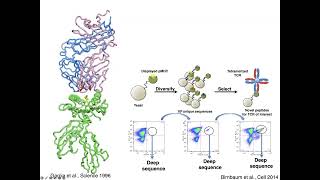
Just to tell you a little bit at a high level what I'm interested in, what my lab is interested in. We're very focused on cell surface receptors and how they engage their ligands, how they signal, And really for me, what I'm interested in is modulating cell physiology using perturbation approaches. And that's rooted in structural biology where we use structures as molecular blueprints to engineer molecules that modulate cell physiology. And so we have quite a few different areas that we work in in the lab. As Chuan mentioned, immunity and infection, regenerative medicine, and then what I call applied protein engineering. So they all sort of have this common theme of what I've talked about, modulating cell physiology using engineering approaches. So today I'm going to focus on our T-cell receptor work. And so With regards to immunology, the lab has two parts to it. One is we study the antigen-specific phase of immunity, the T-cell receptor peptide MHC interaction. And then we also have a very active program looking at cytokine signaling. And I'm going to really focus on the T-cell receptor today because I haven't really given a dedicated talk on the T-cell receptor in a long time. I thought today would be a good day to just put all of our recent studies together in one talk and really drill down on that. I'm going to be giving another talk at NIH in a few weeks, the Walls Lecture, and that's going to be on the cytokines. If any of you want to hear about that, you can just watch the NIH meeting website and they'll have a Zoom link for that. I just want to disclose here my involvement with 3T Therapeutics. Which has commercialized some of the things I'm going to talk about today. So just to start off here, this is where it all started for me. This is the structure of the T cell receptor recognizing the peptide MHC complex going back to 1996 when I was at Scripps. And just to give a little bit of a quick tutorial, the T cell receptor sees the peptide, The peptide has these up-facing residues, which generally the CDR3 loops of the T-cell receptor read out. That's where most of the diversity is concentrated. And the CDR1s and 2s generally position the TCR on the peptide MHC. And we spent the first— when I started my lab at Stanford, we spent the first 10 or 12 years or so, really digging deep into the structural aspects, the details of this recognition process, because it's an incredibly interesting process. This is a composite surface here, where the T cell receptor recognizes a combination of the peptide residues and the MHC residues. And there's really nothing like this in nature. And it's amazing how 25 or so years later, this structure is still yielding fruit. We're still reaching higher levels of understanding of this structure. And it's become particularly relevant now with cancer immunotherapy and T-cell immunotherapy and antigen discovery now, sort of going back to some of the structural principles that really we've studied for a long time. So at around 2012, you know, with a very good understanding of the structural principles for how the TCR sees the peptide MHC, we did something that I think is really important, that which is we invented a technology. And I think for any scientist, you know, if you want to stay relevant, you know, you've got to invent new technologies to different ways of studying the problem. And so we, we came up with this peptide MHC, yeast display peptide MHC library platform. And the idea here is that we can display peptides linked to MHC on the yeast surface, and we can make very large libraries of the peptides presented by the MHC molecule. And we can physically select these using recombinant T cell receptor molecules that are multimerized because of the low affinity. This took our ability to study the fine details of T-cell recognition to a new level. Works really well. You can see as you iteratively select this library, you enrich populations. Then in 2014, really the game-changing— this was Michael Birnbaum, who's now at MIT, probably many of you know his work. Michael really integrated deep sequencing into this pipeline here. And that was, you know, totally transformative thing for this, for these libraries. Because then what we could do is we could really take a given T cell receptor and really ask about its cross-reactivity in an unprecedentedly granular way. And so this is one example where we screened this multiple sclerosis-derived T cell receptor from Kai Wucher-Fennig that recognizes myelin basic protein. And Michael made a library in DR15 presenting this randomized peptide. And you can see how we came up with this substitution matrix here, which is basically this weighted positional matrix after crunching all the deep sequencing data. This is from many thousands of peptides. And we came up with this very clear motif that, that we hit on myelin basic protein as our top hit from a completely unbiased screen of this T cell receptor. And that was a huge surprise that we could recover the cognate ligand for a T cell receptor from an unbiased screen. And not only did we recover myelin basic protein, but we recovered microbial mimics and human mimics that could play a possible role in autoimmunity. But that really wasn't what we were so much interested in. It was that the specificity of T-cell recognition was far more specific than we thought. The fact that the cognate ligand was so dominantly selected. Since then, we've done a lot of things with these libraries. A lot of people that I'm going to mention today have come through. We've used these libraries for two major kinds of, I'd say, two different applications. One is what I call probing dark matter, which is that what does a given T-cell receptor see? Most T-cell receptors are orphans. The dark matter is what are T-cell receptors recognizing? We've been doing a lot of TCR ligand hunting. And I'm going to spend maybe the first third of the talk really going through some of the studies that we've done on different systems, looking for T cell receptor ligands. And then the second kind of unanticipated use of these libraries was in deconstructing T cell receptor signaling and triggering. And this, this is a— was a complete surprise. Some work that I'll talk about from initially Leah Seibener in my lab and then recently Shang Xiao. It's been very interesting how this one tool, this one platform has opened new doors for us along many different avenues. For any junior people out there, what I can tell you is the best thing you can do is invent a platform. It becomes like the source of new data and new insights, new information that you can just go on for a long time with. Okay, so back to— let's just go through some of the applications we've used in the ligand hunting approaches. So Marvin G. in my lab was a student who came here and he got interested in using these peptide MHC libraries for looking for tumor antigens, for cancer antigens. So we developed a collaboration with Mark Davis, where working with Arnold Hahn, isolated TILs from colon adenocarcinoma. Then through some of the first paired sequencing of TILs, identified expanded T-cell receptors that then Marvin screened on HLA-A2 libraries and then developed a computational pipeline for taking the screening data and predicting human proteome. Antigens. And we've also done that recently with Mark Davis in a lung cancer model as well, and the paper is cited there. Sorry. So this is just a quick summary of the study. Marvin got these expanded T-cell receptors from 2 patients with Arnold, and then we got all these paired sequences. Marvin screened 20 expanded TILs and got hits on 4 T-cell receptors. 2 of them gave peptide antigens that were patient-specific, but we also found 2 peptides that were shared between these 2 different patients. And so this was really sort of a mind-expanding exercise that we could screen TILs and identify tumor antigens. And so what's happened with this is that Marvin Gee and Leah Seidener have gone on and started this company called 3T, where they've industrialized this process and have now screened hundreds of TIL-derived T-cell receptors and found just a tremendous number of new tumor antigens. And so they're trying to really use this to develop therapeutics. Now, another interesting application of these libraries was with Mark Davis and postdoc in his lab, Naresha Salekrama, also involving Ricardo Fernandez in my lab. This was more in the autoimmune sphere. And so what the interesting thing that Naresha observed is that, you know, in this EAE model, which I think many people watching know that, you know, when you immunize with MOG, you get this expansion of CD4 T cells. And, you know, these can be stained with MOG tetramer. So these are the MOG-specific CD4s. But you also get this expansion of CD8s that seem to be almost in synchrony with the CD4s. But these are not MOG-specific. These were, you know, sort of, we didn't really know what they were doing. So Naresha cloned out some of these T cells that were expanded and were orphaned. And working with Ricardo and Shinbo screened screened these T cells. And so, yeah, so here you can see that these CD4s, they are using IFB MOG tetramers. You know, they're recognizing MOG, but the CD8s were not. So Naresha screened them on these H2-DFB libraries. And, you know, two of the T cell receptors gave a clear motif indicating a hit. And you can see that the cells were stained with peptides with these tetramers. And when he immunized mice with MOG together with these surrogate peptides, it suppressed the EAE clinical score significantly. And so, what Naresha then did was he characterized these CD8 T cells. And it was interesting, they were LY49 positive. So, LY49 is a murine NK receptor. And the expanded CD8s seemed to be LY49 positive, expanded with MOG with this peptide that was reactive with these TCRs. So, the peptide was like an adjuvant helping to expand these. And And these cells had an effector-like phenotype, you know, expressed granzyme. So, you know, they were cytolytic T cells. And he found something super interesting, which is that these CD8s, they suppressed activation of MOG-specific CD4 T cells. So you can see when you mix the Ly49-negative CD8s from these mice they don't suppress the CD4s. But the Ly49 positive— sorry, let me use my pointer here. The Ly49 positive T cells suppressed the proliferation of the CD4 MOG-specific T cells. So it's this kind of CD8 suppressor phenotype that has turned into a big deal. And let me just show you some more data here. So, this is now immunizing mice with either MOG or the surrogate peptide. And you can see that again, the— sorry, this is an adoptive transfer experiment. The Ly49 positive CD8 suppressed the EAE. So, you know, this was originally in the mouse and Mark Davis' lab has gone on to show that CD8 KIR-positive T cells in humans are doing the same thing. They seem to directly suppress the pathogenic CD4 cells in autoimmunity and an infection. It's turned out to be a big discovery and suppressor CD8s and autoimmunity and could become another therapeutic axis. But what I think is neat is that we got there by just, like I said, screening the dark matter, where these CD8 T cells had no known ligands. We found these ligands and they had completely unexpected activities. Another collaboration we had in our autoimmune system with Diane Mathis and Christophe Benoit at Harvard and Chao-Ran Li was pivotal in this. Chao-Ran had been working in the Mathis lab on these on these visceral adipose tissue CD4 regulatory T cells. Very interesting Tregs that are in fat tissue and they seem to be playing a role in inflammation and possibly in obesity where the adipose tissue is inflamed. But the ligands weren't known and even the extent to which these CD4 Tregs were antigen-specific. And so Ricardo Fernandez in my lab worked with Chauron to screen one of these visceral adipose Tregs. And Ricardo managed to— this was a very tough project because IA-B is a tough one for yeast display. The scaffold is not very good, but Ricardo managed to identify a whole range of surrogate peptide agonists that we're able to activate transfectants of this T-cell receptor to different degrees. And these peptides induce proliferation of these so-called fat Tregs to different degrees depending on the potency of the ligand that we used. And when one of these peptides immunize mice with an adjuvant, you can see it expands very nicely these FOXP3-positive CD4 Tregs in vivo. And so what Chaoran went on to do here is an adoptive transfer model. He made a transgenic mouse of this TCR, took these T cells, put them back into this high-fat diet mouse, and showed that the peptide again, could very nicely expand those T cells, and it actually had some effects in the high-fat diet mice in protecting against diabetes. It improved insulin sensitivity. And so, you know, these metabolic Tregs that Diane Mathis is working on are super interesting, and so, you know, in the absence of ligands to study their function, it's very difficult to know what they're doing. So this was, you know, kind of another interesting application of the yeast peptide MHC library technology. Just very quickly here in the kind of infectious disease realm here, we worked with Andrew McMichael and Geraldine Gillespie in Oxford, They had these T cell receptors, these so-called elite controller T cell receptors against HLA-B57 presenting HIV GAG peptide. And we screened these against the B57 library and came up with a very strong motif. And that motif predicted a couple of microbial peptides that are human pathogens that activated these T cells equally well as the HIV peptide. And in fact, when Geraldine got the bacteria and fed them to the T cells, they were also able to activate— sorry, fed the APC, they were also able to activate these T cells. So, you know, we think that maybe these elite controller T cell receptors, T cells, at some point, were sensitized by microbial mimicry of possibly some pathogen antigen, and so it gave them an advantage in controlling the HIV GAG peptide. Lastly, on the ligand identification front, I just want to finish with a story that was really spearheaded by Xinbo Yang in my lab, also in collaboration with Geraldine Gillespie and Andrew McMichael at Oxford. And it's kind of the perfect system for our yeast display because— so ankylosing spondylitis is one of these classical problems in immunology because it was recognized over 50 years ago that ankylosing spondylitis is almost exclusively HLA-B27. And so, you know, one of the problems in finding T-cell receptor ligands is that when you get the T-cell receptor out of the T-cell, you don't know its MHC restriction. So it's kind of a lottery. You're hoping that the MHC allele that you're screening against is the right match for the T-cell receptor. So it can be very labor-intensive, but here, we should be able to narrow down the MHC restriction to HLA-B27. And so B27, it's this inflammatory disease that affects many different systems in the body. For instance, they get arthritis, they get psoriasis, anterior uveitis in the eye. And it's been very controversial what's causing this. And so this is the structure of the HLA-B27 molecule. And there have been two sort of opposing viewpoints here. One is that the disease is due to some misfolding of the HLA-B27 molecule that in some way causes autoimmunity. And the other hypothesis has been that it's a T-cell-mediated disease, that there's some peptide on these tissues that the T cells are recognizing presented by B27. And it's been relatively unresolved. It's been quite controversial. And so we started collaborating with Andrew McMichael and Geraldine, and then Wayne Yokoyama and Michael Paley came into the picture. And we just had this fantastic collaboration where the Oxford group isolated these T cells from ankylosing spondylitis patients. The WashU group isolated T cells from these uveitis patients. And they sequenced the TCRs. And you can see here are the ankylosing spondylitis T cells. They had these expanded T cells that they sequenced. And the same with the uveitis. So we really got the super high-value TCRs to screen. And what was super interesting was that all these TCRs had this— they were AV21 BV9. Chain pairing. But the CDR3s have this, what we call a public motif. It's not totally public. I mean, it's got polymorphisms, but a very similar CDR3 from two different anatomical sites with anterior uveitis. Sorry, with ankylosing spondylitis and anterior uveitis. Shinbo really worked incredibly hard screening these TCRs. And this is showing you an example of when it works, you get this positive population and you know you've got a hit. And here are examples of 3 different TCRs where we know that we had clear hits on our library. And then I'm not going to get into the methodology, but there's a computational pipeline where we crunch all those peptide mutants into this substitution matrix to then predict what peptides from the human proteome might be. And these are now 5 TCRs that Xinbo screened, and you can see the list of peptides predicted by these motifs are basically the same. And Xinbo went on and he made T-cell transfectants of 12 different T-cell receptors, 7 of them from myocalcin spondylitis, or no, so 9 of them from myocalcin or more, and then 2 of them from the anterior uveitis patients. And he tested them for reactivity with both the self and the foreign peptide, and the microbial peptides are bolded. And you can see that almost all of these TCRs are cross-reactive to some degree with these peptides. And to make it simpler for you to see, Here are the different ankylosing spondylitis TCRs. Here's the anterior uveitis. And depending on the peptide, we either get extreme cross-reactivity or more limited cross-reactivity. But basically, they're all seeing something roughly similar. And this was super exciting. And there's even this microbial peptide from Salmonella in here that is recognized by essentially all of the TCRs. And, um, What Shinbo did, a very clever experiment, was that— so B2705 is pathogenic, but people with B2709, which just has a single polymorphism from 05, or maybe there's a few more than that, but the polymorphism of interest is right in the floor of the peptide binding groove. Shishinbo asked if the protective allele presented these peptides to the TCR. And the protective allele did not present the peptide to the TCR as opposed to B2705. And Shinbo also went on and he solved crystal structures for about 7 of these different T cell receptor complexes, and they superimpose virtually identically. And this is now showing you the CDR3s and the peptide. And the superpositions are so close that it almost looks like one structure. It's really an unprecedented atomic superposition, and the peptide, remember, has different sequences, but it presents kind of this common motif here. There's a proline at P8, there's an aliphatic at P7, and it's kind of this motif that is the, is the kind of the molecular mimic motif that makes all these peptides recognized by these TCRs. So it was a fascinating finding and really, really is a compelling argument that ankylosing spondylitis and the associated spondylopathies are caused by peptides being presented by these tissues in the lesions of these tissues. And I will just mention a preliminary experiment I wanted to show you here from Michael Paley at WashU, where Shinbo's provided him with tetramers. Of the Shigella peptide, the Salmonella peptide. And in patients, he sees— he's now staining blood from patients, and he can see that there's an enriched tetramer staining versus healthy patients. So this is super exciting and really starts to go in the direction of a very compelling argument that we may have found the autoantigens. For this disease. Okay, so while I'm here, I wanted to just tell you a little anecdote related to this, of a collaboration that we had with Bill Robinson here at Stanford. And really, this was really almost entirely carried by Tobias Lanz in his lab, a German postdoc who collaborated with a member of my lab, Kevin Jude, to do some structural work. But I just want to show you, I asked Tobias for a couple of his slides so I can show you the whole story that came out because it's related to the ankylosing spondylitis story in terms of the structural findings. Here Epstein-Barr virus is also associated with autoimmunity. All these different autoimmune conditions, there's a big literature on association with viral infections including EBV. What Tobias did was that he got antibodies out of the cerebrospinal fluid of multiple sclerosis patients and he used these protein arrays. He found that one of these antibodies bound to the EBNA, peptide, to a peptide from the EBNA genome. And he also found that the same antibody cross-reacted with glialCAM, which is a protein expressed on the myelin sheath of oligodendrocytes. And so he showed that if you immunize mice with the EBNA peptide, and you test the same serum for cross-reactivity, you get gliocam reactivity. So there's kind of a molecular mimicry going on here. And he even showed that this ebnapeptide can cause EAE. So what we worked with him on was trying to get a structural understanding of this. And Kevin Jude worked with Tobias And this is the structure of the EBNA peptide bound to the antibody. And this is the structure of the GlyAlCAM peptide bound to the same, bound to the same antibody. And you can see it's just a clear example of molecular mimicry. And, you know, you can read the paper that I cited in Nature last year. But the idea is that, you know, EBV was originally recognized by an antibody that then underwent somatic hypermutation. And in the course of the somatic hypermutation, it picked up this cross-reactivity with gliocamp. And they have more functional data, compelling data here. But, you know, I think this EBV story with MS on the molecular mimicry And this ankylosing spondylitis story with the T-cell receptor, really, I've been a skeptic of this molecular mimicry hypothesis for autoimmunity for a long time. And I've got to say, even looking at this data, I'm becoming a believer. It's very hard to dismiss these kinds of results. Okay. So moving on from ligand hunting, I want to move to an important question, which is that, you know, we spent all this time screening different systems for different peptides, and, you know, I've shown you, but the problem that keeps coming up when we find hits is what do we do now? And, you know, that's especially true for identifying tumor antigens. So you found your peptide, you're feeling good about things. Proving physiological relevance is just incredibly hard. And I got tired of looking at all these hits that we had in the lab where we're stuck, especially in humans, proving physiological relevance. Is that peptide presented on the tumor? It's very difficult. Like RNA-seq data, gene expression data only goes so far. It's not really predictive of whether an antigen is processed and presented. So I wanted to try and solve this problem in a way that I know how, which is using protein engineering. And so this is a, you know, another body of work that Shinbo worked on with another postdoc in the lab, Daisuke. And the idea here was to use TCR mimic, or what I prefer to call MHC-restricted antibodies, as probes for the relevance of peptide antigens, tumor antigens mainly. And, you know, this technology has been around for a long time. In fact, at NIH, I mean, I think some of the first antibodies to peptide MHC were made. And I just started to realize recently just how important this technology is because there are not many other ways where you could really think, how can I prove whether a particular peptide is expressed on natural tissue? But if you have an antibody to the peptide MHC complex, that solves your problem. The problem with this technology is that truly peptide MHC-restricted antibodies are very hard to make. The reason is back to the structural principles I talked about at the beginning, which is that the T-cell receptor sees this composite surface of a peptide and an MHC, It binds in this very stereotypical diagonal orientation where the CDR3s are really maximally reading out the peptide. But T-cell receptors have evolved over hundreds of millions of years to bind to peptide MHC in the perfect way. Antibodies have not. Antibodies can bind any part of the peptide MHC. One of the persistent problems with this technology has been that peptide, the so-called TCR mimic antibodies, they have off-target cross-reactivity because they bind either the, they bind the MHC too tightly. And so they're not truly peptide dependent. And so, you know, this really, I thought about this a lot. And, you know, going back to those original findings, you know, from 25 years ago on the structure, we thought, well, let's see if we can get antibodies to have MHC restriction like the T-cell receptor. And the way we went about that is by looking— if we started with an antibody that saw the peptide MHC in a way almost exactly like a T-cell receptor, instead of having to make a new antibody every time against every different peptide you could just re-engineer this antibody, preserving the MHC context, but diversifying the peptide context to reselect against a new peptide MHC and maintain the perfect binding orientation. And that was the idea here. But when you look at crystal structures of TCR mimic antibodies with peptide MHC, and there are a few that have been solved and published, a lot of them bind in these weird kind of ways. They bind more off to the MHC, more off to the side. And that's really bad because that means that that antibody has a tendency to really bind to the MHC regardless of what peptide is bound. So you really want this TCR-like docking footprint. And we found a structure, we didn't solve it, we found a structure of a T-cell receptor that bound in this perfect TCR-like docking geometry. So then what, what, what, what Daisuke did was he then identified the residues on the antibody that are touching the peptide, and he just made a library of only those residues, leaving the MHC-binding residues alone. And then he reselected this library against different peptides presented by the same MHC. And here showing you, you know, just simply the display of the antibody on yeast, you can see that it binds Kfb-Ova, which was our murine target. This is just a classical synfocal Kfb complex. The antibody is specific for Ova-Kfb. But when we reselect the library on a tumor antigen from B16 called TRIP2, You can see we can re-engineer that antibody to bind to TRPP2 and not bind to OVA. And, and, and Daisuke did an Alice scan of the peptide. It was still binding in the center, just like, just like the antibody was binding to OVA, and did some affinity measurements. These are pretty respectable affinities. It binds to cells loaded with TRPP2 specifically. It does not bind to the same cells loaded with OVA. And we use this for some really interesting experiments. So now that we had an antibody that could see the tumor antigen, we went back to B16 cells, which is one of the most common B16F10 tumor model, and started asking, what is the density of the tumor antigen on these cells? And so we had to use single molecule spectroscopy. We collaborated with Jakob Pieler because the antigen density is so low that you can't even detect it by FACS. When you fluorescently label the antibody, you can't see anything. But you can see individual peptide-MHC complexes on the cells by single molecule. And like when you add gamma interferon, expression goes up, you add the peptide and so on. And we are able to calibrate with Jakob Pieler, the biophysicist we work with, the number of peptide-MHC complexes presented on the surface of the B16 versus the efficiency of ADCC by this antibody. And you need to get up on the order of something like 1,000 peptide-MHC complexes on the surface of the cell before you start getting efficient ADCC. And that's a big limitation for anti-tumor antibodies that are supposed to work by ADCC. For a low-density antigen, that's going to be a really ineffective drug. So Daisuke working with Leon Su in my lab took this TCR mimic antibody and reformatted it as a BiTE instead of an antibody. And now that BiTE is able to lyse the cells both in the presence and the absence of the peptide. This is just unloaded B16. So the BiTE is super sensitive. We also made a CAR out of it, and the CAR is also able to lyse the B16 cells without peptide loaded. And so we're kind of having a fun time in the lab now playing around with different formats of this antigen-specific B16 TCR mimic antibody, sort of asking the question, which is the most sensitive? What is the lower limit of antigen sensitivity that we need for effective anti-tumor therapy? And so that is work being done by several new postdocs in my lab along with Matthias. We're starting to use that with and ask these questions. Okay, so we— now that was— it was good. It's good to be a mouse, but we wanted to do this for humans. And we did the same thing. There was a structure of a antibody to HLA-A2 presenting the NY-ESO peptide. And it was a very nice starting point. It had a very TCR-like binding geometry. And we were able to rapidly re-engineer that to not only bind NY-ESO, but MART1, GP100, and KRAS. And you can see we get specific binding to each one of these antigens and very little cross-reactivity. To the other antigens. And so here again, potent ADCC killing, it's very specific for the MART1 peptide and not NYESO. So the system works pretty well. And I'll just finish up here. Shinbo went on and solved the structure of the NYESO antibody now binding to MART1. And it generally preserves this kind of ideal docking geometry. But there's a real focus on the peptide and away from the MHC helices. So the library has really concentrated the new interactions on the peptide, which we think is going to contribute to its specificity. Okay, and finally, just closing the loop, Shinbo went back and he rescreened this antibody on our peptide MHC libraries to ask if we had picked up any cross-reactive peptides in the process of the re-engineering, and the MART-1 peptide was our top hit. So it worked really nicely. Okay, so just kind of fantasizing here, thinking out loud for a little bit, if we could get really fast at making tumor-specific antibodies so that like a patient walks in the door, has their tumor sequenced, identify their neoantigens, and then rapidly use a platform like this to make therapeutics to the dominant neoantigens, you know, you could possibly envision like real-time therapeutic treatment of individual patients using a platform like this. You know, it's a kind of Star Wars kind of thinking, but, you know, it's good to have, it's good to think like that occasionally. Even though right now this is not even remotely practical. I'm going to transition here in the last part of my talk back to T-cell receptor signaling mechanism. Really the main issue I want to talk about here is some insights we made into T-cell receptor triggering. The point, the punchline ahead of time is what we call TCR turbocharging. That is, we can take T-cell receptors against tumor antigens and we can greatly increase their sensitivity and their target killing potency in a way that does not involve affinity maturation. The story starts like this. And this is a really important statement. T-cell receptor peptide MHC affinity does not always predict bioactivity. And there are T-cell receptors that have high affinity but signal poorly, and there are T-cell receptors with low affinity that signal very strongly. What got our attention into this area was the identification of T-cell receptors with very high affinity that signaled either very little or not at all. And we call these non-stimulatory T-cell receptors. And what we found is what earmarked these T-cell receptors as being non-stimulatory was that when we did mechanical force measurements of the TCR peptide-MHC interaction, they exhibited characteristics of what's called a slip bond, whereas the stimulatory TCRs exhibited what's called a catch bond. And we're going to get into this in a little bit. I'll explain what better what this means in a little bit. And so this phenomenon, the ability to decouple affinity from signaling strength has allowed us to overcome this problem that T-cell receptors isolated from tumors are often weakly reactive to self-antigens. And we can do that without having to affinity mature them. Okay, so where this all started was this paper from 2018. Leah Sibener was the first author. Where we identified many examples where we had tetramer-positive binding to T cells, but those T cells were not triggered by the antigen. And I'll show you what that kind of data looks like. So for example, here's an HLA-A2 HCV peptide, which is tetramer-positive. And then the same T cell is activated by the HCV peptide. It makes sense. We also have tetramer-positive T cells that are not activated by the peptide. And the same through— here's the Hy antigen presented by A2. Tetramer-positive activation, tetramer-positive no activation. And we found— and this is from Mark Davis' collection of tetramer-isolated T cells. A large proportion of T cells that were tetramer positive, but stem negative. And that really got our attention. Because sometimes the best way to solve a mechanism in biology is by starting with a system that's broken and figuring out what's broken than it is by staring at the natural mechanism and trying to figure out what's working properly. So with these non-stimulatory But binding T-cell receptors, and here's an example. Leah took a B35 HIV TCR. Both of them are tetramer positive. Both of them are activated by anti-CD3, but only one of them is activated by antigen and the other is not. And the affinities are are similar. They're both T-cell receptor, they're both in the range the T-cell receptors signal, one's a little higher than the other. But these 17 micromolar is a respectable affinity. And so Leah solved the structure of both the stimulatory and the non-stimulatory T-cell receptor complexes. And there was really nothing remarkable about, but they both look perfectly normal garden variety T-cell receptors. The footprint was fine. We thought maybe there'd be some structural anomaly to explain the non-stimulatory one. It wasn't structure. Really, the turning point came when we went back to the peptide MHC libraries and Leah identified mutants of the HIV peptide which now activated the T-cell receptor compared to HIV, which was nonstimulatory. So we now had agonist peptides for the nonstimulatory T-cell receptor, and which allowed us to really directly ask what's different from the on, from the off state. And here we collaborated with Brian Eivewold at Utah who had been, you know, were looking at mechanical triggering of the T cell receptor for a long time. And what Brian found was that our non-stimulatory peptides showed characteristics of the slip bond, whereas all the agonist peptides showed this very characteristic catch bond behavior. So what is a catch bond? Um, it's a, it's a, it's, it's a, it's a, uh, a very, uh, unusual kind of thing where, um, let me, uh, see if I can— well, let me go back here. Uh, this is a very nice little, uh— okay, so it's like this finger trap where you get your fingers in there, you try to pull it, and there's a resistive force. You pull and the resistance increases. That's the catch bond. And so T-cell receptors do the same thing. As they disengage, there's a transient prolongation of the bond lifetime as it disengages, which gives rise to this increase in bond lifetime. So it turns out that all the agonists we've looked at show catch bonds and all the non-agonists show slip bonds. And so, okay, so I want to make a point here that the affinities, the T-cell receptor peptide MHC affinities of the agonists are virtually the same as the non-agonists. So this difference is not just simply higher affinity. And that's very important that we could turn a T-cell receptor on or off in an affinity-independent manner. And so what that finding allowed us to do was to think about engineering therapeutic T-cell receptors to tumor antigens in a way that bypasses the propensity for high affinity T-cell receptors to have off-target cross-reactivity. And a prime example of this is a famous clinical trial an unfortunate clinical trial of a T-cell receptor against HLA, against a cancer testis antigen, MAJ-A3. And the parent TCR had very low affinity. So it was affinity matured for MAJ-A3. And in the clinical trial, they picked up an unanticipated cardiovascular toxicity. And it turned out to be a peptide presented by HLA-A2 in cardiac tissue, which was mimicked by the, by MAJ-A3 and cross-reacted by the high affinity TCR. So Xiang Zhao in my lab went back to the parent MAJ-A3 TCR, the low affinity version, And he said, instead of affinity maturing, how about if I make a library we call catch bond fishing, where these are amino acids that are buried in the CDRs, and I go fishing against the ligand. But instead of, instead of selecting for high affinity, we select for tetramer low, so low affinity, but activation positive. Normally people are looking in this quadrant, we look down here. You can see through sequential rounds of sorting, he was able to enrich a population of low affinity, but much more potent activation mutants of that parent TCR. Here's some properties of those TCRs. This is now— Xiong titrated the MAJ-A3 peptide against the wild-type TCR, and you can see it barely— it doesn't hardly activate the TCR at all. Here's the high affinity therapeutic TCR. Here are Xiong's catch bond mutants. And these are physiological affinities. Now, when you Do the same experiment against the titan peptide, you see the therapeutic TCR cross-reactivity and only a couple of our catch bond TCRs cross-react with titan at very high concentration. Most of them are completely clean. And here, Xiang measured the affinities of all these catch bond TCRs for MAJ-A3. Here's the high affinity TCR, here's the wild type, And you can see we're still in that physiological regime. We're not in the high affinity regime, and these are going to be much less prone to cross-reactivity. And for example, we can show that the signal, the potency of activation by the TCR is directly related to the bond lifetime of the catch bond. So he went on to, you know, ask about about the activation and target killing potential of these T cells on both a high-expressing line and an intermediate-expressing line. And comparing with the therapeutic TCR to wild type to our catch bond mutants, you see in many instances were more sensitive to the antigen than even the high-affinity TCR, or at least in most cases comparable. And so you might ask, well, did you pick up any spurious cross-reactivities in the catch bond engineering? So now we go back to our peptide MHC libraries, which Shinbo screened, and very satisfying to see the therapeutic TCR, we recover MAGE. Well, we also picked up the Titan cross-reactivity from the screen, but our catch bond engineered TCRs we don't see any activation by either titan or the other peptides. So this is something now, this catch bond engineering is something that really, you know, we think a lot about in the lab now. And, you know, I kind of made this image with a graphic artist, you know, because this is kind of the way I think about it. These catch bonds are kind of like lightning bolts. They're different than than high-energy bonds that give rise to high affinity. These are very transient interactions that form as the TCR is disengaging, but have tremendously powerful effects at triggering the TCR to signal. It's an engineerable quantity, has actionable value. We can engineer catch bonds now. It's really, it's a completely new engineering parameter for T-cell receptors other than affinity. And so we're doing this now on a whole range of anti-tumor T-cell receptors and even extending the concept to other kinds of immune receptors to see if we can do the same thing. So I think with that, I'm going to end. I've mentioned along the way, I've tried to mention everybody who was involved in these studies. I probably didn't hit everybody, but you've probably heard these names today, and apologies to anybody who I might have missed along the way, but I will stop there.
More from Joshua







Scientist Stories: Hans Waterham, The value of phenotypic assays in peroxisomal disorders




Scientist Stories: Joel Zlotogora, Genetic carrier screening programs for reproductive purposes