Scientist Stories: Christina Woo, Chemical Biology to Understand Small Molecule Activity
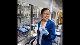
In this seminar, Associate Professor Christina Wu (Harvard Chemistry & Chemical Biology) outlines her laboratory’s chemical‑biology strategies for probing and reprogramming post‑translational modifications (PTMs) to understand protein function and to target the traditionally "undruggable" proteome. She first describes nanobody‑fused O‑GlcNAc transferase and O‑GlcNAcase enzymes that can be directed to specific substrates, enabling precise writing and erasing of O‑GlcNAc modifications. She then shifts to her work on immunomodulatory drugs (IMiDs) such as thalidomide, lenalidomide, and pomalidomide, which act as molecular glues by binding the cereblon (CRBN) thalidomide‑binding domain and recruiting neo‑substrates for ubiquitination and proteasomal degradation. Wu’s group developed a suite of photoaffinity labeling (PAL) tools—including the PALBOX cyclobutane‑based probe and a photolenalidomide analogue—that enable high‑resolution target identification in cells. Using these probes, they confirmed CRBN and IKZF1 as canonical IMiD targets and uncovered enrichment of EIF3I, suggesting novel CRBN‑mediated sequestration. To explore CRBN’s endogenous function, the lab hypothesized that CRBN may recognize either a metabolite or a PTM degron. Structural and chemical analyses led to the identification of C‑terminal cyclic glutamine (cycloglutamate) and cyclic asparagine (cycloasparagine) imides—post‑translational modifications formed via intramolecular cyclization—as previously unappreciated degrons that bind the CRBN pocket. By incorporating these cyclic imides into dipeptide degraders (e.g., JQ1‑FcQ, JQ1‑FCN) they demonstrated cereblon‑dependent degradation of the BRD4 target, with rescue upon CRBN knockout or competition with lenalidomide. Biophysical ternary‑complex assays and structural modeling showed that the cyclic imide ring mimics the three‑hydrogen‑bond motif of IMiDs, while the adjacent amino‑acid side chain adopts a distinct orientation, explaining the lack of IKZF1 recruitment. The work establishes cyclic imide PTMs as natural degrons for CRBN, expands the chemical‑biology toolbox for PTM manipulation, and opens avenues for targeting the undruggable proteome via engineered molecular glues.
Chapters
So today, it's my greatest pleasure to introduce Christina Wu. Christina is an associate professor at the Department of Chemistry and Chemical Biology at Harvard University, and an affiliate member of the Broad Institute. Christina's research focuses on the design of small molecule and protein approaches to alter post-translational modifications and the signaling outcome they produce. Kristina obtained her BA in chemistry from Wellesley College and obtained her PhD from Yale University. For postdoctoral training, Kristina joined the laboratory of Caroline Bartosi, our inauguration speaker at the series, where she developed a mass-independent chemical glycoproteomics platform for the identification of non-template post-translational modifications. Subsequently, Cristina joined the faculty at Harvard University in 2016. Her research has been recognized by multiple awards, including Bayer Early Excellence in Science Award, the NIH DPI Aventur Award, and the Ono Pharma Foundation Breakthrough Science Award. It is great pleasure to have you, Cristina, and we are very much looking forward to your insight into a cereblum function.
Okay. Great. Thank you to the organizers for the introduction and the opportunity to share our work here today. It's really been a pleasure being able to listen and learn from this community over the last few years and now be able to share some of the emerging work in our group, really using chemical biology methods to study the thalidomide-binding domain of cereblum. Of one. So my group broadly is very interested in studying how we can read and rewrite chemical modifications to the proteome. And we think about this as two sides of one coin, where we think about how enzymes are naturally regulated by post-translational modifications, chemical modifications that alter the physiological function of these proteins in cells. And the reason why we're so interested in how post-translational modifications are regulating protein function is that we think that if we can understand this, we can then use chemical biology methods, including chemical probes, protein engineering, and proteomics to develop small molecule mimics or chemical adapters that can alter the enzymatic function, perhaps towards endogenous protein regulation, and also towards new targets such as neo-substrates, and in this way, begin to target the, quote unquote, undruggable proteome through chemistry. We also have taken a significant amount of inspiration from how small molecules act and how these kinds of small molecules can already chemically adapt protein function and take that information back to both characterizing the quote unquote undruggable proteome, but also use it to inspire our understanding of physiological function of these proteins. So my group is broadly interested in two post-translational modifications. One is in the form of glycosylation, nutrient sensor in the cell in particular called O-glcNAc, where to date, we've primarily developed a protein engineering tool called a nanobody O-glcNAc transferase and O-glcNAcase that allows us to redirect these writers and erasers of O-glcNAc to desired target proteins. And going towards the future, we're very interested in taking this protein engineering method to a more small molecule approach. In addition, we also have now very significant effort in targeted protein degradation focused in on another post-translation modification here, ubiquitynylation, which broadly sends a signal of protein degradation when modified. And in this area of our work, which I'll tell you about in more detail today, we really took inspiration from a class of molecules that I think is of no mystery to this community, the immunomodulatory drugs, and studied them both from the context of their mechanism of action, but also brought us in new directions in terms of the physiological functions of cereblon. So in our target protein degradation efforts— or sorry, O-glycan acetylation efforts, I wanted to just really briefly highlight the protein engineering approach that we took in terms of developing methods to write and erase O-glycan from target proteins in cells. This approach, because there are no yet discovered ligands for the writer and eraser of O-GlcNAc to recruit it, we instead co-opted a and developed a protein engineering approach using nanobodies, which are just small antibodies that we can fuse to the writer, O-GlcNAc transferase, and in this way recruit it to the desired target protein to enhance glycosylation in cells. Conversely, we also developed an O-glycnacase-nanobody fusion protein for target protein deglycosylation of O-glycnac in cells. And we're now using these tools both to study the functions of O-glycnac on these target proteins of interest, as well as develop more small molecule versions in the future. So the system I wanted to really focus in on and tell you about today are the immunomodulatory drugs. Which are the first in class was these compounds here, the thalidomide, followed by more recent developments in other compounds like, and derivatives like lenalidomide and pomalidomide. Thalidomide probably needs no major introduction to this community, but was first established as a sedative back in the 1950s, subsequently found to have horrific birth defects effects. And then— and withdrawn from the market. However, continued use of these compounds subsequently discovered their anti-inflammatory effects, immunomodulatory effects, and ultimately, with the development of newer analogs like lenalidomide and pomalidomide, the ability to have anti-cancer effects and life-saving properties in certain forms of hematopoietic cancers, including multiple myeloma and del(5)qMDS, amongst others. And so one of the questions that really intrigued me and my group as we initiated our efforts, as well as I think many other scientists in the world, was this question here of how do the immunomodulatory drugs have so many biological activities? And I think of the many— while these compounds are likely pluripotent and have multiple targets, one of the major breakthroughs really came in in 2010 by Itohano and coworkers, we discovered that these compounds interact with a protein called cereblon. Cereblon being part of a broader E3 ligase complex, it was subsequently discovered that when the immunomodulatory drugs interact with cereblon, they don't just block cereblon's endogenous functions, they actually recruit new substrates or neo-substrates to a ternary complex mediated by these small molecules. And by this induced proximity, promote ubiquitynylation and proteasomal degradation of specific neo-substrates. So E3 ligases like Cereblon, they can recognize degrons, which are signal sequences on substrates. And small molecules like the immunomodulatory drugs can chemically induce these degrons on these neo-substrates like IKZF1, IKZF3, amongst others. And additional targets of the IMIDs and the physiological function of Cereblon itself remain to be discovered. And these were some of the questions that we initially sought to resolve in my group or address. And so we began a program using chemical biology methods, really initially inspired by the power of chemical proteomics to characterize targets inside of a cell. And so we were initially focused really in on characterizing cellular targets with molecular glues by chemical proteomics with a real emphasis on photoaffinity labeling chemistry. We then subsequently developed probes for lenalidomide and used these to characterize the novel targets from cells. And the story I really want to share with you today is focused in here on emerging work from our group on the discovery of a degron for the lenalidomide-binding domain of cereblon. So in the interest of time, I'll just quickly tell you about some of the photoaffinity labeling work that we've done, both in terms of characterizing alkyl diazerines and how they preferentially label acidic residues in a pH-dependent manner. This work was done by Alexander West, who was really initially characterizing how the photoaffinity labeling chemistry can allow us to access binding sites in cells to give us higher resolution on how these types of photoaffinity labeling platforms can give us structural information. And what Alex found was that when diazirines interact with proteins and cells in a cellular context, en route to their formation of carbene, they also form a diazo intermediate. And this diazo intermediate is long-lived enough to react with acids in a pH-dependent manner. Alex has now developed a new compound that we call PALBOX, which is chemical biology speak for a photoaffinity label conjugated onto a cyclobutane butane ring. And this reagent, we're very excited to share with the community as well, because we hope that it'll be able to address some of the target identification studies using photoaffinity labeling in the future. In addition, we've also taken a few different molecular glues, including rapamycin, zinglifarin A, and the immunomodulatory drug lenalidomide, and functionalized them with photoaffinity labeling. And what's been really a pleasure for us as we've gone after these different targets is that in the cellular context, often we can— well, the compounds often have a known target. In a cellular context, we can visualize what those targets are using this photoaffinity labeling chemistry. And it leads us in new directions and new biology. And so if you endeavor to pursue a photoaffinity labeling or any kind of chemical proteomics platform, the very first thing that you want to do, for example, with taking an immunomodulatory drug towards a target identification approach is to develop a probe. And this can be more or less challenging depending on the compound. Fortunately for me, I had a set of fantastic graduate student and postdoctoral pair, Zeelin, or Lindsay, and Hugo Amako, who undertook this effort to take lenalidomide and develop a photoaffinity labeling version that we termed photolenalidomide. And the characterization of photolenalidomide, we first introduced it to MM1S cells, which is a popular model for multiple myeloma. And on this volcano plot, what we observed is that we could enrich both cereblon and IKZF1 as target proteins from— in a cellular context, allowing us to be fairly confident that these kinds of chemistries can also capture protein-protein interaction complexes in the cellular context. In the course of this work, we also evaluated Hep293 T cells stably expressing cereblon. And what we found was that cereblon is also being enriched. However, another protein called EIF3I was being enriched to a more substantial degree. We then took a deeper look at EIF3I. And in a study that is now currently on BioRxiv, Lindsey took this further to characterize how lenalidomide sequesters EIF3I from the broader EIF3 complex. The EIF3 complex is the eukaryotic initiation factor 3 complex. And EIF3I itself is part of the subunit between subunits EIF3B and EIF3G. And what Lindsey discovered is that when lenalidomide interacts with cereblon in the cell, it recruits EIF3I out of this complex and sequesters it into a complex with cereblon. And so, we think this has translation to some of lenalidomide's antiangiogenic effects, but also really highlights the advantage of a chemical proteomics approach for performing direct target identification by chemical proteomics. So, in the course of this work and our thoughts about how the immunomodulatory drugs are functioning in cells, We also became more and more intrigued by another competing hypothesis that perhaps the blockage of ZARIBONE's endogenous function by the immunomodulatory drugs may also be playing a role in the overall phenotypes of these compounds. And if I paraphrase that question, the kind of question that we were really trying to ask is shown here. Is thalidomide mimicking the natural recognition mechanism of cereblon? And as we embarked on asking this question in more detail, we came up with two specific hypotheses. We thought that perhaps— I just told you in detail that thalidomide and phenylalanine interacts with cereblon and recruits a neosubstrate, or one of multiple specific neosubstrates, through an induced degron. We thought that perhaps cereblon may be acting as a metabolic sensor where a metabolite may be serving that role that recruits Cerabon to various target proteins depending on metabolic levels. We also hypothesized that perhaps Cerabon is recognizing a post-translational modification, and that the only difference— that would recruit these substrates. And the only difference between the post-translational modification and metabolite models was the presence or absence of a covalent bond. So the— in order to pursue this kind of question, we were really looking for a degron, either a chemically induced degron or one that's substituted directly on the protein. And again, degrons are these signal sequences that induce substrate ubiquitination and degradation. And multiple studies have found examples of both the metabolite and post-translational modification forms of degrons. Including multiple versions of this kind of metabolite recruitment in the context of plants. Perhaps one of the most famous versions of this is in the form of auxin, which is a plant hormone that is recognized by TIR1 and recruits AuxIAA factor for degradation, causing plants to grow towards the sun. Alternatively, post-translational modifications, such as oxidation of proline on HIF-1 alpha, acts as an oxygen sensor for recognizing recognition and degradation by VHL. And in the absence of oxygen, HIF-1α is no longer degraded, resulting in angiogenesis. And a number of degrons or signal sequences have also been found just constitutively present on the termini of proteins, either on the N or C terminus of these proteins, recognized by their respective adapter proteins. And so, if we take a closer look at the immunomodulatory drugs like lenalidomide and how it interacts with SARA1, Well, here's a two-dimensional view or linear view of cereblon. And what we can see is that the thalidomide binding domain, also called Cul-T, is found at the C-terminus of cereblon. If we look closer at one of the crystal structures obtained by Eber and Toma and coworkers, what we see is that with pomalidomide now, the thalidomide binding pocket or Cul-T domain is coated by these beautiful 3 tryptophan residues shown in purple. And pomalidomide interacts with the backbone of this protein through 3 hydrogen bonding events. The isooxindole or thalamid fragment of lenalidomide or pomalidomide, if I take this crystal structure and I rotate it by 180 degrees and now show in IKZF1, is resurfacing. What you can see is that pomalidomide is resurfacing the thalidomide binding domain, such that now new substrates like IKZF1 can dock in. And this 3 hydrogen bonding event is so essential that a very common control in the field is that by methylating the glutamine functionality, this actually blocks the interaction with cereblon, rescuing the recruitment and blocking the degradation of transcription factor IKZF1. For example. And so as we thought about how the thalidomide and lenalidomide were interacting with the thalidomide binding domain of SERAB1, we came up with a few hypotheses of what could be the structural homologs that thalidomide is mimicking. And so again, here is the key glutaramide of thalidomide that's conducting those 3 hydrogen bonding interactions. We first came up with uridine and related metabolites as possessing the ability to conduct that similar binding pattern analogous to how thalidomide interacts with cereblon, thinking that this looked kind of like a Watson-Crick ringland base pair, and perhaps these kinds of metabolites could serve as a ligand for the thalidomide binding domain. We weren't the first to think of this. Many publications have reported that the various uracil metabolites can indeed interact with the thalidomide binding domain of cereblon, But whether or not this recruits a structurally defined degron has remained to be defined. In the course of our efforts to evaluate metabolites, we also came up with a second hypothesis that came back to post-translational modifications. We started to hypothesize this because we were making and synthesizing photolenalidomide analogs. What we realized was that when we were synthesizing these compounds, We were actually synthesizing the red fragment of thalidomide from an amino acid. That amino acid was glutamine. And so, for the more chemically oriented members of our audience, what you start to see is that this is a dehydrated glutamine amino acid with thalamide as a common protecting group that's used in peptide synthesis. And so, we started to more seriously investigate glutamine derivatives and post-translational modifications as well. Well, we came up with one post-translational modification that occurs on the N-terminus of proteins that through the action of glutamyl cyclotransferase forms this pyroglutamate post-translational modification. We thought perhaps this type of structure would still be able to execute the same 3-hydrogen bonding motif as thalidomide. We also took inspiration from work from inteins, which is a form of protein self-splicing that's primarily found in bacteria, archaea, and fungi. Few viruses, where a side chain asparagine— I'm just showing the penultimate cleavage event of inteins as they excise themselves out of the protein. And what this chemistry has been defined to show is that the side chain asparagine, and occasionally glutamine where N equals 2, the nitrogen on the side chain amide will collapse down, attack the backbone of the protein, resulting in cleavage and excision of the, of the X-teen and revealing the N-teen with a post-translational modification. In the case of asparagine, a succinamide or a cyclized asparagine, and in the case of glutamine, a cyclized glutamine or glutaramide. And so as we came back to probing these various hypotheses as ligands of cereblon, We eventually realized through extensive studies that we were not just asking one question here on the slide, we were actually asking two. And the first question we were asking was, what is the structural homolog that thalidomide or lenalidomide could be mimicking on SARA1? But the second question we were asking had to do with the identity of the green protein or the endogenous substrate. And as we contemplated really combining these two questions together, It was a combinatorial challenge to answer them both at once. And so, what we eventually realized and took advantage of was a number of discoveries from the target protein degradation community, and particularly inspiration from using target protein degradation to provide a functional readout in cells, allowing us a mechanism to separate these hypotheses or questions. And so targeted protein degradation can occur both in the context of molecular glue degraders like thalidomide and lenalidomide. But in addition, through pioneering work from Craig Crews, Rita Shea, and many others since then, you can take E3 ligase ligands now, for example, like thalidomide, and conjugate them onto other ligands to redirect them to target proteins of interest like GQ1. Through the context of a linker. JQ1 now gives us a way to control for the target protein that we're interested in studying for degradation, such as in the form of BRD4. And now through this PROTAC or this degrader molecule, we can have a mechanism to screen for protein degradation and thus a functional readout for engagement of the thalidomide binding domain in cells. And so what we ultimately did was to substitute the thalidomide ligand with some of the chemical hypotheses hypotheses on the previous slide. And this work was really beautifully executed by the fearless efforts of both Saki Ichikawa, Hope Flaxman, and Wencheng Su in my lab. So we took thalidomide from this degrader molecule, and we substituted it with all of the different hypotheses I showed on the previous slide. And what we ultimately and eventually discovered was that we could take thalidomide and substitute it with a dipeptide. Again, this dipeptide is 2 amino acids where we have a glutaramide functional group, a dehydrated glutamine that we call cyclic glutamine or CQ. And if we're thinking about this in the context of the protein, this is an N-1 position that's substituted with a phenylalanine. We chose phenylalanine initially largely because we, for synthetic ease, but also because we thought perhaps it looks a bit like how thalidomide thalamide functionality of thalamide looks. And we took both of these compounds into the cell and compared them. We were very excited to discover that BRD4, the target protein of JQ1, was rapidly degraded by our JQ1 FcQ dipeptide degrader, and at equivalent levels as DBET6, the parent molecule. And this was rescued by MLN4924, a nedolation inhibitor of cholinesterase ligases, like the E3 ligase that Cereblon is substituted on. If we next asked whether this was cereblon-dependent, and indeed it was, if we knock out cereblon from the cell or knock down cereblon from the cell, we see BRD4 levels are rescued by both compounds and unaffected by both compounds. In addition, this is dependent on the thalidomide binding domain, as we can compete the degradation that we observe with our dipeptide degrader by both lenalidomide and the Boc-protected dipeptide. And if we epimerize any of the 3 stereocenters in our dipeptide degrader, what we see is rescue of the degradation of BRD4, indicating that this natural stereochemistry of the peptide is important for degradation. Now importantly, we next asked this follow-up question. This glutarmide chemistry formation is relatively rarely observed in cells. Or to date. However, intinc and the formation of sexenomides has been slightly more prevalent. And that largely has to— comes down to the chemistry of formation and the kinetics of the cyclization between 5-membered rings and 6-membered rings. So we next asked whether or not a cyclic asparagine or CN residue could act as also a functional degron or candidate degron for engagement of thalidomide binding domain in cells. And indeed it can. So if we compare BRD4 levels between this JQ1-FCN dipeptide degrader now, we see a dose-response nanomolar concentration degradation of BRD4 equivalent to dBAT6. This is also cereblon-dependent, as well as competed by lenalidomide or the Boc-protected FCN dipeptide. And degradation of the— of BRD4 is time dependent and complete between— in about 2 hours, which is similar to the timeframe of degradation of any of all the other compounds I've shown to date. So we next took a closer look at how these dipeptide degraders are engaging the thalidomide binding pocket, first in the context of an alpha screen looking at the ternary complex formation between cereblon and the BD2 domain of BRD4. And what we observed was that classic Koch effect from dBetv6, that was actually a stronger ternary complex based on the area under the curve for the JQ1-FCEQ and the JQ1-FCN degraders. Now, if we took a closer look at how these ligands may interact with the thalidomide binding domain, we modeled in the dipeptide versions of these compounds overlaid onto pomalidomide. What we saw is that this glutaramide ring or aspartamide ring can still interact similarly to the backbone, with the backbone of cereblon, equivalent to how the immunomodulatory drugs act. However, this peptide at the amino acid at the N-1 position is in a significantly different orientation than the thalamid on pomalidomide. And if I show now IKZF1 in this structure or this model, we can see that the phenylalanine amino acid likely intersects and conflicts with IKZF1. So while we didn't expect IKZF1 and some of the canonical beta loops to interact with these dipeptides on their own, we thought perhaps these molecules may recruit other neo-substrate or endogenous substrates that may be relevant. And we took a concerted look at this, leading us to ultimately conclude that the dipeptide alone, either through the FcQ or the BACH-protected FcQ, CQ on its own doesn't seem to behave as a molecular glue. We conduct— we carried this out both in the form of co-immunoprecipitation studies from HEK293T cells overexpressing IKZF1. Whereas expected, we can see IKZF1 co-immunoprecipitating in the presence of pomalidomide. But in the presence of the dipeptide, we don't see IKZF1 co-immunoprecipitate. And we also didn't see any additional proteins. Proteins. We also took a look at this in the context of global proteomics, where we saw IKZF1 get significantly degraded, as expected, by pomalidomide. As expected, IKZF1 was relatively unaffected by the BACH-protected FcQ. And we did not see any additional proteins that came out across these studies. So, this led us to really take a step back and to think carefully about what we knew.
We—
I just showed you that we could take a thalidomide-based degrader and degrade BRD4 in the context of Cereblon and replace the thalidomide functionality with a dipeptide. We figured that that dipeptide alone probably wasn't recruiting neo-substrates or endogenous substrates to Cereblon, that perhaps this peptide or amino acid glutaramide and aspartamide is acting as a post-translational modification. As we thought about how this post-translational modification could form, let me just remind you of the chemistry. The hypothesis is that the side chain asparagine or glutamine amino acid residue can attack the backbone and thus release or cleave a protein. To release a fragment and thus reveal this C-terminal cyclic imide as a digron. This C-terminal cyclic imide is now a digron that's a substrate for Cereblon, allowing its recognition by Cereblon for proximity induction, recruitment, ubiquitinylation, and proteasomal degradation. And as we thought more carefully about what kinds of substrates could be carrying this C-terminal cyclic imide digron, We started to wonder if perhaps we weren't looking for one substrate, but for a multitude of substrates. And we took a few inspirations to come to this hypothesis. So, we began to wonder if models for physiological function of cereblon may come from other models of protein damage. And so, just to remind you of the chemistry here, the side chain asparagine or glutamine attacking that backbone, revealing the cyclic imide degron, If this is a peptide bond or a protein bond, this would form a— this would represent a form of protein damage. And cereblon itself is a protein that was very— it was initially discovered by Higgins and coworkers back in 2004 as associated with neurological mental retardation. And specifically associated with point mutations in the thalidomide binding domain. And subsequent studies with a separate point mutation from Shareen and coworkers showed that the loss of the thalamide binding domain in specific was associated with severe mental retardation, seizures, and self-mutilation. And so we began to ask this question, is the C-terminal cyclic imide an overlooked form of protein damage? And that this protein damage is removed by cereblon, and perhaps that's why we haven't seen it yet too. As extensively as to date. And one of our major inspirations came out of another field of different post-translational modification called spontaneous deamidation. Spontaneous deamidation has very similar chemical and biological hallmarks as what we were looking at with the cereblondiguron. With spontaneous deamidation, asparagine residues are attacked by the backbone of the protein to the side chain asparagine amide. This results in deamidation and the formation of an internal cyclic imide intermediate. This internal cyclic imide intermediate can then go on to be hydrolyzed. It's rapidly hydrolyzed to an aspartate if water attacks from one side of this, um, cyclic imidate intermediate. But if water attacks from the other side of the cyclic imidate intermediate, it affords an isospartate. And this isospartate gives an unnatural kink in the backbone of the protein, which can turn— which turns out on buildup to be quite deleterious. Fortunately for us, we have a protein called PIMT, and many higher organisms have this protein. PIMT stands for protein L-isospartyl methyltransferase. And what PIMT does is it methylates this isospartate carboxylate. The methyl ester is then promoted to reform the cyclic imine intermediate such that over time it can disproportionate to the spartate, resulting in the natural protein backbone. And what's been shown in the field of spontaneous deamidation is that loss of this PIMT enzyme or in other words, a loss of the ability to correct the isospartate modification, is in fact quite deleterious, and especially in the brain. And these mice that have lost PIMT have several phenotypes that are very similar to the loss of the thalamide binding domain, including seizures and intellectual disability. If we take a look at the brains of these mice, we can actually see these brains are on loss of PIMT are engorged or enlarged, indicative of incorrect processing of protein turnover. So, to connect the observation of the C-terminal cyclic image to some of these hypotheses, we first needed to establish whether or not these are actually modifications that are occurring in the proteome. And if you've done a lot of mass spec, you probably, have also encountered these kinds of species because they can be quite challenging to avoid in situ on a mass spectrometer. So, we took two steps to establishing that these type of cyclic imine modifications are underappreciated. The first was to analyze a series of global proteomics datasets for these C-terminal modifications. And here we took advantage of the Clinical Proteomics Tumor Analysis Consortium and the data that they've made publicly available and looked across a number of different tissues from human patients who have had cancer for the C-terminal cyclic asparagine or C-terminal cyclic glutamine for these post-translational modifications. What we found is that several hundred proteins are carrying both of the— one or the other of these modifications. And if we take a look at the actual modification site, we can find this across several thousand sites. Interestingly, if we take these sites both on the asparagine or the glutamine site and in the context of the full protein before its excision, we can see a consensus sequence start to emerge between these between these— from these data. We took a close look at the consensus sequence as well as the types of proteins that were emerging quite frequently from these data. And then pursued a characterization of this in the context of our own datasets to ensure that these modifications were indeed forming and present in our hands. And where we think actually one of the major challenges that this modification as a form of protein damage may actually may actually play a role is not necessarily, or in addition to the cyclic imidagron itself. But this modification, if it's not intercepted by cereblon in the appropriate timeframe, can actually go on to be hydrolyzed. And this hydrolysis product is what we think can also lead to accumulation and buildup in the absence of cereblon. We first took a look at how fast this is hydrolyzing. And we found that it's hydrolyzing in about 17 to 25 hours with the half-life of hydrolysis is in this timeframe, which is well within the recognition timeframe of cereblon and most of its substrates that have been characterized in the context of targeted protein degradation of a few hours. And so, we first characterized whether— validated these C-terminal cyclic amide modifications by mass spectrometry. And we initially focused in on red blood cells because we realized that in our global proteomics analyses, quite a few modifications on hemoglobin were emerging. And we thought perhaps that's simply because red blood cells are around for 120 days, and they don't have cerulopon to remove forms of protein damage, this form of protein damage. And indeed, red blood cells from 2 different donors do not show cerulopon levels, but do have very high levels of hemoglobin. And upon closer analysis of hemoglobin, we were able to find a number of different modifications that align closely with the frequency that we are observing them in the CPTAC analysis. We then took peptides and took a closer look at these peptides, both in the context of the red blood cells as well as in the context of cellular digests that we obtained in our own group. And we ensured that these peptides have separate— are both hydrolytically suscceptible to hydrolysis, indicating that they were present at the start of our analyses on the mass spectrometer. And additionally, characterized them by retention time, which shows that they were also bona fide species prior to analysis on the mass spectrometer. Finally, we showed that these peptides also have the same retention time as isotopically labeled standards. Standards, both for the red blood cell lysate as well as an actin site that we had observed in HEC293T cells. And I should say, it took— we purchased these peptides, both the light and heavy peptides, initially as standards to control, to characterize and ensure these modifications were present in our mass spectrometry analyses. And it took us a little bit of time to eventually realize that we could use these peptides, the parent peptide in particular, to actually monitor for the next phase of the— of what we needed to establish was that these modifications can form rapidly on peptides and proteins. And so we took both the hemoglobin peptide and the actin peptide, and we monitored them as a function of time and pH. And what we found is that these peptides, both the cyclic amine, the cyclic asparagine, and the hydrolyzed form, can form rapidly as a function of time and pH. And importantly, what we observed is that the cyclic versions of both the hemoglobin peptide and the actin peptide both formed rapidly within about 2 days, and then seemed to plateau. In the case of both HBB and the case of AcB. It reached a relatively low steady state level. But what we found is that the hydrolyzed form can really build up significantly over time, indicating that perhaps if cerulopon is not intercepting this intermediate, the hydrolyzed form will significantly build up in cells. Finally, we needed to take— now that we had established that these modifications are forming in cells and are forming, and modified modifications that have been previously overlooked. We next needed to establish that the C-terminal cyclic imid is in fact engaging Cerubin, and that leads to proteasomal degradation. Having worked with post-translational modifications, this is no small feat, especially if they're occurring on a substoichiometric level. And so we turned to a sort tagging approach first developed by Heidi Plou and coworkers, to install a C-terminal cyclic imid to GFP as an engineered protein to first show these points. We took GFP-LPETG and sort tagged it with C-terminal cyclic imids, as well as various controls, and then took these chemically defined proteins on for in vitro ubiquitylation and electroporation and flow cytometry to characterize this both in vitro and in cells. And what we saw with GFP is that using in vitro ubiquitination system, where we took these GFPs carrying different C-terminal cyclic imids, was that we could observe the formation of a ubiquitination event, and that this ubiquitination event was competed off by the presence of lanolidomide. Importantly, if we take the GFP and we electroporate it into cells, we can also observe rapid degradation of the GFP within 6 hours, either with— so the GFP carrying the C-terminal cyclic imide, either the CQ or the CN version, is rapidly degraded. And this is competitively inhibited by lanolidomide. In addition, this is really quite specific for the C-terminal cyclic imide where we see rapid degradation in the presence of the cyclic imide, but the acyclic C-terminal glutamine or asparagine does not— is not susceptible to serum bond-dependent degradation in these timeframes. And finally, to characterize the formation of these modifications and their dependence on cereblon in cells, we used quantitative proteomics to monitor for the peptides carrying this post-translational modification as a function of letalidomide treatment for 2 days. And using— looking at this volcano plot just showing the peptides carrying these modifications, what we see is a global increase in these modifications over time. This is true both in HEK293T cells as well as in MM1S cells. And if we take a deeper look at the actin site that we had peptide standard for, and by selected ion monitoring, we see both that the cyclic imid modification is increasing upon lenalidomide treatment, and this, importantly, the hydrolyzed product is as well in both cell lines. And so all of this led us to conclude that the physiological function of cereblon may be to remove this C-terminal cyclic imide modification. And this C-terminal cyclic imide modification is an overlooked form of protein damage that would otherwise accumulate if in the absence of ceruloplasmin. So hopefully I've gotten you excited about using chemical biology methods to elucidate both important questions in how molecules work using chemical proteomics with lenalidomide to reveal sequestration of EIF3I, as well as using ideas from target protein degradation efforts to feedback on and learn more about the biology of the thalidomide binding domain of Cereblon, and that these C-terminal cyclic imids act as degrons of this protein. However, there's still a good amount to do as we move forward, and my lab is now very much focused on studying and understanding the formation of this post-translational modification with specific questions of where, when, and how do these C-terminal cyclic imids form in biological systems, and secondarily, concerted efforts on looking at the substrates in more detail, including which proteins are carrying these modifications, how do they contribute to the mechanism of the imid, What are their implications beyond the engagement of cerebellum? With that, I just have to thank my lab and especially the cerebellum and digraph team whose work I shared today. Saki Ichikawa, Hope Flaxman, who's not in this picture, Wenqing Zhu, Nandini Balavodju, Hannah Lloyd, and Daqing Chen all played major roles, including support from a good friend and collaborator of ours, Matt Pratt and Bin Yu Wang over at USC. I also have to thank these sources for funding, and I thank you again for the opportunity to speak here today, and I'm looking forward to our discussions.
More from Joshua







Scientist Stories: Hans Waterham, The value of phenotypic assays in peroxisomal disorders




Scientist Stories: Joel Zlotogora, Genetic carrier screening programs for reproductive purposes