Scientist Stories: Dagan Wells, Karyo and Meio Mapping for Human Embryo Selection
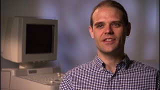
In this 35‑minute lecture, Dagan Wells (Speaker A) provides a comprehensive overview of preimplantation genetic diagnosis (PGD) and its evolution from single‑cell PCR to genome‑wide SNP‑based carrier (karyo) mapping. He explains the IVF workflow, embryo biopsy at day‑3 (cleavage) and day‑5 (blastocyst), whole‑genome amplification, and the challenges of allele dropout and contamination. Carrier mapping uses hundreds of informative SNPs around each gene to track parental haplotypes, delivering >99% diagnostic accuracy for single‑gene disorders and simultaneously detecting the majority of aneuploidies (≈75‑85%). Clinical metrics are presented: ~92% definitive diagnoses, ~8% no‑call rate, ~15% of cycles with no embryo transfer, ~5% pregnancy loss (vs 15‑20% expected), ~70% live‑birth per transfer, ~60% per cycle, and a work‑up time of 1‑2 weeks. The talk also covers the biological insights gained: recombination patterns, meiotic vs mitotic origins of aneuploidy, maternal age effects, segmental abnormalities, and clinic‑specific mosaicism. MyoMapping, an extension of carrier mapping, analyzes the oocyte and its two polar bodies to resolve maternal meiotic errors with higher resolution. The lecture concludes with a discussion of preimplantation genetic screening (PGS), its past controversy, recent randomized trials showing benefit, and how carrier mapping provides partial PGS information while retaining haplotype origin data. A Q&A session addresses false‑positive/negative rates of carrier mapping versus NGS‑based PGS (VARI‑seq) and the ongoing debate about PGS efficacy.
Questions this video answers
Carrier mapping detects at least 75‑85% of the aneuploidies that a dedicated PGS method like VARI‑seq would find, with near‑zero false‑positive rate for meiotic trisomies because the haplotype change is present in all cells. False positives are only a small concern for monosomies or mosaic cases that may resolve, and overall the false‑positive rate is lower than sequencing‑based PGS.
Yes, PGS remains debated, largely because older studies used obsolete technologies. Recent randomized trials (six to date) consistently show benefit when comprehensive, blastocyst‑stage screening is used. The ability of carrier mapping (and MyoMapping) to distinguish meiotic from mitotic errors provides mechanistic insight that supports the clinical value of PGS, though the debate about cost‑effectiveness and patient‑group applicability continues.
Chapters
So just during the course of this lecture, over the next 35, 40 minutes or so, I'll try and hit a few different things. I'd like to talk a little bit about preimplantation genetic diagnosis, which I know will be familiar to many of you, but I'll just briefly outline exactly what that is and why we do it. I'll talk particularly about the technology known as carrier mapping, which is, for us running a PGD laboratory, been quite revolutionary in terms of what it's allowed us to do diagnostically. I'll also consider about why chromosome abnormalities may be particularly important when considering fertility treatments such as in vitro fertilization, IVF, and I'll describe how methods such as carrier mapping and the related method of myomapping can provide some scientific insights into the basic biology of gametogenesis and embryo development. So to start with preimplantation genetic diagnosis, I guess the first question, what is it? You can consider it really to be an alternative to routine prenatal testing for couples who already know they're at very high risk of transmitting a particular inherited condition. So it's not something you would use just as a routine screen in a population. You would use it for patients who already have been identified at being at high risk of transmitting a Mendelian disorder. The vital part of PGD is that the couple are going to have to undergo an IVF treatment cycle. So this usually involves the stimulation of the ovaries to produce several eggs instead of the usual one per month. So you collect several eggs and you then fertilize them with the partner's sperm to hopefully produce several embryos. You're then going to biopsy these embryos to obtain genetic material to allow you to do a test. Now, years ago, it was done something like this. Here you have an embryo being held in place by a pipette that you can see just down here. You might be able to just about make out the membrane around the embryo. That's the zona pellucida. And this video is probably about 20 years old. And in those days, we would breach a hole in the zona pellucida using a weak acid. These days, this is done with a very precise laser. Of course, you have to make a hole in that membrane in order to be able to extract one of the cells. Hopefully this will run. You'll see the zona pellucida just go there. We'd also make a much smaller hole these days as well. The embryologist is now just changing over the pipette, and they're going to go in and remove one cell. This embryo is about 3 days old. It's at the cleavage stage, composed of between about 6 and 10 cells at that stage. All the cells are totipotent. In other words, they can produce any of the tissues of the body at this point. So the embryologist is just gently teasing out that cell, and that will be taken away for analysis. The embryo itself will go back in the incubator. They're extremely sensitive., and need to be kept under very precise conditions, so it will be safely put away. These days, more often this testing is done at a slightly later stage. This is a 5-day-old embryo, a blastocyst, and what's normally done these days is some of the tissue from the trophectoderm is taken. So here again, you can see the embryologist pulling at some of these cells. They're also using a laser, which you can't see visually, but Every now and again you'll see the embryo flicks slightly. That's when the laser hits the cell junction, and they're just trying to pull away some of these cells. It looks like quite an invasive, quite a brutal technique, really. But these embryos at the blastocyst stage are very robust. There's the embryo after biopsy. Always looks a little bit sad. There's the tissue. But believe me, it recovers very nicely. These embryos are very robust. And if you look at it a few hours later, it will look as though nothing happened to it. There's been a big preference now for analysis of embryos at this slightly later stage, just because the embryos are more robust, so the risk of damage is lower, and because typically you can get about 5 cells for analysis rather than just 1. Now, as a geneticist working in PGD, all of the methodologies we have are designed to work on 1 cell. So when you have 5 cells, it's like this amazing luxury of DNA that we have. Everything works even more robustly. Hopefully you're going to detect some unaffected embryos, and they will then be transferred to the uterus of the mother, and hopefully you're then going to end up with a healthy pregnancy. Traditionally, PGD has been a very rapid process. The embryos, as I say, biopsied either on day 3 or day 5. They usually need to be transferred no later than the morning of day 6, and so there's a very a very narrow window of time to do the analysis. Oocyte retrieval is usually referred to as day 0. Fertilization has happened by day 1. Biopsy day 3 or 5. Transfer usually about 24 hours later. So not only are we having to do a test often on a single cell, but we have to do it within about 24 hours. So it really does push some of the genetic technologies to their limits. These days, thankfully, the methodologies for cryopreservation of embryos have got really, really good. And so Very often now clinics will biopsy the embryos and then cryopreserve them, and that of course takes a lot of the pressure off both the embryology lab and also us in the genetics lab. There's less time pressure. The key thing about PGD, the main reason why people would do this rather than conventional prenatal testing, is that it avoids the entire issue of pregnancy termination. Patients may choose this for various reasons. It may be for their own personal ethical reasons that they have an objection to pregnancy termination, maybe for religious reasons. It may be that they've gone down the traditional route of prenatal testing before and they've had negative experiences. You know, perhaps they've had a couple of affected pregnancies, a couple of terminations, and don't feel they can go down that route anymore. Interestingly, from the infertility perspective, most of these patients are perfectly fertile. They're only undergoing IVF to access PGD, and so they're an unusual patient group for those of us involved in IVF clinics. Of course, we also get those patients who are coming for PGD because they've been doubly unlucky. They're unlucky enough to be infertile and need IVF to conceive, and they're unlucky enough to have a single gene disorder being segregated through their family. Obviously, for a family like that, it doesn't make sense to struggle with IVF, sometimes for years, finally achieve a pregnancy, and then find it's affected and maybe need to consider termination. For them, of course, PGD makes a lot of sense. Today, we've done PGD for more than 300 different single gene disorders, and really, as long as the underlying basis of the condition is understood, it's possible to design a PGD test for almost any condition. The accuracy, I think, is very good, especially considering the limitations that we're under. Typically, accuracy rates are over 98%, not 100% though, that must be noted. Rapid tests usually available. The only, I guess, limitation has been that traditionally PGD has involved a customized test, most patients have ended up having a unique protocol designed just for them. And the reason for that is, of course, that the off-the-shelf kits that you can get for diagnosis of some common disorders really aren't designed to work in single cells. The typical methodology has been something like this. You biopsy the embryo, you do a PCR— in fact, usually it's a multiplex PCR— you'll amplify the mutation site, and you can detect detect that in any of the vast number of different ways that you're all very familiar with. That allows you to make your basic diagnosis. The problem is, as any of you who've worked on low input quantities of DNA will know, that when you get down to the single cell level, the phenomenon of allele dropout becomes very common. If you do a direct PCR of a mutation site, you can probably expect about 5% of those to suffer allele dropout. In other words, you have a— maybe a normal copy of the gene, an abnormal copy, but you only succeed in amplifying one of the two, and so you only detect one of the two. So for that reason, you cannot, when doing PGD, rely on the mutation site alone, and consequently, we always look at linked polymorphisms. So maybe if we have the mutation site there where the green— where the blue line is, we'll test a polymorphism above and below. In fact, typically, we'd probably detect We look at maybe 10 polymorphisms in the vicinity of the gene, informative ones of course, and that gives us the kind of backup redundant test that you need in the face of things like allele dropout. These polymorphisms are usually short tandem repeats, so of course they're hypervariable in nature, which also gives us a simple DNA fingerprint for the embryo, which helps us with the other big problem of PGD, which is contamination. Obviously, if you're doing an amplification from a single cell, if any other DNA gets into your sample tube, that's going to be a major problem. So while doing a fingerprint doesn't help you avoid contamination, at least it lets you know when it's occurred, so you know not to trust the result from that particular embryo. So those methods have been really used for over 20 years in PGD, and they've been extremely successful. Successful, but the problem is that the customization adds significantly to the waiting time that patients experience and also to the cost of the procedure. It's very intensive on the laboratory side in terms of design and optimization. Now, the timing is very important because, for a start, IVF becomes less and less successful the older the person is, and so it's important really to do the test as soon as you possibly can. In some countries, there may be national health or insurance funding that covers PGD, but usually only up to a certain age. And so if your patient falls over that age threshold, they may no longer be able to access the service. And finally, for a couple to actually decide to go down the route of PGD is not a trivial decision. IVF, with all of its uncertainties, it's an emotional roller coaster for the patients. And so once they've come to this quite momentous decision, they, they really want to get on with it. They don't want you to tell them, okay, that's great, now wait 6 months while we develop a test for you. The other issue with conventional PGD technologies is that while they're very good at detecting an affected embryo, they don't really detect anything else. And there are many other genetic problems which are very common in our embryos,, and could— would ideally be detected. And the chief amongst these is aneuploidy. Of course, chromosome abnormalities, as you're all well aware, are the principal cause of miscarriage, probably causing at least two-thirds of all miscarriages. They're also actually responsible for most IVF implantation failures. When an embryo is transferred during an IVF cycle and fails to produce a pregnancy, the most common reason for that is that it's chromosomally abnormal. And then finally, of course, chromosome abnormalities are associated with various conditions such as Down syndrome. Now thankfully, all of these limitations have been overcome with the introduction of carrier mapping. If you— you could describe carrier mapping perhaps as a more generic approach to PGD. It's based on a genome-wide SNP array, and essentially you're going to track the inheritance of polymorphisms rather than mutations. It also allows us to see some aneuploidies. So I'm sure this audience is all familiar with the concept of a SNP array. So essentially what we're doing is getting a unique DNA fingerprint or haplotype for each region around each gene and following their inheritance through the family. So perhaps a mother with a dominant mutation, we'll have a look at the polymorphisms either side of this. In reality, with carrier mapping, and we're usually looking at a couple of hundred polymorphisms in the vicinity of each gene, type the father and affected child too, and then get results from the embryo. And of course, what we're really looking for is indications of particular parental haplotypes. So for example, a B at this third position and an A at the penultimate position is only seen on the father's green chromosome, so that chromosome must be there. The A at the second position, B at the last position, only seen on the mother's blue chromosome, so that must be there, and since that one was in the mother and the affected child, that must be the one carrying the mutation. Obviously, for this audience, the concept of good old-fashioned linkage analysis is all very nice and familiar. Of course, we can also get results like this. Here again, we have results suggesting the presence of the paternal green chromosome. Happily, this time, no evidence for the mother's blue chromosome. Her pink chromosome seems to be there, so this embryo should be unaffected. But unfortunately, there's also evidence for the father's yellow chromosome. So while unaffected, this embryo is, of course, trisomic, so we wouldn't want to transfer that embryo. This is what the actual data output looks like using the BlueFuse software that we use for this. Here's a normal embryo, and you can just see all of the different SNPs lined up in the order they appear along the length of each chromosome. This is the log R ratio. It's essentially telling you the amount of DNA bound to each probe. And then below it we have the actual SNP calls. So homozygous AA, if you like, at the top, BB at the bottom, and the heterozygous calls in the middle. Those of you who use this kind of technology on large amounts of DNA will, of course, usually see things where the AB calls are very tight along the middle. This is a little bit more noisy because, of course, it's after whole genome amplification from a single cell. The software also does a very nice job of giving you a sort of graphical representation of the parental chromosomes, and it shows them something like this. Here's just one chromosome that I'm showing you here, and what we see along the top, all these little dots, are results for individual informative SNPs, so it's only showing you the informative informative ones, and it color-codes them according to the parental chromosomes. So you can also quite nicely see where a combination occurs. So you've got one paternal chromosome here, all the way down to here, and then there's been a recombination, and then a second recombination. So it gives you that kind of recombination information as well, which can be quite interesting. Errors can look something like this. This is a trisomy 21, and you might be able to just about make out that there is a little bit more DNA being detected on chromosome 21 quantitatively, and then qualitatively as well, you can see this sort of splitting of the AB calls, because actually they're not AB, they're either AAB or ABB. The other thing is you get this characteristic pattern when the software tries to tell you what parental chromosomes are present. Here you see this kind of, mixing of the two maternal chromosomes, which would normally either be just green or just yellow. So you get this very characteristic pattern. A loss looks something like this. Again, you can see less DNA on that particular chromosome, chromosome 18 here, and of course there's no heterozygous calls at all because everything has to be hemizygous effectively. When you actually look at the graphic for the chromosome, you see there are no SNP calls along its length. So the actual accuracy of this kind of linkage approach for PGD is extremely good. Our actual clinical accuracy rate is well in excess of 99%. Again, I think very good considering the limitations that we're under. We get a definitive diagnosis from the majority of embryos, about 92%. That leaves about 8% 5% that don't get a diagnosis. About half of that is due to just amplification failure. Either the cell didn't get into the tube, or the DNA in that cell was too degraded for analysis. Or the other, I guess, probably about 4% of cases are due to a recombination just too close to the gene or within the gene of interest, and so we're not confident to make a call. Those embryos can still often be rescued diagnostically it's just that we then have to go and do a direct mutation detection for those. About 15% of the patients who begin this process end up with no embryo transfer. Either that's because all of the embryos were diagnosed abnormal, or because the embryos had not developed, so a limitation of the IVF process essentially. The pregnancy loss rate is extremely low given the average maternal age of these patients. Patients, which is in the mid-30s, you would expect about 15 to 20% of conceptions to fail to go to term, but we actually only see a loss rate of about 5%. So that, that's very encouraging, and it's almost certainly due to us not transferring aneuploid embryos that would normally have produced those losses. The birth rate per embryo transfer is almost 70%. So If a normal embryo is discovered, the chances of it producing a healthy, unaffected child are really very good. Per cycle, it's about 60%. That's taking into account that some patients get no embryo transfers. It's actually likely to be a little higher than that. That's because some embryo— some patients have more than one unaffected embryo, so they'll transfer one and freeze the other unaffected embryo. So sometimes they may not have got pregnant yet, but they may still get pregnant in the future from frozen embryos. The workup time for carrier mapping is typically a couple of weeks, although we have done workup in just 24 hours before, so that is possible, compared to a conventional PGD, which would take, you know, 2 or 3 months in the quickest labs, could be a year in the slower labs. As I mentioned, you can detects the majority of aneuploidies. It probably doesn't detect all, but certainly at least 3/4 of them will be picked up in this way. And interestingly, it will give you a confirmation of the parental origin of the embryo. Now, in a modern IVF lab, the chances of mixing up embryos from different patients are almost zero. But should it ever happen, you would immediately know it, because the embryo's genetics wouldn't match that of the prospective parents. Several publications have come out on this if you're interested. Largely off the back of this kind of advance, we've seen PGD referrals increased by about 500% since 2014. I think this is down to a few different reasons. One is methodologies like carrier mapping that have just made that process so much easier and so much more streamlined. I think another part is improved patient awareness. You know, you go back 15 15, 20 years, and most patients never heard of PGD as an option. I think there's also increased acceptance of PGD by the clinical genetics community. I remember when I was first doing PGD about 25 years ago now, yet there was some reluctance to really discuss PGD as an option with patients, not because the genetic tests were inaccurate, but more because the perception was that IVF was just simply too inefficient. Sufficient, why send a perfectly fertile couple down the IVF route when the chances of taking home a baby after IVF were so low? But of course, IVF has got better year on year, and I think most people have reevaluated that position over the last few years, knowing that actually there's a very good chance of a pregnancy following IVF now. The other final thing is that we're seeing more and more patients coming through after under having preconception carrier screening, so patients who would probably never have even known they were mutation carriers in the past, but have undergone carrier screening and found that, sure enough, they and their partner both carry a recessive mutation in the same gene. That's still a small fraction of our total referral base at the moment, but I note it simply because it's gone from zero to an appreciable fraction over the last couple of years. I'll just say a few words about why it's important to detect aneuploidy in IVF and PGD cycles. And really it all comes down to the fact that in human embryos, perhaps more than any other species, chromosome abnormalities are extremely common. Of course, they're extremely common in our eggs. That's where most of them are coming from, and therefore in the embryos too. And of course, it's a problem that increases greatly with advancing maternal age. Of course, we're all familiar with this kind of shape of graph from the incidence of Down syndrome with advancing reproductive age. But actually, that's really just the tip of the iceberg, because in these early embryos, all of the chromosomes really follow this kind of pattern. You can, in an early embryo, easily see a monosomy 1, for example. Of course, you'll never see this in a miscarriage or in a prenatal sample, because these abnormalities are so lethal that they are unlikely to survive to implantation. So I guess a couple of things to take away from a graph like this. This is actually showing you the percentage of abnormal embryos that we would see on day 5 in an IVF clinic. The first thing is that actually it's common even in the younger patients. So for these patients in their sort of early or mid-20s, 20s, at least a quarter of the embryos are chromosomally abnormal. But sure enough, it does get much worse with advancing age, so that for the patients over 40, it's more like three-quarters or more of the embryos which have an aneuploidy. And in fact, beyond the age of 35, the aneuploidy rate is increasing by about half a percent per month. So really, every month can be important, especially for these older patients. Now, these aneuploidies are almost invariably lethal to the embryo. And so not surprisingly, as the aneuploidy rate goes up, the chances of a successful pregnancy go down. And of course, when as a scientist you see a graph like this where the two lines look almost a mirror image of each other, the most likely explanation is that what you're seeing is cause and effect. So in other words, the increasing aneuploidy rate with age is responsible for for the decline in IVF success rates with advancing age. That gave birth to a whole concept in IVF of preimplantation genetic screening, which is distinct from PGD in that you're doing a genetic test not for someone with a known risk factor, but for really any IVF patient in order to help identify chromosomally normal embryos. The idea is in an IVF cycle you generate several embryos, maybe half or more could be chromosomally abnormal. And rather than transferring one not really knowing whether it has the right number of chromosomes, because certainly the morphological analysis that's done in all IVF clinics will not identify aneuploidy— the aneuploid ones can look just as good or even better than the normal ones— why not do a genetic test that allows you to specifically transfer chromosomally normal embryo? It's not going to guarantee that you get a pregnancy,, but it will eliminate what is the single biggest factor in embryo implantation failure and miscarriage. In theory, this should allow patients to have much reduced risk of aneuploid syndromes—Down's, Edwards', Patau's, Klinefelter's, Turner's. It should have a big impact on the miscarriage rate, since the majority of miscarriages are caused by chromosome abnormality, and you simply won't be transferring those embryos anymore. And of course, course, it should give you a greater chance that the embryo that you transfer is viable and can sustain a pregnancy. Has been controversial. About a decade ago, there were several randomized controlled trials, I think about 10 in the end, and none of them were able to show that this was beneficial. But this seems to have been mostly due to technical limitations, both on the embryology side and on the genetic side at the time. And over the Over the last 5 years, there have been actually now several randomized controlled trials using modern technologies, and each one of them has shown that PGS can be beneficial. I'll just show you very briefly some uncontrolled data. The blue bar shows the increase in pregnancy loss rate that you see in IVF cycles. This is from thousands of cycles collected by the SART data registry in the United States, showing that for the patients under 35, there's about about a 10% loss rate. For patients over 40, it's more than a third of pregnancies that are lost. These are pregnancies that are measured at a very early stage, so typically about 2 weeks after embryo implantation. If you do PGS, you really don't see a change with age. So all of that increase in loss rate is all chromosome-related. So take chromosomes out of the equation, you don't see that anymore. Similarly, in terms of embryo implantation, again, as we saw in that earlier graph, a big crash really in the implantation rate with advancing age. If you transfer a chromosomally normal embryo, you don't see that. And a chromosomally normal embryo from a 42-year-old really has about the same chance of making a baby as a chromosomally normal embryo from a 22-year-old. The difficulty in that older age group is actually finding them. You, you'll do many cycles where you look and all of the embryos are abnormal. This has led to a very big increase in the utilization of this kind of chromosome screening in the United States. So today we think probably about a quarter of all IVF cycles have chromosome analysis, and that's driven partly by the increased evidence for this, this approach, and also because the costs of this kind of analysis have been falling. So this is really why things like carrier mapping that allow you to do PGD but also give this chromosomal analysis are very important. It's really disappointing when you have a, a couple that you've got to know quite well, and you're so keen for them to have a successful PGD cycle, and you find an unaffected embryo using a conventional PGD method, and you transfer it, and they become pregnant, and then a couple of months later they suffer a miscarriage, and it turns out to be due to a chromosome abnormality. Now, thankfully, with methods like carrier mapping, that kind of outcome has pretty much gone away. So the interesting thing about this sort of approach is that it does give you some basic biological information as well, which can be quite interesting to analyze. For example, you can see all the recombination sites around the genome using carrier mapping. You can see the aneuploidy, but you can also see exactly which parent it came from, the mother or the father, by tracing these haplotypes. You can even tell the difference between whether an aneuploidy arose in meiosis 1 or in meiosis 2. Finally, you're collecting a huge amount of SNP data, so down the road there's the possibility of a retrospective analysis to do genome-wide association study or something like that. The way that it works in terms of looking at the stage, the meiotic stage at which an abnormality occurs, is something like this. Here's how it, of course, it should go, which I know you're all well aware of. You have the two homologues, they've both replicated to have two chromatids each. You may get some recombination that goes on between these as well. Meiosis I, you get separation of the homologues. Meiosis II, the separation of the chromatids. Chromatids. So that's how it should go. If there's an error in meiosis 1 so that both of the homologues go to the same cell— maybe this could be the egg that they're in— then when you go to the second meiotic division, you'll end up with a situation like this. So here the chromatids have segregated appropriately, but here the chromosomes have not. Now, if we do carrier mapping, The thing is to look around the centromere. Around the centromere, recombination is suppressed, and what you'll see in this instance is evidence for both of the different centromeres from the yellow chromosome and the green chromosome in this case. So if we zoom in, this is the centromeric region. Remember, these are the individual informative SNPs along the top, and you can see there's a mixture of yellow and green ones there. So this is telling us that there's been an error in meiosis I. And again, you're seeing this characteristic mixture of green and yellow all the way along the chromosome, which tells you that there's both copies present. If you had an error in meiosis II, so meiosis I goes fine, but then you get a failure of the chromatids to divide in meiosis II, the difference here is that both the centromeres are identical. So sure enough, when you look at carrier mapping, Here's the centromeric area over here of this particular chromosome. All of the SNPs are from the same chromosome. So that's how you can actually tell the difference between the two. Now, we've had a bit of a look in some of the embryos that we've diagnosed for reasons of single gene disorders, and look back at some of the chromosomal problems that these have had. So looking at the errors, about 170-odd errors that we were looking at, As expected, the vast majority of them are derived from oogenesis. Of course, this is what we expect, more than 85% typically. If we look at how many of these are trisomies and how many are monosomies, we get a pretty even split in the maternal side, but the interesting thing is in the paternal, we get far more losses. And actually, most of these are not whole chromosome losses, but segmental abnormalities. We've mapped out these segmental abnormalities that we see across the genome, and they don't sort of randomly distribute. They are actually concentrated in certain areas. Some of these actually correspond to known fragile sites in the genome, but others are apparently novel sites. We think this may be related to deficiencies in the embryo culture. Of course, we all know that we can induce fragile cytes by depleting certain things from culture medium, and it may be that embryo culture medium needs further optimization since we are seeing quite a lot of chromosome breakage going on. We see mitotic errors and also meiotic errors, and not surprisingly, just as we would expect, the meiotic errors increase with advancing female age. The mitotic errors do not. Seem to— these are occurring primarily in the first 2 mitotic divisions and really underlie the very high level of genetic instability during early human development. Those first 2 or 3 divisions, they occur without genome activation. So really the whole embryo is being driven by maternal RNA, maternal proteins that have laid down in the oocyte. It may be that during those stages, there is either a rigidity in cell cycle checkpoints and other mechanisms, or even an absence of some of them perhaps. And so they seem to quite often suffer mosaicism and chromosome breakages, which are not resolved in the normal way. So some of the insights— segmental abnormalities and chromosomal mosaicism, very common. As I say, most arising shortly after fertilization prior to switching on the embryonic genome, which happens around the 4 to 8 cell stage. We are interestingly, you know, we're a genetic service lab essentially. We get samples from many different IVF clinics around the world, and the interesting thing is the rates of segmental abnormalities and the rates of mosaicism are not the same from all clinics. It seems that there could there could be a clinic effect that influences the frequency of these abnormalities. So that's kind of interesting. It suggests that there is an opportunity for further optimization of the IVF process, which should improve patient success rates. I'll just finish up by saying a few words about MyoMapping. I was just asked to say something about this, and I have to stress this isn't my work. This is mostly the work of Alan Handyside's group and Christian Ottolini. It's really, I guess, an extension of carrier mapping. It's very much similar in principle. The idea is, though, to focus on female meiosis only. And rather than looking at the embryo, you're looking at the three components of female meiosis: the egg itself and the two polar bodies produced during the first and second meiotic division. So you collect all of those, you do a whole genome amplification, amplification. It's usually multiple displacement amplification-based. You're then going to use a genome-wide SNP array to identify informative polymorphisms, look at the inheritance of haplotypes through meiosis, and mostly focus on identifying sites of recombination. It does have some advantages over a routine carrier mapping approach, which is applied to embryos, in that you don't have that paternal background having the paternal chromosomes there means that you get less informative maternal SNPs. So if you're just focusing on the mother's side, then using this kind of MyoMapping approach allows you to get more informative polymorphisms and therefore higher resolution. It's also possible to detect recombination that has been associated with monosomy. When we're looking in the embryo, we can't really tell what's happened during meiosis when a monosome has been produced, because the chromosome is simply not there. So we don't know whether it recombined or where it recombined. In this instance, because you have the other products of meiosis, you can see exactly whether there was any recombination on those affected chromosomes. Not surprisingly, what they've seen is that most of the errors are actually caused by premature separation of the sister chromatids. And this has really been understood to be a principal mechanism of aneuploidy generation since the work of Ross Angel a couple of decades ago now. The idea is that instead of the chromatids separating in meiosis II, they separate early in meiosis I, and then the individual chromatids will segregate essentially at random. Although what they found quite interestingly is that it didn't seem to be entirely random random segregation. In fact, more often than not, instead of, you know, maybe ending up with 3 chromatids here and 1 chromatid there, or, you know, these combinations, more often than not they actually did still end up balanced. So it suggests there are some other mechanisms going on there that may perhaps tether some of the chromosomes together. Um, what they also saw was that when you've got the egg and the second polar body, more often than expected, if one of them has failed to recombine, that one tends to get pushed to the polar body. So there seems to be a preference for retaining recombined chromosomes in the egg itself, which is kind of an interesting observation. So just to summarize what they found there, they found more recombination in female meiosis than male meiosis. Again, that's not an unexpected finding, that there was already extensive evidence that that would be the case. The recombination rates varied interestingly a lot between individual patients, and even within a patient, there could be significant differences in the recombination rates in individual oocytes. On average, those eggs that were chromosomally abnormal had less recombination than those that —chromosomally euploid— had a little more recombination. They found 135 chromatids that had no evidence of recombination at all along their lengths in meiosis II. In theory, those should be equally distributed between the eggs and the second polar bodies, but twice as many of these recombined chromosomes were found in the eggs— sorry, twice as many many of these unrecombined chromosomes were found in the second polar body compared to the egg. So, you know, there seems to be some kind of way of driving out these unrecombined chromosomes, although the mechanism behind that is not understood. So just to conclude, these kind of genome-wide SNP methodologies, I mean, really in a way they're old-school genetics, and they're taking us back to the days when when we were doing genetic diagnosis just based on linkage analysis before we even necessarily knew what the causative gene of the condition was. So in a way, it seems very old-fashioned, but actually they've been quite revolutionary in this little niche area of genetics where we're doing PGD. It's really helped to provide very accurate tests, and it's helped also a lot with the whole pipeline that delivers PGD, making it a a lot less labor-intensive, more well-optimized. It's shedding some interesting light on the origin of aneuploidies, and importantly, it's telling us about these around the time of conception, really before there's been any selection against the aneuploidies. When we look at miscarriages or prenatal samples, we're really getting a very narrow view of what's going on because so many of the other kinds of aneuploidy, which is so common early in development, have already been eliminated by then, just not available for us to see. He's doing a lot to clarify the relationship between recombination and aneuploidy. Of course, the relationship's been known for quite some time. If you don't have recombination, that's a risk factor. If you have recombination too close to the centromere or too close to the telomere, you may have a risk risk factor, but it's giving us some more detail on that. And finally, I think as these methodologies evolve, moving on to sequencing-based methods like the haplorhythmic, which I'm sure we'll hear about next, I think we're going to get even greater power to really start delving into not just the diagnostic potential, but also a better understanding of the mechanisms that are underlying these kinds of incredibly common abnormalities. Anomalities. Just to acknowledge the rest of my group based in Oxford, who produced quite a lot of the clinical data that I showed today, and thank you very much for listening. Thank you very much for this fantastic talk. The floor is open for discussions. Are there any questions?
Yes, please. Thank you for your talk. Ekaterina Pomerantseva from Moscow. You mentioned that you see with karyomapping 15% less of aneuploidies than by regular BGS, like VARI-seq, I believe. And what about comparison with VARI-seq in terms of false positive? If you compare results for aneuploidy obtained by karyomapping with 20 for Sure or VARI-seq How many are not confirmed?
So yeah, I would confidently say that with carrier mapping, we're detecting at least 3/4 of the abnormalities that you would detect with a PGS method focused only on chromosome abnormality detection. In reality, it may be a little bit better than that. It may be up to perhaps 85%, but we definitely miss some, and the ones that we miss are not the meiotic abnormalities, abnormalities, but the mitotic ones. And that's because the carrier mapping is really designed and optimized to provide a qualitative view. So although I showed examples where you can see a quantitative difference on the amount of DNA from a particular chromosome, that is not super reliable. I would never trust that as a primary diagnosis. What you can trust with carrier mapping is is what it's telling you about the inheritance of haplotypes. So anything that's come from meiosis is going to cause a change in the haplotypes that you see there. So those are detected probably with near to 100% accuracy. However, as you know, mitotic abnormalities are very common in human embryos during those first couple of divisions, and so those are the ones that might get missed. Things like VeraSeq or the other next-generation sequencing methods or array-based methods, array CGH, detect those very well, but of course then you don't have the information on origin. So I'm not really proposing carrier mapping as a PGS method. It's a PGD method that has— delivers some PGS information.
But how accurately in terms of false positive?
Because what you're talking about is about false negative. Sure. So false positives are close to zero with a carrier mapping approach because, again, what you're really seeing, or at least for trisomies, it's close to zero because what you are seeing there is a meiotic error, therefore it's present in all the cells. The false positives that you can get in PGS are usually related to a failure to detect mosaicism or detecting a mosaic abnormality when actually the embryo is going to end up being completely normal. So if you're looking only at meiotic abnormalities, then actually you can be very confident. The place where you might run into a problem with a carrier mapping approach is if you had a monosomy. If the chromosome simply isn't there, you don't really know whether it was a meiotic or mitotic problem, or you can say it's not there. So it would be a small false positive rate associated with that, perhaps. Not false positive technically— what you're— what you've seen really will be in the cell, but it may not be in the final fetus that would be produced, because a lot of those mosaic abnormalities actually resolve themselves. But it's still a very low fraction. I mean, when we do PGS using a sequencing-based approach or an array-based approach, it's only a fraction of just a few percent of embryos that can actually survive that. So it would be even lower with carrier mapping. Thank you. Sure.
Second question, Hido, I believe. Yes, thank you. Hido de West, Maastricht. Thank you for your highly informative presentation. I was struck in a way by your point that PGS has been controversial. And I have, well, some information and my impression looking at the literature, talking with your colleagues, that PGS still is. Controversial. So apparently among experts there is some dissent about the quality of the data suggesting that this is really evidence-based, no longer experimental, PGS. At the same time, I think you present some highly interesting new data about scientific capacities to make a distinction between the origin of aneuploidy in terms of mitotic and meiotic origins. Can you comment on this? Do you agree that there is still some sort of controversy, and can you further clarify as to whether your new data about meiotic or mitotic origin might help to strengthen the data supporting the evidence that, yes, this will do the job? And yeah, thanks, Peter.
That's absolutely right. So it has been— I mean, it's probably been the most divisive thing in the whole field of IVF. I think you could argue, couldn't you? We have to draw a line under the studies that were done a decade ago. All the technologies have been obsolete for the best part of 10 years. No one has been doing those technologies, or no one should have been doing those technologies for many years now. So we have to look at the data that we have right now. And right now, I believe there's 6 randomized controlled trials which all show benefit for PGS. All of the trials have flaws, as all trials do. 4 of them, I think, are now in the literature. 2 of them have been presented at meetings, so I guess we have to treat those carefully for the time being. But they all point in the same direction. So I agree, there is still controversy in the field. There are occasions when an embryo embryo has been given an abnormal diagnosis and has been transferred anyway, and very rarely these have produced completely healthy babies. And that's really keeping a controversy going, because it's clear that in a sense it's not a 100% diagnostic methodology. There is a screening element to it. But certainly the weight of the data at the moment is all pointing in one direction, and it's saying that if you do comprehensive aneuploidy screening, you look at all of the chromosomes, and particularly if you do it at the blastocyst stage, then it seems to be a positive, beneficial way to go. You know, you can make arguments about the cost-effectiveness. You can make arguments that maybe occasionally a viable embryo could be discarded, and I think we will continue to see the debate going on. Also, there isn't a lot of data about individual patient groups. So there are, you know, traditionally PGS is applied to patients with recurrent unsuccessful IVF treatment, recurrent miscarriage, or advanced reproductive age. But we don't have trials necessarily in all of those groups to support the use of it for those. So it's an ongoing story. All we can say at the moment is that the pendulum has swung towards favoring PGS based on the evidence that exists in the literature. Literature. But yes, I think it's not— I don't think the argument has been won there. It's still ongoing.
More from Joshua







Scientist Stories: Hans Waterham, The value of phenotypic assays in peroxisomal disorders




Scientist Stories: Joel Zlotogora, Genetic carrier screening programs for reproductive purposes