Scientist Stories: Daniel Mucida, Microbiome & Communication Between Our Nervous & Immune Systems
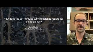
In this Rockefeller University webinar, Professor Daniel Mucida presents a decade‑long research program focused on how the intestinal microbiome shapes communication between the gut’s immune system, its epithelial and neuronal cells, and the central nervous system. He begins by contrasting the classic view of immunity as pathogen resistance with the gut’s need for tolerance toward dietary antigens and commensal microbes. He describes the structure of the intestinal epithelium, the dense network of intra‑epithelial lymphocytes (IELs), and the gut‑draining lymph nodes that initiate oral tolerance. Using live‑imaging, he shows IELs constantly scanning the epithelium and altering their behavior during Salmonella infection, linking their activity to pathogen clearance and suggesting a role in colorectal cancer surveillance. He then turns to neuroimmune interactions: enteric neurons differ along the proximal‑distal axis, and germ‑free versus specific‑pathogen‑free mice reveal that the microbiota profoundly remodels neuronal gene expression and function. Pathogenic infections can cause selective loss of excitatory neurons, leading to prolonged motility defects reminiscent of post‑infectious IBS; fecal microbiota transfer restores neuronal regeneration, implicating microbiota‑derived signals in disease tolerance. Muscularis‑resident macrophages sense neuronal neurotransmitters and activate a protective program that shields neurons from inflammation‑induced death. The talk expands to translational themes—oral tolerance therapies for food allergy, defined bacterial consortia versus whole‑fecal transplants, taste‑bud‑like epithelial cells forming synaptic “neuropods” that convey nutrient status to the brain, and the possibility that lingering SARS‑CoV‑2 antigens in the gut drive long‑COVID antibody evolution. He also discusses the hygiene hypothesis, evolutionary perspectives on allergy prevalence, and the current limits of microbiome diagnostics and probiotic interventions. The presentation ends with a Q&A covering these topics, highlighting both mechanistic insights and future therapeutic directions.
Chapters
Greetings, everyone. I'm Rick Lifton, President of the Rockefeller University. I'm delighted to welcome you to this afternoon's webinar. Today we will hear from Professor Daniel Musida, who will give us the inside story on how the microbiome influences communication between between our nervous system and our immune system. This annual fall program brings together many groups of the university's benefactors and friends. We're pleased to have the participation of trustees, members of the Rockefeller University Council, and the Committee on Trust and Estate Gift Planning, as well as supporters of our Women in Science and Parents in Science initiatives, foundation colleagues, and other university associates. Normally, of course, we'd be welcoming you to campus. However, the continued threat of the Delta variant and breakthrough infections leads us to continue to hold programs like today's presentation as a webinar via Zoom to ensure the safety of everyone. Some of you may have joined us for the first webinar of the academic year held on September 14th, which was a session of our virtual discussions with genuine experts on COVID-19 vaccines and variants. As we reported in that program over the last year, Rockefeller scientists have joined with investigators worldwide working to understand the biology of the SARS-CoV-2 viral life cycle and to contribute to the development of vaccines, monoclonal antibodies, and small molecules that prevent or treat COVID-19, as well as to understand the development and duration of immunity after both natural infection and vaccination. And understanding of how variant viruses increase transmission and evade the immune response to vaccination. Our scientists have also discovered one of the principal mechanisms explaining why some people may have no symptoms following infection while others progress to severe disease and death. Your philanthropic support has enabled Rockefeller researchers to quickly undertake these new initiatives as they confront the continuing challenges of the virus. This vital work will continue as long as COVID-19 remains a major threat. Your ongoing contributions also help to sustain our broader research and educational efforts. Philanthropy has always played a critical role at Rockefeller, where support of the most gifted and creative scientists has led to an exceptional record of accomplishment. Since our founding, 26 university faculty have been recipients of the Nobel Prize. Including 3 in medicine in just the last 10 years, including last year's prize when virologist Charles Rice received the 2020 Nobel Prize for his key contributions to the development of a cure for hepatitis C, a major cause of death from chronic viral infection. Rockefeller is a world leader in scientific discovery because it has always invested in audacious and original scientists. The university recruits only the most creative investigators and provides them with the scientific freedom and the financial resources to pursue novel and important research. Today's speaker, Daniel Musida, is a prime example of such an investigator. Daniel's fascination with immunology stretches back to his college years in Brazil in the late 1990s when he contrasted the role of intestinal microbes and diet in stimulating the development of the immune system. As a PhD student and a postdoctoral fellow, he went on to study how the immune system, which evolved to reject foreign antigens, does not normally mount an immune response to novel proteins in food, allowing its absorption and use for cellular energetics and metabolism, but does not— but does respond to pathogens. Daniel was recruited to Rockefeller in 2010 as an assistant professor, founding the Laboratory of Mucosal Immunology, and launched several lines of research with powerful implications for human disease. His overarching goal is to understand how the immune system is able to respond efficiently to pathogens while maintaining tolerance to food and roughly 100 trillion innocuous and beneficial microbes we each harbor in our intestines. Maintaining the delicate balance of the digestive system depends on the complex interactions of microbes, gut cells, immune cells, and neurons. Many have considered it folly to try to understand the gut's activity by experimentation in living animals, but Daniel has proved to be a fearless, brilliant, and incisive experimentalist, and his studies have provided fundamental insights into the normal and disease-related activities of the GI tract. Recently, Daniel's research has revealed unexpected cellular and molecular mechanisms that underlie the so-called gut-brain axis. It's fascinating to consider that the human digestive system contains about as many neurons as the spinal cord. Daniel's group has shown, for example, that intestinal microbes and intestinal neurons exchange signals that are vitally important for the absorption of nutrients. This intriguing crosstalk even has a role in feeding behavior. Daniel and his group have also studied inflammatory pathways and shown that certain types of immune system cells can shield neurons in the intestine from damage caused by inflammation. Daniel's research is providing insights into the origins of many gastrointestinal pathologies, including food allergies, colon cancer, and chronic inflammatory disorders. The understanding gained from his work can inform new approaches to the prevention and treatment of these and many other diseases. This past spring, Daniel was promoted to professor with tenure here at Rockefeller, and just last month he was named an investigator of the Howard Hughes Medical Institute. HHMI supports 250 to 280 of the world's most innovative biomedical scientists, providing them very generous unrestricted funding to pursue high-risk, high-reward projects for 7 years with the potential for continued support. Every 3 years or so, they conduct a highly competitive national search for new investigators, and this year Daniel, along with Rockefeller colleagues Vanessa Ruda and Daniel Kronauer, were 3 of the 33 biomedical scientists selected to become HHMI investigators. This prestigious appointment reflects the high regard in which Daniel's scientific achievements are held. As you listen to Daniel tell us about his research, You are welcome to submit questions at any time. You can do so by typing them into the Q&A function at the bottom of your screen. At the end of the presentation, we will give Daniel the opportunity to answer as many questions as time allows. It is now my great pleasure to turn the screen over to Daniel Musida.
Thank you, Rick. It's always difficult to speak after your introductions because they are very often better than what we can present. But thanks for the very kind words and I'm very happy to speak to you today and also thank you, thank you for your support to the university. Basically, I want to today use this time to go over different topics of the laboratory that we have been developing the past 11 years since I joined the university and focus in particularly on the topic of neuroimmune communications, as Rick mentioned in his, in his introduction. So I will start with the way I actually teach immunology to the students here at Rockefeller. And sometimes I make analogies to this comparison here with the immigration system. So basically what we learn in textbooks is that the main function of the immune system is to provide resistance against pathogens that are invading our body. And that I'm not here to disprove that, but the focus of the laboratory is the intestine. In the intestine, the vast majority of the non-self interactions as Rick just pointed out, are not with pathogens, but rather with likely beneficial microbes from the microbiota as well as beneficial nutrients that are coming via the diet. And these are also part of something that we call non-self. However, against this non-self from diet and microbiota, we must develop a form of tolerance rather than develop resistance, which would cause damage, of course, to our body. So that's one aspect of the laboratory. And the second aspect is that our own immune system very often causes damage to our body, and that is called inflammation-induced damage. That is the case, as you probably know, about the pathology of COVID-19, that is not necessarily the viruses that are causing the damage in the body, but rather an inappropriate immune response triggered by the virus. However, there's another aspect of this relationship between the body and the host and the pathogens, is that the body has also acquired evolutionarily mechanisms that help us to deal with this inflammation triggered by a pathogen, and that's something called disease tolerance. So the overarching question of the laboratory is basically how does a gut immune system balance resistance against pathogens and these forms of tolerance. I will go over an image, a video of the intestinal tissue just to start introducing about the questions that the lab is asking. If you slide open the intestine and look at the luminal side where the food, microbiota, etc., is present, what you encounter, it's really a very rich system and very complex. Here we're looking as if you have a tube inside intestine like an endoscopy. What you see in the intestine is these structures that are villi structures that actually help the body increase the absorption of nutrients. What is special in the intestine is that the separation of this environment where the bacteria and the nutrients and the dietary components are present in the body only takes place with a single layer of highly absorptive cells, the epithelial cells of the intestine. There is a large component of non-self interactions with the diet and microbiota. This stimulation, it's only separating us. We are only separated from this to the core of the body by a single layer of cells. That's very different, for example, than the skin, which has several layers of cells separating the outside of the body from the inside. If you look at this scheme representing the intestinal tissue, I guess here you can start appreciating the complexity of the system as we study it. In the proximal part of the intestine is where you mostly absorb the nutrients from the diet. As you go distally towards the large intestine, you increase the density and the complexity of the gut microbiome. In addition to this proximal to distal differences, There's also several difference in terms of layers of cells that are populating the intestine, and I will go over that today with you. One topic of the lab that we are particularly interested is how do we get food allergy or how we avoid it by means of something called oral tolerance. We study that because by studying the initiation of the responses that are in this region of the intestine, the proximal intestine, we may understand mechanisms that are physiological and are in place to avoid us to develop food allergy, and also what happens when these mechanisms fail and then we acquire food allergies or celiac disease. In this particular topic, the place that we are focusing our studies are these organized structures, lymphoid structures called lymph nodes. In particular, we are interested in these gut-draining lymph nodes because that's where the initiation of the immune response against what is happening in the luminal of the intestine, including immune response against food, takes place. And here is just one example of how these structures are organized. This is in an animal intestine. And what you're going to see as I start the video are these structures in red, these round structures, which are the lymph nodes. And you're also going to appreciate the intestinal tissue here in red here. So, what you see here are the lymphatic vessels that are connecting the intestine to the gut-draining lymph nodes. And also, you can appreciate by, for example, staining the insulin in green, that these tissues are surrounded by additional tissues that can influence what is happening in the intestine. So, by focusing our studies in these gut-draining lymph nodes and how the initiation of the immune response takes place, We hope to understand the process that induces tolerance to the food as well as what happens when they fail and that leads to food allergies. Another major topic of the laboratory, as you could see by the initial video I showed, is the intestinal epithelium, and that is a particular fascinating site. It's fascinating because, as I mentioned, it's a single layer of cells that are highly absorbent, They are in contact with this vast amount of nutrients as well as microbiota and bacteria, as well as pathogens. In addition to this area which is vast, the intestinal epithelium has these structures called brush border or microvilli, which further increase the area in which the intestinal tissue is exposed to the lumen. For example, in humans, it's estimated that the total area of the intestine alone It's about 300 square meters or a size of a tennis court that we have here at Rockefeller. It's a massive area exposed to the environment, which is in this case, the lumen. A huge amount of antigens and stimulation from bacteria, as I mentioned, and diet. Not surprisingly, in this area you have also associated with it a lot of immune cells. A particular cell type that we are interested in the epithelium are these cells represented here in green. These are called intraepithelial lymphocytes. They are lymphocytes that are located right at the epithelial surface of the intestine. In the past several years, we have found, for example, that these cells play an important role in the resistance or in the immune defense against enteric gut pathogens that are invading the organism. But we also studying how these cells can can acquire some forms of dysregulation that can lead to additional pathologies. For example, these cells are the major cell type thought to be associated with celiac disease or gluten intolerance. They are also related to the pathology of several types of inflammatory bowel diseases. I'm going to give an example of what we study in these cells and how we approach this question. Here what we did was to use fluorescent dye to visualize these intraepithelial lymphocytes in a live animal. On the left side, you're going to observe how the cells are moving in the, as I said, in a live animal. And on the right side of the screen, you're going to see what happens when you have exposure to a pathogen. So just to point out first that these cells, as you can see here, in each of these structures, this kind of surrounding structures, this one is is one villus of the intestine. These green cells are moving constantly around the epithelial cells which are here in purple. That was actually a surprising observation because initially people thought that these cells were static, they are not moving. We observed they are moving quite a lot and they are able to really scan the entire epithelium of the intestine. What happened after a particular pathogen exposure, in this case, is a foodborne pathogen Salmonella, As you can see here is that these cells drastically change the way they move in the epithelium. Now what they are doing is they are going very close and between two neighboring epithelial cells and they are scanning these epithelial cells. We found that this movement of these intraepithelial lymphocytes is essential for the resistance for us to clear this bacteria in our intestine. There's another aspect that we found in this initial study is that this these lymphocytes that are residing in the epithelium, in a space of 2 hours, they are able to scan the entire 300 square meters in humans, a bit less of that in mice, area of the intestine. They are constantly scanning this immense area that is exposed to pathogens and other organisms. What we are wondering and studying now, and I think this is going to be part of the lab focus in the coming years is whether these cells also may play a role in preventing or regulating the development of colorectal cancer. And that's because colorectal cancer initiates by a transformation exactly of the epithelial cells. Since the cells are surrounded by these intraepithelial lymphocytes, it really makes a lot of sense, and that's kind of what we found in our initial studies, that the function of the cells may also regulate the initiation or progression of colorectal cancer. As I mentioned, the main topic of the presentation today, it's another major interest of the lab, which are neuroimmune interactions in the intestine and how this regulates gut resistance against pathogens as well as those forms of tolerance I've been describing. In this image here, you can see just the staining of the neurons that are located within the intestine. Here you have both neurons that the cell body of the neuron is located in the wall of the gut. This is part of what people call the enteric nervous system, and many people also name this as a second brain. But you also have here neuronal processes that are coming from outside the intestine and reaching the intestine which communicate what is happening in the intestine to the central nervous system, including the brain. There are many questions we are trying to understand here. As you can imagine, it's a fascinating place to study that as well because you have a huge amount of stimulation from the bacteria present there. You have a large concentration of immune cells and you also have, as Rick mentioned, a really large collection of neurons. Which like the immune cells are able to sense the environment and communicate with neighboring cells what is happening in such environment. There are many questions we are addressing in this aspect here. The first question that we addressed and still trying to answer this question in the lab is how can the neurons located in such place which is constantly bombarded by bacteria, most of these bacteria non-pathogenic, how can these neurons survive in such an environment that is highly inflammatory in a physiological manner? To address this and other questions in the lab, what we do is to compare these neurons in regular mice that we call specific pathogen-free. These are the mice we use in the laboratory in a regular condition with mice that are devoid of any bacteria that live in bubble and they don't have any commensal bacteria or no gut microbiota present, and these are called germ-free mice. And in the work that preceded the work that we do in the laboratory, what people have found is that there's really large effects of the commensal bacteria, both in the neurons that are located within the gut, and it changes the way they are excited, for example, when they fire, but also the presence of the gut microbiota influences what is happening in the brain. There are many studies showing, for example, the effect of the gut microbiota composition in neuropsychiatric disorders and neurodegenerative diseases, including autism, Parkinson's disease, multiple sclerosis, and other types of disease. There's a major influence of the gut microbiota in these diseases, and you probably have seen that in the major press in the past few years. But it's really not clear how this is happening. That's part of the question that the lab is interested in answering. I'm going to give an example of how we approach this in the coming slides and bear with me, I'm not going to go using too many jargons in this presentation. What we did was first to compare the neurons that are located in different regions of the intestine. As I mentioned in the beginning, the proximal part of the intestine, for example, the duodenum, is primarily tasked with food absorption, nutrient absorption, and it's in this location that is very low concentration of the gut microbiota. As you go to distal parts of the intestine, including the ileum and the colon, there is increased density and diversity of the gut microbiota. For example, in the colon, there's about a thousand-fold more bacteria than in the duodenum. You also have a reduction in this capacity of absorbed nutrients. There is a really reciprocal role between the proximal part of the intestine near the mouth with the distal part of the intestine, the large intestine, in terms of the presence of the gut microbiota. What we found is that if you isolate, and it doesn't really matter what the axes are saying here, but Here we are looking at overall profile of these neurons located in different regions of the intestine. What we found is that neurons located in the distal intestine or in the colon, in the ileum, and in the duodenum, they are quite different from each other, which was somewhat surprising because that means that even though they are enteric neurons, they are neurons located within the intestine, they are very different from each other in terms of function. Our main question is, if you have neurons that are different from each other, what is the impact or influence of the gut microbiota that these neurons are sitting really close by in the function of these neurons? What we did was to compare the same profile of neurons isolated from animals that lack entirely the gut microbiota. They are called germ-free mice, as I said before. What we found is that if you isolate neurons from the proximal part of the intestine from germ-free mice, the duodenum, they are very similar to the neurons located from the regular mice that have microbiota. However, when you look at neurons isolated from the distal part of the intestine where you have increased presence of the microbiota, in germ-free mice, they don't separate from each other. Both in the ileum and in the colon, these neurons resemble the neurons that are from the duodenum, from the proximal part of the regular mice, suggesting that the gut microbiota is exerting a major influence in the function of the neurons as you go from the proximal part of the gut to the distal part where most of the microbiota is present. That's just an example of finds that we did by comparing— that we obtained by comparing mice with or without the gut microbiome. Okay. So the next step is we observed that the physiological stimulation by these bacteria present in intestine is able to modulate the function of the neurons. But another question is how can these neurons— how can they respond when you have a foodborne pathogen? For example, Salmonella infection. That's what we looked next in the lab. As a side note for this question, I must point that in humans there's an example of this happening, and that's called post-infectious irritable bowel syndrome. Basically, it's thought that after enteric infections, after infections of the intestine with pathogens, and that includes several different pathogens including Shigella, Salmonella, and Campylobacter, this triggers a state of— I think many people are familiar with that— it's called IBS, irritable bowel syndrome, which includes visceral pain, altered fecal output, which includes, for example, diarrhea and constipation that lasts many months after the pathogen has been eliminated. So that's a state that infections trigger and lasts in many cases half a year or 1 year in these patients. And that's something that we could recapitulate in mice. And what we found is that different types of pathogens, including those that are mentioned here that are foodborne, for example, they are able to really deplete and kill subset of neurons located in the intestine. Here I'm showing an example of one subset. These are particularly excitatory neurons that are reduced after an infection. You may be asking, what happens if you lose neurons in the intestine? What we found, and that really is very similar to what is observed in patients, is that losing neurons in the intestine post-infection results in a delayed motility of the intestine and delayed peristalsis. Or the transit time. And what was interesting is that the delayed motility, that is, and the delayed peristalsis that is then acquired post-infection lasted several weeks after the pathogen was gone. So this Salmonella, for example, here in about 1 week was no longer in the organisms. However, the delayed motility lasted up to 10 weeks after the Salmonella has been cleared. Suggesting that a single infection, it's able to really cause loss of neurons in the gut that is persistent for really a long time after the pathogen is cleared. And I'm happy to discuss this, the impact of this later in the Q&A. Okay, I want to first mention another aspect of this, which is a question that we ask: how long does this last? As far as we measured, we did not see the neurons that died after the initial infection return to normal. Very different than the brain, the gut is able to regenerate the neurons that is located within the intestine. However, after infection, we did not see this happening. The question that we wondered in this study was whether the infection would affect the composition of the gut microbiota. This changed composition of the gut microbiota would perhaps prevent the return of the neurons to the steady state, to the normal levels. That's what we tested. We did something that you guys probably are familiar that has been in the press recently. What we did was we infected two groups of animals with a foodborne pathogen, Salmonella. But in one group, we performed a fecal microbiota transfer giving these animals the microbiota pre-infection, the normal microbiota. If you do that, replacing the microbiota, we allowed the neurons to regenerate and return to the steady state levels similar to these levels that the animals had before the infection. We concluded that the presence of the normal gut microbiota regulates also the regeneration of neurons in the intestine. Okay, in the last, you know, 5 to 10 minutes or so, I want to address the question of tolerance to pathogens again, particularly because that's where we include the immune system in this picture that I was presenting so far. So the question that we addressed is what are the mechanisms that the body acquired over the evolution to deal with the inflammation triggered by pathogens. In other words, what are the mechanisms that are involved in disease tolerance in regards to neuronal loss in the intestine? What we found over a series of studies in the past 5 years or so is that a particular subset of immune cells called macrophages, particularly the ones that are residing in the muscularis layer of the intestine, that's exactly where the neurons are also located. They mediate disease tolerance by preventing the neuronal cell death after an infection. You can see these cells here in green. These are the macrophages that we found to be mediating the tolerance. They are surrounding and wrapping around the neurons located in the gut. This is an intestinal tissue of an animal. You can see how these cells are really interconnected. We found that this interconnection between the macrophages and the neurons, it's crucial for the cells to prevent the damage that is caused by inflammation in these animals. I'm not going to go too deep into these studies, but I will mention how this is taking place. Basically, what we found is that you have neurons in the gut that are surrounded by these cells called macrophages. Macrophages can sense neurotransmitter released by neurons. And upon sensing these neurotransmitters, macrophages, they turn on a protective program. The surprising thing is that this protective program that these macrophages turn on is not a program that is related to killing the bacteria, but rather they turn on a program that helps them prevent the death of the neurons during an infection. By this means, macrophages would mediate a prevention, really protect the neurons that are surrounded by them in the course of foodborne pathogen infection, for example. Basically, what we get from this is that we believe that this inflammation-induced damage, it's rather relevant to at least one human pathology, which is, I mentioned before, the IBS. But we believe that this information that we are learning from the intestine could be translatable or transferred to the brain in conditions when you have inflammation in the brain region, in the CNS. What we are doing in the lab now is to identify gut-brain secrets that impact not only immunity, how we deal with pathogens, but also impact the physiological state of the organism or how we deal with the pathogens, for example, but not in terms of clearing the pathogens, but rather how we deal with the inflammation that these pathogens trigger in our body. I think basically by covering different aspects of the lab, I think Basically, I gave you some glimpse of how we address the question of tissue cells, including neurons or epithelial cells, for example, interacting with the immune cells. It's a very important aspect of the lab is that we don't study the immune system as if it's in a vacuum. The tissue that the immune system is present, it's highly influential in how the immune system perform a particular function. We believe by understanding these tissue cells, immune cell interactions, we are able to really uncover novel pathways of resistance and tolerance in the gut. For example, in the aspect of the inflammation-induced damage in the body, I think this is quite relevant for infection, for irritable bowel syndrome, as I mentioned before, colorectal cancer, which is a form which you require a form of resistance as well as neurodegenerative conditions, as I mentioned a few slides before. Additionally, while studying tolerance to food and tolerance to the gut microbiota, I think we are able to understand how pathologies such as food allergies, celiac disease, irritable bowel syndrome, and different forms of inflammatory bowel disease, IBD, as well as additional neurological conditions, are regulated by the immune system located in the intestinal tissue. Finally, I want to just thank the generous support that the lab has obtained over these past 11 years. Many of this support are from people that are in the audience today. I'm very thankful for everything you guys did for the university and for the lab. It's something that I can't emphasize enough, how supportive the university has been for the junior faculty and has been in history to the junior faculty, really allowing us to explore really novel frontiers of research. For example, my lab was not involved in the study of neuroscience before, and now we are deeply involved in this neuroimmune interactions that definitely would not be allowed possible if it was not for the support of the university as well as the structure of the university. Which lacks departments, allowing, for example, my lab studying immunology interact with several different labs that are expert in the world, experts in neuroscience. And actually some of these labs are mentioned here in the acknowledgments. This is the lab in the pre-pandemic. I think hopefully we can take a picture like that in the coming weeks as things return slightly to normal. It's not going to be as sunny as this one, but In any case, I think we are towards the tail end of this pandemic. I'm happy to take questions that you guys may have from the presentation or discussions that what I presented may trigger, and I think Rick is going to mediate that. Thank you for your attention.
Thank you, Daniel. That was a spectacular presentation. We have a lot of questions. I'll start with thinking about the microbiome and its two aspects of it. Its communication to the body. How does the microbiome, do you think, influence not just neurons but this pathway toward the brain? What do you think the signals are that are being received by the brain and what does the brain do in return?
Yeah, so I think this is a major question for the field, and there are multiple ways that this can happen. I think the most direct way that people have proposed is that neurons themselves can sense bacteria or bacterial products. So because the gut is innervated by neurons located outside the gut that communicate with the brain, if neurons can directly sense bacterial products, and transmit this information via sensory neurons and interneurons to the brain. That is a possible way, which is basically a neuronal circuit, which is possible. We have actually found ways that this is communicating. Another way is that bacterial products and metabolites can gain access to the circulation. Via the circulation, some of these products may cross the blood-brain barrier and directly access the brain. That also has been shown, different metabolites that are able to do that. In a third way, which is actually the one that we are focusing on, is that perhaps the initial sensor of the microbiome are the gut epithelial cells, and gut epithelial cells are directly connected to the sensory neurons. Via communication with the epithelial cells that may transmit to neurons what is happening in the lumen, That's another pathway that the gut-brain may talk to each other.
Great. There are a lot of questions about food allergies and tolerance. I'd be interested in your thoughts on some of the details as to how the immune system distinguishes proteins in foods that are beneficial for absorption and nutrition from proteins that obviously are pathogen-related, then should stimulate the immune system?
So it's a question that addresses many, many aspects, but first I would just raise a point of view of a colleague at Yale, Rozan Medzitov, who is also a friend. Rozan sees food allergies not necessarily as an off-target of the immune system as we are used to learning in textbooks. But he proposes, and I think he has substantial evidence for that, that allergies, they represent an evolutionary selected mechanism to deal with noxious substances and to get rid of them. Not surprisingly, a form of allergy is how we deal with, for example, helminths. It's exactly the same type of response. Even taking into consideration that point of view, which I share, I think that helps understand the question that you raise is, you know, why you develop allergy, for example, to egg more commonly than you develop allergy to wheat, for example, you know, what distinguishes this? And one point of view to understand that is that the composition of, for example, several different kinds of nuts in terms of lipid composition resemble more to our organism noxious substances than the ones that have different composition of lipids. Not surprisingly, when the initiation of the immune response to those substances take place in the one I mentioned, the gut draining lymph nodes, if they have this composition of lipids that some nuts have, your body is more likely to develop a response to eliminate that, then would have to develop a response that you don't need to eliminate, like a harmless food protein. So I think the solution is in that direction. We don't have all the answers to this, but our studies, as well as those that we do in collaboration with Ruslan himself, I think are pointing in this direction.
Several people have asked questions about trying to tame food allergies with oral tolerance therapies.
Does—
what is, what is your view of that? And if that direction is pursued, you know, do you need to do this forever at some level, or how do you— interested in your thoughts.
Yeah, I mean, the first point, uh, which is the main challenge of using oral tolerance as a therapy, is that autotolerance, as we learn in immunology, must be developed before inflammatory response has been developed. In other words, how you induce tolerance in mice and how we think it happens in humans, you have an exposure to a dietary protein, and that induces a form of T cell, of lymphocyte, that actually we have described called regulatory T cells in the periphery. These regulatory T cells will prevent the development of allergy when you have interaction that could be more inflammatory. What I'm trying to say is it's much more difficult to induce tolerance once the allergy is already established. That said, it's interesting to look at two examples, I would say, to your question. One is a recent example in Israel, when they followed kids that are allergic to peanuts and they compared those kids that you just follow what, you know, people have, pediatricians and others have suggested for decades, which is to avoid peanuts as you are young versus kids that have been exposed to peanut butter and, you know, chronic forms of ingesting peanuts. And those kids that are, you know, they have susceptibility to allergy by family that have ingested peanuts are much less likely to later develop food allergy than those that actually avoid completely peanuts during early childhood. So that's an example. And the second, I would say, it's what they call desensitization, which is actually what you propose. It's sort of trying to develop tolerance after an allergy has been established. And there are mixed success on that, but there's actually a therapy that's used now worldwide, right? You expose the patients to this allergen orally in a given dose chronically. It's not clear, kind of you also asked that, how long you have to take. But nevertheless, this exposure, it's known to kind of suppress allergy.
There's been a lot of interest in recent years in fecal transplant for a particular disease called C. difficile, which raises the issue of how do antibiotics change the microbiome, and in the setting of C. difficile infection, what's the role of fecal transplant?
Yeah, so I mean, one of the, you know, leaders in this research is Eric Painter, who was here, our neighbor at Memorial, now is at Chicago, and did really beautiful work on this, but what you're saying is true in both aspects. So you have basically use of antibiotics that lead to antibiotic-resistant bacteria or to an intestine that is more permissible for C. difficile infection if you are in a hospital setting. And what people have thought is that, OK, because you have changed the composition of the gut microbiota with antibiotics, If you provide the fecal microbiota transplant to those patients, you may prevent the disease or the expansion of the C. difficile, which releases a toxin and is very toxic. I think everybody's familiar with the success of this therapy, but also with the challenges of this therapy, because the main challenge is that you are taking a pool of bacteria that you don't know exactly what they are. And giving to patients to try to prevent— normalize the gut microbiota. So I would say that if you have to do that because it's a very serious infection, right now, you know, this has been— people have been doing that. But ideally, when our research with microbiome advances, and that's the direction that we are going, we're going to be able to define What bacteria species or bacterial products that are able to mimic what a full transplant is doing? Because in that case, I think the risks are much reduced in terms of just giving a pool of bacteria we don't know exactly what they are, and maybe the effects are enhanced by really giving a single bacteria. In fact, Eric Painter, who I mentioned earlier in the response, has uncovered, for example, the role of secondary bile acids, exactly in this C. difficile response. For example, by modulating the bacteria that are able to influence secondary bile acids, he was able to mimic a whole fecal transplant setting. I think that's the direction that I wish the field is moving. I think the field is moving to this direction.
One of the members of the audience raised the question of eventually, and I very much appreciate your thinking about defined bacterial products rather than just everything that's found in somebody's gut. Yeah. That might you be able to reverse the neuronal loss in IBS with fecal transplant?
Yeah, we wish that this is the direction that we'll take. What we are doing in the lab is exactly what you mentioned and what I think the member of the audience asked. What we are doing is we provide animals with infection that leads to loss of neurons. Then we can very specifically control what bacteria we put back to see what bacteria are leading to the regeneration of the neurons. More than that, we wish to define what in this bacteria is doing this change in neuronal recovery. Because then it's kind of what ideally you want to use in patients if you go to clinical trials after this research. But that's exactly the direction I would like to see our, you know, basic science going into.
Daniel, the videos that you showed of intraepithelial lymphocytes migrating and flossing the epithelial cells was quite amazing. Which T cells are these and what signal— are they returning to lymph nodes? Are they transmitting signals back to the immune system? What are they— what is their effector function?
Yeah, so let me just say something that I think I should have said in the presentation, and I think it's interesting data. So if you just count the number of these interferon lymphocytes in human histology from biopsies or in mice, and you can estimate by the number of cells you have there, there are more T cells and they are mostly T cells by 99%, 95%. There are more T cells in the epithelium of the intestine than in all other tissues put together. So they are really a major T cell subset that people have mostly ignored. There's not many people studying them. And they are— I think you are familiar, they are both gamma delta T cells and they are alpha beta T cells, but they are not the same as you have, for example, in your lymph nodes, in your spleen, or in your circulation. The composition is very different than you have in the other lymphoid tissues. And you ask whether they move around, they go back to the circulation. These cells normally don't move around. They are residing in the epithelium. And very rarely they leave the epithelium, but they are able to communicate really intrinsically with the neighboring epithelial cells. And what we found is quite interesting on that regard as well, is that these T cells do not see the antigen, the pathogen, but rather the epithelial cells that are in touch with them, they are perceiving the pathogen and communicating to the neighboring T cells, the intrapithelial lymphocytes, what they see. And that is crucial for the change in behavior and movement that we showed here, and is also crucial for the function in kind of eliminating the pathogen. Another aspect of that, which I briefly mentioned in the presentation, is that if an epithelial cell that is infected with a pathogen can communicate with the T cell what and tell the T cell what to do, you would imagine that during colorectal cancer, that you have transformed the epithelial cell, some sort of communication with these T cells is also happening. And in our work that is unpublished from the lab, that's what we find. We find that if you modulate these intraepithelial lymphocytes before colorectal cancer developed, you actually can modulate to have more or less colorectal cancer arising in these animals, suggesting that this scanning behavior of these cells also may have really a relevance for the development of cancer in the intestine.
Fascinating. One of our listeners comments that taste receptors like those that are found on the tongue are found in intestinal epithelia and wonders whether this may be part of the axis that communicates from the gut nutrient status to the brain and may contribute to regulation of satiety.
Fascinating question. In fact, the audience got exactly right. In fact, it has been shown that there's a paper from a colleague at Duke University, Diego Bohorquez, and I'm happy to share the paper with this person if you're interested. But paper published in Science in 2018 showing exactly that. The taste buds in the gut are a specific subset of epithelial cells. I briefly mentioned this. These cells are in close contact. Actually, they make what they call neuropods, pseudopods, and they are making synapses with sensory neurons that are in contact in the gut. The sensory neurons, so the epithelial cells can communicate, including glucose sensing and satiety with the sensory neurons that go to regions of the brain that regulate appetite. It was a very fascinating finding because they can modulate the activation of this epithelial cell in the gut, and B and C, observe the neurons firing in the brain, is another way of communication. That is the third way I mentioned when you asked the first question. It's completely correct, the assumption. In fact, there's a paper showing exactly that.
Turning to COVID-19 for a moment, it has been an interesting question as to whether there is Well, first, our colleague Michelle Nussenzweig and others showed that the evolution of the immune response to the SARS-CoV-2 virus continues for a surprisingly long time after the initial infection, which has led to speculation about there being stores of locations, perhaps in the gut, where the virus or pieces of the virus are continuing to be produced, leading to this evolution of the immune response. Interested in your thoughts on this.
Yeah, I would— I don't have much to add. I basically agree. I have followed Michelle's findings. So basically what they did was to compare the B cells which produce antibodies to the virus, the coronavirus, over time in patients, a long time after the initial infection has been cleared. And what they found is that if you compare, you know, patients at 6 months as well as 12 months after the original infection, the antibody response keep improving. And the only way— another person in Rockefeller is studying this, Gabriel Victor, at the Geminal Center, which is these structures that make the B cells better over time. The only way that this can happen is if you have pieces or the entire virus present in the organism. And Michel has found with a colleague from Mount Sinai, Sarad Mehendru, that that he actually showed the EM, electron microscopy, high resolution, that indeed you can find in several of these patients in biopsies pieces that resemble really pieces of the virus. It's not clear if the virus is intact. In other words, whether the virus is able to in a long time replicate inside the organism. But for sure you have, there's an evidence that you have pieces of this virus staying for a long time after the infection. And this can be what is causing this improved antibody response. One thing that I'm not saying it's happening, but one could speculate that this is a beneficial aspect of that. But you may have a detrimental aspect of this. For example, in case of long COVID, these long-lasting symptoms. In fact, at Rockefeller on Friday, we're going to have a person that specialist in COVID, Akiko Iwasaki, who studies long COVID. It's possible that these pieces of the virus that remain a long time in the intestine of patients after the virus is gone may keep triggering the inflammatory response that may be inducing these long COVID symptoms. So that's a speculation of this that could be a detrimental side of this, the same observation.
There's, there are a number of questions coming in online again about food allergies. And one of the questions relates to the incidence of asthma has gone up over time. And it's been noted that people who are raised on farms and around farm animals have much lower levels of asthma than people who are raised in so-called cleaner environments, uh, and speculation that there's increased incidence of food allergy, and might that have a similar origin to a change in our environment?
Yeah, I mean, this is, uh, that so-called hygiene hypothesis, uh, it has been revisited many times. And the recent, uh, the most recent version of hygiene hypothesis, which I happen to agree is that the exposure to different kinds of pathogens creates a state in the intestine that is less prone to induce a type of response that would otherwise trigger an allergy. This type of cells that they believe this dirty environment is triggering are these called the cell that I mentioned earlier called regulatory T cells, which we helped describing several years ago as a cell that mediates the suppression or prevention of food allergy. I think it makes sense. What is less clear, of course, that goes to the controversy of the hygiene hypothesis, is whether, you know, the allergies in developing countries are correctly assessed. Because, of course, you may be underreporting allergies and they also could be raising in developing countries. I know that, for example, in Brazil, But from the allergies, which is developing country allergies, has also, you know, have been increasing quite drastically in the past, you know, decades or so. It could be still part of the hygiene hypothesis scenario. Let's say Brazil is developing and then it's less exposure to helminths and other pathogens. But it could be because you are improving reporting systems. So I think there's more more to be studied on this in terms of mechanisms as well as in terms of epidemiological data for the hygiene hypothesis. But I think it makes a lot of sense, you know, the idea that a very clean environment maybe is more prone for the allergy. I would really, you know, try to incorporate what I mentioned in the first answer, the view of Rosa Metzgerthoff about the evolutionary aspect of allergies into that. And this has not been done yet, how you merge hygiene hypothesis with this view of allergies.
Yeah, another aspect of that that you touched on briefly in your talk is an evolutionary hypothesis that may relate to what kinds of exposures we've had in our ancestral populations that may differ across across the world that you could imagine could lead to different susceptibilities. Is there evidence for substantial variation across populations and food allergies and other reactions along the lines of what you've been discussing?
You are asking in terms of transgeneration?
No, more ancestral populations where, you know, major populations from Asia, Africa and Europe might have different susceptibilities to these different immune disorders?
I mean, I think this exactly is what people have been studying now. For example, because of migration, you take a Japanese population that has been in Japan for millennia, and then you look, you follow them as they go to the US and change diet and change the composition of the microbiota, and the studies that have that followed those migrations have actually supported the view that, of course, a diet will allow for changes in the composition of the gut microbiota that will increase or decrease susceptibility to several of these diseases. But for sure, this is the case, you know, in different types of diet or cultures, you have drastically different composition of the gut microbiota, and this is associated with several of these diseases that we have been discussing in the past hour.
In the last question, I'll try to condense. There are a number of questions about the microbiome. How do we measure the microbiome? Are there biomarkers for a healthy microbiome versus one that's not healthy? What is the role of probiotics in trying to rectify microbial composition of the gut?
Yeah, so I would say that there are many companies that measure your microbiome. I would be hesitant and say that this profile is healthy and this profile is unhealthy. There we need to get much more resolution to say that. This was a mistake of the field, I would say, 10 or 15 years ago, is to say, well, if you have more Bacteroidetes, you get more fat. If you get less Bacteroidetes, you get more diabetes, and so on. I would say this has been really revisited as you get higher resolution. It's very important, these studies that we discussed in this Q&A, of understanding the metabolites. But there is another aspect, which is the same bacteria in a particular environment may produce certain metabolites, and this can shift if you have changing nutrients, for example, of the same bacteria. So there's a lot to understand of the composition. Of course, the place we are today, it's really, you know, really advanced compared to where we were 5 years ago. But I don't think we are in a position that a company can say— I mean, I'm sorry for those who have companies— but that can say, well, your composition is saying that you are more prone for diabetes. We are not in this place yet. But certainly the increased sequencing capacity of the species and as well as strains of bacteria Particularly the increased capacity of metabolomics tools that allow us to understand different metabolites that are in the bacteria or derived from the bacteria will eventually lead to this biomarker question that you asked, as well as healthy composition of the microbiome and how to shift that. In regards to probiotics, it's not something that my lab studies. What I can say is that probiotics in general, The main thing of probiotics in terms of limitation is that probiotics in general are not able to colonize the intestine. As you drink a Yakult, Japanese yogurt, or you drink a probiotic pill, even those that have really high density of microbes, they are normally passing through, they are not staying. Of course, I think there's potentially more benefit of probiotics if you are under a current antibiotic treatment. They may help preventing, for example, a pathogenic bacteria to colonize the intestine as you are free of bacteria because you take antibiotics. I would say this can be beneficial. But I would say in the future, very likely what they call prebiotic. Prebiotic is different ways of modulating the microbiota rather than giving the bacteria to you. For example, by changing the diet, by changing the substrates that a particular bacteria can thrive or succumb. I think that's a better direction because it's going to be more controlled than necessarily probiotics. But, you know, it's possible that, you know, the companies that are developing probiotics also develop bacteria that are able to stay in the intestine as you ingest. But normally that's not the case in these days.
Well, Daniel, we're at time. I want to thank you for an extraordinary discussion, a wonderful presentation. The audience obviously was extremely engaged. And I want to thank everyone for their insightful questions and for making you just put on full display the breadth of your scholarship and intellect. So thanks to everyone for your participation today. Thank you so much for joining us. And I also want to thank everyone everyone for your generous support. All of us here at Rockefeller hope that you will stay safe and be healthy. Thank you very much for participating this afternoon. Good evening.
More from Joshua







Scientist Stories: Hans Waterham, The value of phenotypic assays in peroxisomal disorders




Scientist Stories: Joel Zlotogora, Genetic carrier screening programs for reproductive purposes