Scientist Stories: Dennis Lo, Cell free DNA in Plasma
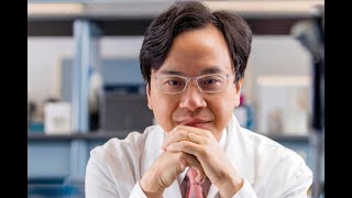
Dennis Lo describes his discovery of cell-free fetal DNA in maternal plasma and how it led to non-invasive prenatal testing with over 99% accuracy. He reveals that fetal DNA fragments are shorter than maternal DNA and carry a 10-base-pair pattern reflecting nucleosome structure. The enzyme DNase III shapes these fragments, and its signature appears in cancer cells too, offering a potential biomarker.
Chapters
Welcome to the ESHG Award Session. My name is Joris Veltman, and together with SBC co-chair Alex Raimond and ESHG President Maurizio Ghenuardi, we will be chairing this session and we'll start with the Mendel Lecture. The laudation for this Mendel Lecture will be given by Professor Maurizio Ghenuardi. Maurizio, the floor is yours. Thank you, Joris, and I'm delighted to introduce the speaker, of this Mendel Lecture. Each year, the Scientific Program Committee selects for the Mendel Lecture a distinguished scientist who has provided fundamental contributions to the field of human genetics. This lecture was established in 2006, and since 2009, it has been named the Mendel Lecture. This year, the awardee is Professor Dennis Law for his pioneering research on the biology and diagnostic applications of cell-free nucleic acids in plasma. Uh, he discovered the presence of cell-free fetal DNA in 1997, pioneering non-invasive prenatal diagnosis and providing the grounds for a turning point in prenatal medicine that now heavily relies on this technology. He also applied this approach to the detection of circulating tumor DNA by urine liquid biopsy. Dennis Lo is the director of the Li Ka-Shing Institute for Health Sciences in Hong Kong and professor of medicine and chemical pathology of the Chinese University of Hong Kong. He is a recipient of several awards, among which the King Faisal International Prize in Medicine in 2014, the Future Science Prize, Branch Life Science Prize in 2016, and last year he had his first virtual award at the Association of Molecular Pathology. This is the the second virtual award for Professor Luo. He's a fellow of several prestigious institutions, including the Royal Society, the US National Academy of Sciences, and the Academy of Sciences of Hong Kong. ESAG is proud that the Mandel Award will be added to this prestigious list and is honored to host Professor Luo, to whom I now leave the podium. Please.
I'd like to thank the ESHG for inviting me to deliver the Mandel Lecture this year. It's my great honor and privilege to deliver this lecture to this distinguished audience. The title of my talk today is Cell-Free DNA in Plasma: Coming in Different Sizes and Shapes. So this is my conflict of interest statement. So over the last few years, been a lot of interest in the use of extracellular DNA in plasma for a variety of molecular diagnostics. And two areas particularly stand out. On the left there, we have non-invasive prenatal testing for the detection of fetal DNA in maternal plasma. And the second is in cancer liquid biopsy. So my talk today, I'm mainly going to quote example from NIPT, but will also occasionally use examples in cancer. So first, for fetal DNA in maternal plasma, it's a phenomenon which was first described by our group in 1997, and we will show that during pregnancy, a fetus would release its DNA into blood plasma of the mother.. And we first used the Y chromosome from male fetus as a marker. So we noticed fetal DNA entered maternal circulation very early on in the first trimester. And actually by the 10th week of pregnancy, the concentration reaches some 15%. And then after delivery, the DNA is cleared very quickly. So in 2008, we developed a protocol whereby we try to non-invasively screen for risk of Down syndrome in a pregnant mother. So we do so by sequencing millions of DNA molecules in plasma, and then we map each of those molecule back to chromosome from which it come from. And then the bottom here, we can plot the proportional representation of different chromosome in plasma.. And as a result, in the first large-scale trial of this technology published in 2011, we showed that the diagnostic accuracy of this approach is over 99% accurate. And because it's so accurate, the test was rapidly introduced into clinical practice. And actually, to date, this technology is available globally in many countries around the world, and millions of tests are done every year. Now, for example, in China alone, 1/3 of all pregnant women actually undergo NIPT. So it is one of the most rapidly adopted genomic tests worldwide. So for the last few years, we've been very interested in studying the biophysical characteristics of circulating DNA, and one of those is size. Now for example, you measure the size of fetal DNA in the maternal plasma, you will see a distribution in blue here. And the x-axis is the size and the y-axis is the frequency. But on the other hand, if you profile the total DNA, which is mainly from mother, you got a distribution which is in red. So you can see from here that the fetal DNA is a little bit shorter than the mother's DNA. And also, instead of having a smooth distribution, you can see a series of peaks, one at 166 base pairs, and after that, Every 10 base pairs is another peak. So to understand what this peak all means, we have to remind ourselves how the long, 2-meter-long DNA is being packaged in the chromosome in our cells. It does so by interacting with a variety of components such as histone protein to form nucleosomes. So here we can see that the DNA DNA coiling around some core histone, and then there's a linker region, and then go into the next nucleosome. So it's eternal, this middle region is 146 base pairs, and the linker region is 20 base pairs, and the two together is 166 base pairs. So what we're proposing is actually this first peak is the core plus the linker, and this second peak is just a core with a linker. And interestingly, this is one of the fundamental difference between circulating fetal and maternal DNA plasma. In the fetal DNA, somehow lacking in the linker region. And then how about these 10 base pair periodicity? So we think it has to do with the fact that DNA is a double helix and is coiling around the core histone, and every 360-degree turn of double helix is 10 base pairs. So this is telling us some secrets or hints about how the DNA is being cut. So we've been very interested in studying and learning more about the players which are involved in the fragmentation of cell-free DNA. Now we think it's likely to be an enzymatic process, probably mediated by some nucleases. So if that's the case, one way we can tackle this is to actually knock out the nuclease gene selectively in mouse models. Now for example, you would take some blood from a wild-type mouse and then you create any Illumina sequencing library from it, then actually you would see a predominant band at 300 base pairs, which is 160 base pairs of the mononucleosome, size plus 140 base pair as the sequencing adapter. But if you look in the literature, then many people regard the DNase 1 to be a predominant nuclease in our blood. So when we knock out the DNase 1 gene, we initially expecting that we might see some sort of size change. But surprisingly, even when we knock out DNase 1 gene, you basically see the same mononucleosomal band. And actually we feel so surprised by this that we decide to actually document this in a negative report back in 2018. Now if it is not DNase I, then what other nuclease could play a part? So when you look at the literature, then one other nuclease actually come to mind, and it's a nuclease called DNase I-like 3, and some people also call it DNase III. Is gamma. Now interestingly, this nuclease actually likes to cut chromatin. Now because circular DNA is basically circular chromatin, so that might be a good candidate. Now interestingly, when we knock out DNase I-like III gene, then we start to see actually the dinucleosomal, tri-, or even tetranucleosomal peaks. When we do the double knockout DNase I and I-like strain, then basically the same sort of peaks. Now when we sequence the DNA molecules up to the limit of the Illumina sequencing, which is about 600 base pairs, you can basically see that the DNase1-like3 knockout mice have longer DNA molecules. So now having established DNase1-like3 gene to be a likely candidate in this fragmentation of certain DNA, we wonder are there some sequence motif that it particularly like to cut. So we decided to actually study the end motif of plasmid DNA. So remember that when we sequence plasmid DNA molecules, we have to polish it, and when the polishing is actually, we'll change the 3' end motif. So basically whatever we're measuring here is only the 5' end motif. So we decide to look at 4 nucleotides and then profile the frequency. Now if there are 4 nucleotides, then the possibility is 256. So if the whole phenomenon is random, then on average one of each motif would be present at a frequency of 0.39%. But interestingly, when we actually look at the motif frequency in wild-type mice, we find that actually some of the top motif is present at a ratio much higher than 0.39%, like this one, which is 1.51%. Now interestingly, when we knock out this DNase1III gene, then we can see the motif frequency changes a lot. And interestingly, actually the first top 6 of those motifs all start with CC, so suggesting that DNase1III likes to cut DNA molecules with the CC CC sequence. So in subsequent work, we decided to group all the 6 CC motifs together. Now in wild-type mice, the grouping of those 6 represent about 7.5% of all the circulating DNA molecules, which is quite a lot. Now you knock out the DNase I gene, then actually motif frequency doesn't change. But on the other hand, if you knock out DNase I-like region, than the motif frequency of those 6 halved. And double knockout is the same. So we wonder whether we might gain additional insight into this phenomenon by looking at a pregnancy model. And so we decided to create a pregnant mouse model in which we have female black 6 mice in which we knocked out both copies of the DNase1-like 3 gene. And then we mix those female mice with male Bubbsy males, which are wild type. So the fetus would be heterozygous, so one copy of DNA is one LIF3 gene, and one which is deleted. So assuming the situation in murine pregnancy is similar to human pregnancy, then we expect the fetal males to actually release the fetal DNA into the maternal plasma. Now just to remind ourselves of some of the basic numbers. So in a non-pregnant situation, as we mentioned just now, in wild-type mice, those 6 motifs represent 7.5% of all circadian DNA molecules. And then if we knock out DNase 1 Black 3 gene, then the motif frequency is half. But in pregnant mice, in which both the mother and baby have the same genotype, so wild-type, wild-type, and mutant, mutant, then the multi-frequency trend mirror that non-pregnant situation. So now come to the interesting bit, is that when you have in the pregnant situation, when the mother and fetus have different genotypes, so you can see in this case that the mother is knockout and the fetus is heterozygous, then you can actually see there's a partial salvaging of the multi-frequency. Now because the mice, the fetal mice, will have the paternal genotype, which is Babsy, so we can specifically look at certain DNA molecules which carries a Babsy genotype.. And you can see that in that situation, there is even further normalization of the motif frequency. So basically, the conclusion of this study is twofold. One is that the pregnant mice will release at least two things in the maternal blood. One is the DNase 1-like 3 enzyme, and this actually results in a partial normalization of a mother motif frequency. And the second is actually because the fetal mice is a source of this DNase 1-like 3 enzyme. So this enzyme will carry out some localized cutting of the DNA inside the fetal mice, and so that's why the mutational frequency of the mice is further normalized. So we think this work is interesting because it's the first link of nuclease biology and circulating nucleic acids. And also suggests that changes in pregnancy, in physiology like pregnancy, might affect it, and pathology like gene deletion or mutation might change it. But of course the data here is generated in a mouse model, so what about the situation in human subjects? So in collaboration with Stefano Volpi, Linda Hierarchi, and Boris Rassis, we have studied some cases of familial SLE, which is caused by DNase I-like 3 gene mutations. Now, so for example, if you look at control subjects, as Minnie mentioned before, the plasma DNA profile is really dominant by this mononucleosomal band with some dinucleosomal structure as well. As well. And if you look at the parents of those patients who have only one copy of the DNase 1-like 3 mutation, then the profile is very similar to normal. But on the other hand, if you look at the patients with two doses of DNase 1-like 3 mutations, they see that the size profile is very similar to those of mice, and you can see basically there is some serious amount of the long DNA. And I won't talk about this short DNA changes now. This has, from previous work, been shown to be due to the antibody effect of the SLE. Now when we look at the motif frequency, you can see for example in healthy individuals, you can see there's a lot of the CC motif there. But when you look at those individuals with 2 dose of the DNase I light frame mutation, you can see there's a massive reduction of those CC motif frequency. But on the other hand, if you look at SLU without this gene mutation, then it's very similar to normal, and so are autoimmune disease and also the parents of the patients. So when you actually plot the motif frequency out, you can see that in wild-type individual, then the motif frequency of those CC motifs approximately 10%, but whereas in the homozygous mutant individual, it's about 5%, so halving. So once again, very similar to the situation in the mice. So in other words, this effect can also be replicated in human subjects. So next question is, how about this plasma DNA end motif? Can we use it as some sort of diagnostic markers? So we have actually decided to take a look at cancer, and actually we survey a variety of cancer gene expression. And surprisingly, interestingly, in many cancer tissues, actually there's a reduction of DNase 1-like 3 gene expression. Now for example, in liver cancer, we've shown it here, this is a liver cancer tumor tissue, and this is an adjacent normal liver tissue next to the cancer. You can basically see there's a reduction in DNase 1-like 3 expression. So if that's the case, we would hypothesize that that reduction might result in a reduction of those CC motifs in plasma. And indeed, you can see that in the plasma of liver cancer patients, there's a reduction of the CC motif. So now with this advent of this CC motif approach to study plasma DNA fragmentation, we think that actually this is going to be a very powerful method to actually take a look at this nuclease-based fragmentation phenomenon in general. So we've shown that in DNase I-like III, but we think it's time to actually maybe have a second look DNase I because previously when we look at the size, we don't see any difference in the DNase I knockout. But how about multi-frequency? And also we can actually take a look at other nucleases like for example caspase-activated DNase and some people also call this DFFB. So DFFB stands for DNA Fragmentation Factor Beta. So the overall study study is quite similar to before. So basically we have wild-type mice and also mice in which each of those nuclease genes is knocked out. But in addition to what we studied before, we also add one more step, and that is we look at blood either freshly taken or which have been deliberately left in room temperature for 6 hours. And the reason why we do that is because from our previous work, we know that leaving the blood in room temperature for 6 hours will allow the DNA from blood cells to leak into plasma. So this allows us to see what happens to the newly released circulating DNA. Now because of this design of this structure, it's quite complex, so we decided not to look into the 256N motifs, but instead just to look at the single nucleotide motif. So just separate the plasma DNA into A, C, T, and G. And this is what we find. Now for example, you look at wild-type mice at 0 hour, you freshly take a blood, then you measure using sequencing and you can see that plasma DNA molecules with different end, A, C, T, and G, have this distribution. But on the other hand, if you leave the blood for 6 hours, then you can see that the frequency changes. So you can see that the newly released plasma DNA are more predominant in A and G, so there's two color. Now interestingly, when you knock out DNA, the DFFB gene, then this phenomenon is gone. So conclusion from this data is basically DFFB like to cut DNA in the A and G end. So we have something like this. Now you can repeat experiment with DNase I-like region and then you can find that it like to cut C more than T. Now you can also do the same thing with DNase I, but with DNase I we also add one additional step which is heparin because heparin will disrupt the histone binding which will make the DNase I activity stronger. And you can do the same thing for other nucleases. So basically by using this approach, we can actually work out some of the steps in the fragmentation process, and this has recently been published in the American Journal of Human Genetics. So up to now we talk about these short fragments of circulating double-stranded DNA, but one question is, is this double-stranded DNA a blunt-end entity, or can it be can some of the molecules be partially double-stranded? So in other words, would they have this sort of a jacket end? Now, if that's the case, to answer this question we need to develop technology which allow us to assay the jacket end. So one way we can do that is to polish this end by polymerase, and during the incorporation and repair, we can incorporate some marker which we can assay. For example, if your assay is a bisulfite-based sequencing, we can actually incorporate an unmethylated C, which will allow us to detect it. And the reason why that is useful is because most of the C in the CpG context in our genome are methylated. And so if there is a jacket end, then you see there's a reduction in methylation density. So we call this technology JAK-Seq, so analysis of jacket ends. Ends by sequencing. And interesting, when we do this, we find that actually plasmid DNA does have a lot of jacket ends. So you can see the jacket index is very high. On the other hand, if you have cellular DNA, which we fragment by sonication, then actually jacket ends are very low. Now apart from actually telling just the presence or absence of jacket ends and the quantity concentration, we can also measure how long the jacket end is. We actually find that, for example, about 12% of circadian DNA are blunt end, and the rest have jacket ends, and the jacket ends could range from 1 nucleotide to over 70 nucleotides. And also interesting, we find that in the pregnancy situation, the circadian field DNA have more jacket ends compared with maternal DNA. And actually this jacket end are more pronounced in the size range between 130 to 160 base pairs, so just in the subnucleosomal to nucleosomal size. And indeed this jacket index predominance in the fetal DNA allow us to actually use the jacket end to actually measure the fetal DNA fraction. So field DNA fraction is actually the number one determinant of the accuracy of NIPT. So question is, what is causing jacket hens? We think it's likely that it might be caused by Nuclease as well. So we once again actually studied this by looking at wild-type mice and also mice with Nuclease gene knockout. You can see that when we knock out DNase I gene, there's a reduction in jacket hens. Index in plasma DNA, but when we knock out DNase I-like region, there's an increase in the JAK2 index. So our working model is like this, so that the DNase I would actually have also quite a high probability of actually cutting just one strand of DNA. So in other words, it's actually one of the mechanisms whereby JAK2N is produced. So if we knock it out, then there's a reduction in JAK2N. Ends. On the other hand, DNase I-like III gene will actually more likely to cut both strands. So if you knock it out, then it will basically further unmask the effect of DNase I. So we believe actually jagged ends are an intrinsic property of plasma DNA, and fetal DNA have more jagged ends than background maternal DNA, and there's some potential with NIP, for example, can measure the field of DNA fraction this way, and we also highlight the role of nucleases in the production of jackknives. And this work was also recently published in Genome Research. So up to now we've talked about this circulating DNA being fragments of DNA, and the study has been referred to as fragmentomics, which is a term which was first coined to describe the fragmentation of peptides, but I think it's equally applicable to fragmentation of DNA. But now, if you think on the other side about integrity of plasma DNA, so how intact can it be? And of course I wouldn't be naive enough to think about a whole chromosome being in circulation, but how about a small one? For example, like that in the mitochondria. And of course mitochondria are the powerhouse of a cell. And has its own genome in the form of a circular mitochondrial molecule of 16 Kb. Now in previous work from our group, we have shown the presence of mitochondrial DNA in plasma. For example, you sequence it, the size of mitochondrial DNA in plasma is a little bit shorter than that of the nuclear DNA. But in this type of work, we extract the plasma DNA and then add on the Illumina sequencing adapter. And then we sequence. So by definition, whatever we're measuring is mitochondrial DNA which are already in a linear form. So the question is, are there circular mitochondrial DNA in plasma? So to answer this question, we have to add on some additional steps. Now for example, let's imagine there are both linear and circular mitochondria in plasma. So one thing we can what we can do is to get rid of the pre-existing linear mitochondrial DNA by using exonuclease V. And then after that, we can actually open the circle by, for example, restriction enzyme, like MSP1 shown here. And then we do the massive parallel sequencing, and then specifically looking for molecules in which both ends are flanked by MSP1 sites. Now to show you some result now, for example, if you have plasma DNA, no treatment of any of those enzymes, then the mitochondrial DNA percentage is very low, 0.001%. But now if you open MSP1, then the amount will increase 10 times. But however, if you first cut with the Exo5 followed by MSP1, then amount will increase about 10 to the 4th times. So those are basically circular mitochondria in plasma. But another way to look at it is that this is no treatment, and this time we open MSP1 and then specifically look for molecules which is flanked by MSP1 on both ends. You can see that in this context, basically exonuclease V doesn't do too much more additional enrichment. So in other words, both of those are circular mitochondrial DNA in plasma. So having shown that both linear and circular mitochondrial DNA exists in plasma, the next question would be, are they of different tissue origins? To answer that question, we can actually use the context of transplantation. So we look at liver transplantation and bone marrow transplantation, and then we can look for polymorphisms between the recipient and donor in the mitochondrial DNA. And then we use enzymatic tricks to look at, see whether those DNA are linear or circular. And interesting here, when we look at mitochondrial DNA of donor origin, in the liver transplantation context, actually most of those DNA are linear. But on the other hand, in a bone marrow transplantation context, then most of the donor-derived DNA is circular. So now we have a very interesting situation that in liver transplantation, then the donated organ would predominantly release linear mitochondrial DNA, but whereas in a bone marrow transplantation context, then the donor cells would predominantly release circular DNA. Of course, we can actually ponder what's the nature of this circular mitochondrial DNA. They could be anti-mitochondrion, which is in plasma, which has also recently been shown to be possible. And then another possibility is that those mitochondria could be protected by certain particles. But even at this stage, this discovery already has certain diagnostic implications. For example, in the liver transplant context, The concentration of liver donor DNA in plasma has been found to be a rejection marker. So rejection associated with increase in the amount of donor DNA. But here you can see that the linear mitochondrial DNA in plasma has a direct correlation to the donor DNA fraction, except that we don't even need to genotype the donor. And also similar technology will allow you to actually detects certain liver pathologies like liver cancer shown here. Now interesting, when we actually look at the number of mitochondria per cell in different tissues, we actually find that it varies a lot. And actually the one with the most mitochondria DNA per cell is the liver, and also certain muscles like heart muscles or skeletal muscle. So one corollary of this work is that the linear mitochondrial plasma might also be a marker for cardiac abnormalities or skeletal muscle abnormalities. But one question which has not been answered in the field is, does fetal mitochondrial DNA present in maternal plasma? Now this is a difficult question to answer because of the maternal inheritance of mitochondrial DNA. So recently we have a study in which we actually look at surrogate pregnancy, and because the pregnant mother is biologically unrelated to fetus, then we can actually differentiate the fetal and maternal mitochondrial DNA. And by using enzymatic steps I described to you just now, we can actually tell whether the fetal mitochondrion present in the maternal plasma and what topology they are, so it's either circular or linear. And interestingly, we can see that the fetal mitochondria in maternal plasma are mainly linear. On the other hand, the mother's side, the mother's own DNA, there is actually a substantial amount which is circular. But of course, that is also consistent with bone marrow work because we know that in background maternal DNA, it has a large hematopoietic component. And this work was recently published in Prenatal Diagnosis. I think this work shows that it's interesting to actually look at the topology of plasma DNA. So recently we've also extended this work to look at non-mitochondrial DNA, which is circular in the plasma, the so-called ECC DNA. So that is another development in its own right. So in summary, hope I've convinced you that plasma nucleic acid represent a treasure trove for molecular diagnostics. With it, the global era of NIPD is already with us. And today we'll touch on some of the emerging concepts in the field, including fragmentomics. We talk about fragment size and motifs. And then we also talk about plasma DNA topologics, talk about circular and linear DNA plasma, and also talk about this intriguing link to nucleus biology. And recently we have summarized some of our thoughts in this area in this review in Science. And finally, I'd like to thank individuals from my group for generating the data which I've shown you today, and thank you very much for your time.
Thank you very much, Professor Luo, for this extraordinary history, exciting science. And we have a few questions. And the first one is by Alexander Hoisin. Who states, amazing work and lecture. Thank you so much. Do you know the DNase enzyme concentrations, if they are similar in other body fluids, urine, tears, cerebral, spinal fluid, and if the motifs are similar or different?
So thank you very much for a very interesting question. We have actually started to look in this phenomenon in urine. Actually, in urine, the highest concentration of nucleus is DNase I. And actually, urine DNA have a very different pattern. It is actually a predominant of T and N. So I think that this is actually potentially an area for further exploration. For example, we find even for example, bladder cancer, also have aberrant nucleus expression. So the DNA from the bladder cancer cells also are different in the urine.
So thank you. Thank you for your reply. And second question by Peter Bram Thijen, who also writes great work. Can we observe a change in the fragmentation patterns in plasma between cancer patients and controls? And this is essentially the— and is the CN frequency an early biomarker for cancer?
Yes. So actually, we profile the gene expression of different nucleases in tumor, and actually over 85% of different tumor types actually have reduction in DNase 1-like 3 gene expression. And actually we have profiled probably maybe 7, 8 different types of cancer plasma and they have actually reduction with the DNase 1-like 3 associated motifs. So I think that with early detection, we're basically trying to combine different signatures of a cancer together to try to push up the sensitivity. So every single little bit will help, including motif and other markers like epigenetics or mutations.
So thank you. Thank you. Juliana Cerqueira asks, as CF DNA maintains the epigenetic signatures of its tissues of origin, do you think that your work might have insights on how to track inherited transgenerational epigenetic marks in the tissues specific manner in humans?
Okay.
So this is a very complex question that opens many other questions. So we can't answer in one go except to say that we have actually recently surprisingly actually showed that the nuclease knock-out model actually would change the DNA methylation status of plasma DNA. Actually, even knock out DNase 1-like 3 gene, the plasma DNA become hypomethylated. And of course, just now I told you that there's reduction in DNase 1-like 3 expression in many tumor and tumor is hypomethylated. So that is a link. And then as you regard to the transgenerational transmission of those epigenetic marker, we have shown that in NIPT context, you can basically detect the methylation status of imprinted genes in a baby. So the answer is yes. And presumably if you have some diseases like diabetes, if they have some DNA methylation changes, then maybe we can also look at that.
So thank you. Thank you. We have a few other questions. We have a question from the co-chair, Joris Veltman.
Please. Loris.
Thank you. Let me first congratulate Professor Lo, of course, with the Mendel Award, and thank you for a great talk. I was just wondering, the reduced expression of the DNAs like 1, like 3 that you see in cancer, can you explain this? Does this benefit the cancer, and how does it achieve that?
Actually, we have been pondering with that. And at this moment, I don't know the answer, but we are collaborating with some other cancer biologists who actually suggested some very intriguing finding that this reduction in DNase 1-like 3 could make the cancer resistant to certain treatments. So hopefully in the audience, if there are cancer experts, maybe we can work more on this.
Thank you. Fascinating.
Thank you.
This is a very interesting hypothesis indeed. Sophia Derdaq asked your opinion about cell-free RNA.
Thank you. We have also looked at cell-free RNA in plasma, and one of the latest is that we use single-cell transcriptomic of the placenta and also of cancer. To actually correlate with what is happening in the plasma. And using this approach, for example, we can identify which type of trophoblast is specifically associated with the release of cell-free fetal DNA and what happened in some diseases like preeclampsia when we find one particular type of trophoblast have aberrant gene expression. So, so I think this is a fruitful area, but I have to warn that this technologically is quite tough because the RNA is really quite unstable. So, but nonetheless, I think it's worth pursuing.
So thank you. Your lecture has elicited a lot of interest. There is another question from Ines Zutter, uh, who asks if there is a correlation with proliferation status of cells, fetal tumor, etc., versus slow turnover.
So I think that, I mean, if you look at the cell turnover, of course, there's a life, birth of cell and also death of cell. So I think that basically this cell-free DNA phenomenon is a measurement of a death of cell. And also I think the nuclease mechanism is basically a type of clearance mechanism.. And so basically with this technology I described to you just now, it will have given us some tools to study this cell death and how well the body is clearing that.
Yeah, thank you. And finally, Laila Al-Khattabi asks that since plasma DNA is a mixture of DNA from different cell types, do you think that it could be a good source to look for mosaicism?
Yes, it is a very good way to look at mosaicism. For example, we have looked at a similar situation. We have looked at tumoral heterogeneity. So we find that if you have a tumor of multiple clones, basically plasma will represent those clones at the concentration in which they are present in the, in the tumor. So the answer is yes. And of course, recently, some interesting work talking about in the placenta, there's also a high frequency of de novo mutations. So I think you can also use the mother's plasma DNA to look at that phenomenon.
Thank you. With reference to the question posed by Ines, I, and also to the somatic mutagen, question. I was thinking, do you have an idea of the contribution of different tissues to cell-free DNA, since this could be an important source, a way to know if you can detect mosaicism?
Yes, thank you. So we have developed a technology whereby we profile about 12 different types of somatic tissues and we look at the DNA methylation status and then we use genome-wide bisulfite sequencing of plasma DNA and then to use the convolution to calculate what tissues produce what. And so basically the answer is just over half of the DNA is from hematopoietic cells. And then you have maybe 1 or 2% from the liver. And then you have some other components, and some other components depends on what disease the person has.
Yeah. So thank you very much, Professor Luo. Unfortunately, we need to come to an end because timing is strict. We need to go to the next presentation, next lecture. So thank you again and congratulations for the award. And hope to see you again at the European Society of Human Genetics. Now we can move to the other session.
More from Joshua







Scientist Stories: Hans Waterham, The value of phenotypic assays in peroxisomal disorders




Scientist Stories: Joel Zlotogora, Genetic carrier screening programs for reproductive purposes