Scientist Stories: Donna Farber, Human Anti-Viral Immunity in Tissue Sites
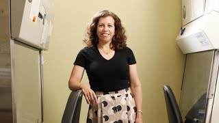
Dr. Donna Farber (Columbia University) presented a lab‑meeting webinar on human antiviral immunity with a focus on tissue‑resident memory T cells (TRM). She described the anatomical distribution of immune cells, emphasizing that most lymphocytes reside in tissues rather than blood. Mouse studies showed lung TRM (CD69⁺, CD103⁺) are sufficient for protection against lethal influenza and can mediate cross‑strain immunity; intranasal live‑attenuated vaccines generate robust lung TRM, whereas injected inactivated vaccines do not. To study human tissue immunity, her lab partnered with a New York organ‑procurement organization, collecting multiple tissues (bone marrow, spleen, lymph nodes, lung, gut, etc.) from >500 donors of all ages. Phenotypic mapping revealed that most memory T cells in mucosal sites are TRM, with rapid acquisition in gut, slower in lung, and sex‑linked differences (more lung flu memory in males, more CMV memory in females). Virus‑specific analyses showed influenza‑specific TRM concentrated in lung and lung‑draining nodes, while CMV‑specific cells were dispersed, especially in bone marrow. Pediatric donor data confirmed early development of gut TRM. In severe COVID‑19 patients, paired airway washes and blood samples demonstrated that higher airway CD4‑TRM frequencies correlated with survival, whereas elevated airway myeloid cells and chemokines (CCL3/4, MCP‑1) correlated with mortality. Post‑infection SARS‑CoV‑2 memory was mapped using AIM assays, revealing lung‑and‑draining‑node TRM and follicular helper cells, with limited circulating memory. Dr. Farber discussed vaccine implications, noting that injectable mRNA or protein vaccines mainly stimulate lymph‑node responses and antibodies, while live‑attenuated or intranasal delivery may be needed to elicit protective lung TRM, especially for immunocompromised individuals. She also addressed waning immunity, unknown protective thresholds, and the need for tissue‑centric studies to guide booster strategies. The talk concluded with acknowledgments and a preview of upcoming work on vaccinated donor tissues.
Chapters
It's just 10 o'clock. We will get started. Hello, and thank you to everyone around the world for joining us for this week's lab meeting. My name is Kristin Aboud. I am the science editor at the Human Vaccines Project, and I will be your moderator today. Since our last lab meeting a few weeks ago, cases of the Delta variant have continued to surge in many parts of the world. In the US, cases are more than 300% higher than this time last year, And children now account for more than a quarter of cases nationwide, which is particularly vexing for parents as school resumes. Globally, some leaders, including the director of vaccine research and development at CEPI, are warning that new vaccines will be necessary in the fight against COVID-19. And while there are numerous vaccines in development, there are substantial barriers to testing their efficacy including the limited availability of licensed vaccines to which new vaccines must be compared in clinical trials. Meanwhile, there are still concerns over the potential for waning immunity in those vaccinated against COVID, which brings us to the importance of today's lab meeting. However, before we begin with today's presentation, just one note. The information presented today includes some pre-published data that is currently under peer review. At the request of our speaker, we ask that you not reproduce or disseminate the data presented. This session will be recorded and made available on our website and our social media channels for you to review. With that, I am very happy to introduce today's speaker, Dr. Donna Farber, the George H. Humphreys Professor of Surgical Sciences and a professor of microbiology and immunology at Columbia University. Dr. Farber's research focuses on antiviral immunity and human immunology. Her laboratory originally identified a specialized subset of memory T cells that take up long-term residence in tissues such as the lung. She discovered that these memory T cells mediate optimal protective immunity to respiratory virus infections. These findings led her to establish a major initiative in translational immunology to study human tissue immunity and its development from infancy through adulthood in multiple mucosal and lymphoid tissues from organ donors of all ages. Dr. Farber leads the NIH NIAID-funded program grants on human immunity antiviral responses and is part of the Human Immunology Project Consortium. She has over 150 publications, is a fellow of the AAAS, and has served on advisory committees for the NIH, the American Association of Immunologists, and multiple editorial boards. During the presentation today, please send me your questions using the Q&A function in Zoom. I will ask our speaker a broad selection of your questions after the presentation, and we will have about 25 minutes for discussion. It is my pleasure to now introduce Dr. Donna Farber. Dr. Farber, you can go ahead and share your screen.
Thank you very much. Thank you for the invitation to speak to all of you today on our studies on human antiviral immunity and tissue sites. Just want to make sure you can hear me okay.
A little louder might be better, but we can hear you.
Okay. So our immune system is distributed across tissue sites and also in circulation, but most of our immune cells are localized and are residing in multiple tissues throughout our body. So we have specialized tissues, so lymphoid organs, which, which have many immune cells. So really most of the immune cells in terms of lymphocytes most of the numbers are in lymphoid organs, and these would be primary lymphoid organs like bone marrow and thymus, and secondary lymphoid organs, which would be spleen and multiple lymph nodes throughout our body. And humans have an estimated 700 lymph nodes that are localized all throughout our body. We also have immune cells that are localized in our mucosal and barrier sites, including lungs and intestines, skin, a lot of— all of your mucosal surfaces. These are specialized, mostly tissue-resident cells that localize in specific structural niches. So here we have cells around the airway and the alveoli of a lung and in the lamina propria and intraepithelial region of the intestines. And then throughout the body, of course, we have blood, and there's many immune cells in blood that are circulating in blood, but these are not necessarily representative of what goes on in tissue. So if you go down and look at what are the different subsets of immune cells that are in circulation and also resident in tissues, for both myeloid cells, which include macrophages and granulocytes— macrophages and dendritic cells, as well as granulocytes, there are tissue-resident macrophages that are really adapted to each individual tissue site, and also tissue dendritic cells, and then there are circulating versions of these cells, but they're not the same as that are in tissue. So a monocyte would be kind of a circulating version of a macrophage, but it's quite different. And then you also have some dendritic cells. There are not many, but there are some that are circulating. For lymphocytes, you also have circulating subsets. So we have many different kinds of lymphocytes that are circulating: T cells and B cells, plasma cells, and K cells as kind of an innate type of lymphocyte. And these are both memory cells and naive cells, and I'll go into that a little further, a little in more detail. These are in circulation. However, there are also tissue-resident lymphocytes. There's tissue-resident memory T cells. There's tissue-resident innate cells such as NK, innate lymphoid cells such as NK cells, innate lymphoid cells. And there's even tissue-resident B cells. So for both lymphocytes and myeloid lineage cells, you have many cells that are in tissues and then cells that are in circulation, but these are the minority of immune cells, and they're not the same types of cells that are in your tissues. So just to drill down, I'm going to be talking really focusing more on T cells, almost exclusively on T cells in my talk today. And just to drill down, what are the different types of T cells and how are they distributed in different— in between certain circulation and tissues so that we know that T cells emerge out of the thymus. So this is where T cells develop. They come into the periphery as naive. So they have a T cell antigen receptor that's, that's clonally distributed, but they're naive. They've never been activated. These naive cells mainly populate lymphoid sites, so spleen and multiple lymph nodes. And it's there that they will meet the antigen and become activated. And this will be by dendritic cells, say dendritic cell that's been infected with a virus, where it's carrying viral epitopes, and present the antigen in conjunction with the MHC molecule here to the naive cell that has the antigen-bearing T cell receptor. And it recognizes that complex of that peptide epitope within the MHC molecule, as well as a second signal that's expressed by the dendritic cell that's induced by an infectious state. So this two-signal recognition leads to naive cell activation. And naive cells will become activated, and they will clonally expand and differentiate to effector cells. And these effector cells acquire the ability to produce different types of cytokines. And there's all sorts of effector cells. There's different kinds of helper effector cells like Th1, Th2, Th17. There's cytotoxic T cells for CD8 cells. So I'm just talking about this in a broader way. But these are sort of cytokine effector cells. These will leave the lymphoid sites. They're going to migrate through circulation and go to different target tissues. And it's there that they're going to coordinate that clearance of the pathogen because that's what their job is. And then after that pathogen is cleared, most of these will die. Most of the effector cells will die. A small proportion will then become— will persist and become different kinds of memory cells. And these can be either effector memory cells. So effector memory cells, again, through circulation, these effector memory cells can migrate both to lymphoid tissue and also to different peripheral sites, or central memory, which express the lymph node homing receptor, and these will go back to lymphoid tissue. Now what the effector memory can do, though, is they can go to many different tissue sites. They can go to lungs, intestines, I list them here, and a proportion of these effector memory will actually take up long-term residence in these sites and don't go— and not go back to circulation, and these will become tissue-resident memory cells. So they're not circulating, and they can be in peripheral tissues, and also they can be formed in lymphoid sites. They have distinct phenotypes. They're retained in tissues. And they have a distinct transcriptional profile. And the phenotypic markers that I'll refer to that are kind of the canonical markers of tissue-resident memory are CD69. That's an early activation marker for T cells but also has tissue-retention properties. And another marker, CD103, mostly expressed by tissue-resident CD8-positive tissue-resident memory cells. This is the alpha E integrin. And it interacts interacts with E-cadherin on epithelial cells. You'll find CD103 expressed by TRM in sites like the intestines or the lungs where they're going to interact with epithelial cells. So what do TRM do? So TRM were initially discovered in mouse models. And we had initially identified these in the lung. There's other groups like Dave Massopus at the University of Minnesota who identified them in the gut and multiple mucosal sites. The group, Frank Carbone, and others, Laura McKay and others at Melbourne, had identified them in the skin and other tissue sites. So TRM, but here I'm showing you TRM in a mouse lung that take up residence after influenza infection. So influenza infection of the mouse, you simply infect it intranasally. It's very much like human infection. It's localized to the lung. It's cleared in the lung. And after it's clear, you will find these tissue-resident memory that express CD69 and CD103. Congregating around the airways. And this is for both CD4 and CD8. And we generated mice that only had— so the only memory they had to flu was lung tissue resident memory, and the— versus mice that had, again, only T cell-mediated immunity, but T cell memory to flu in multiple tissue sites. So the mice that had only lung TRM were fully protected from a lethal challenge, whereas mice with circulating memory were not, and mice with no memory T cells were also not protected. Protected. So what this showed was that lung TRM were actually sufficient to protect against a lethal challenge with flu. And we've gone on to show that, in fact, this is just showing you a diagram, in that lung TRM can protect, can mediate cross-strain protection in flu. So you know how flu varies every year, and so the antibodies that you generate to one type of strain don't protect you to the other type of strain that may come along. In another year or another season. But T cells recognize invariant epitopes of flu and can mediate cross-chain protection. And in fact, these lung TRM will mediate cross-chain protection to multiple strains of influenza. And they do so by— and this is just showing you a diagram. So if you have TRM in the lung, when you challenge in the recall response, when you challenge that mouse with flu, the TRM will begin to proliferate in the lung. They will also recruit and have this enhanced recruitment and proliferation of T cells from the local lung-draining lymph nodes. This would be the mediastinal lymph node in the mouse. So it's this interaction of the lung and the lung-draining lymph node, this very localized kind of protection is really important for T cell-mediated protection. It's very local. And what about vaccines? So we know that for vaccines in mice, so it's hard to know whether you have tissue-resident memory in humans. You really can't look at the lung, but we can do this in mice. So we did experiments. We took the clinical, the human vaccines that are given to people, the flu jab, circulating inactivated influenza vaccine, and the live attenuated Flumist, which you deliver intranasally. So we took these and gave them to mice, and we asked, do they generate tissue-resident memory? And this is showing you lung. This is lung T cells. This is a flow cytometry plot. This is a memory marker, CD44, and this is a TRM marker, CD69. So what's in this box would be the TRM. And what you can see in the unvaccinated mouse, you don't have any TRM in the lung. The mice vaccinated— this is IP, but we also did subcutaneous with the inactivated virus. Inactivated vaccine, again, had a little bit, maybe a little bit of CD8 TRM, not much. But the mice that received the live attenuated attenuated through the nose had a nice population of tissue-resonant memory, and you can see here through the vaccine. And if we looked at the influenza-specific CD8 cells in the lung, which can be protective, we found that you only saw them— this is now the influenza-specific response— you only saw them that were generated from the intranasal virus vaccine. So again, this live attenuated intranasal delivery was able to generate TRMs specific for the virus. And so when you challenge that mouse and we treat them with a drug that actually blocks circulating responses, so we're only looking at that tissue response, and we found that mice that had either were previously infected with flu or that received the intranasal virus vaccine were fully protected from a heterosubtypic challenge. This is a different strain of mouse— of flu. So this would give you that cross-chain protection. They gave it as well as the mice that were vaccinated intranasally were protected just as well as if they had previously been infected with flu, whereas the mice that got the inactivated vaccine were not as well protected, and mice without— vaccinated mice succumbed. So TRM can be protective, but knowing that— and so we know that TRM actually are protected in many different tissues and in many different mouse models from work from a number of groups have shown that in the lung, TRMs can be protective to a number of viruses, to bacteria, to allergens, vaccines, tumors, intestines. In the skin, they can be protective, the TRM, even in the liver, to parasites, and other, uh, in genital surfaces. And so the TRMs are really important for T cell-mediated protection, but what about humans? So humans, of course, we're largely limited to looking at the immune response in circulation, which is really good for looking at antibodies. So we know, and there's been a lot of work done, and especially for SARS-CoV-2, it's probably the most studied antibody response ever, I would say, maybe HIV, that we know that you get neutralizing antibodies, that this can be a correlate of protective immunity, but we don't know for T cells. And so what's circulating in T cells isn't necessarily representative of what might be protective. So in the mouse model, we only had circulating T cells to flu. They weren't protected. So you could see that they had a T cell response, but it wasn't a protective T cell response that was in the tissue. And if you look at the numbers, there's been estimates that only about 2 to 3% of human T cells are in blood. So even just any circulating— any T cell in the body, most of it's not in blood. And of course, TRM are not in blood. So about a decade ago now, over a decade ago, we set up a tissue resource so that we could start to look at What does human tissue immunity look like? What's in our tissues? We've been focusing on T cells. I'm going to show you data on T cells, although we've published data on other immune cells, and we're looking really more globally now at the complete immune response in tissues. But what we have set up is we've set up a collaboration with our local organ procurement organization to get multiple tissues from human organ donors. So these would be brain-dead organ donors whose next of kin have consented to have their tissues donated for life-saving transplantation, at this— at the same time, you can also consent to have tissues or organs used for research. So we have an arrangement with that organ procurement organization, which coordinates organ donation in our New York metropolitan area, to have access to all research-consented organ donors. And so I have an on-call surgeon in the lab who goes to the site where the organs are being obtained for life-saving transplantation, and after all the teams have obtained donate the organs for their patients for life-saving transplantation, we come in at the end and we get all of these different tissues. So it allows us to get multiple tissues from one person so we can unambiguously define how tissue immune responses are different than those in circulation and really start to understand more about what blood is teaching us. And we also— all donors, organ donors, these are free of cancer, they're free of mostly free of chronic disease. They're of all ages of life. So, they're really very representative of sort of the healthy population walking around. So, we get a view and a vision of immune health that we couldn't normally get. So, this is what the diagram is. So, we get, you know, bone marrow. We get all these different tissues. And this is what they look like. So, you can see that, you know, unlike a mouse where every mouse is really looking pristine, humans is a little bit different. So this is— I'm just showing you that we get a lot of lymphoid tissue, spleen, lymph nodes, and the major mucosal sites are lung and intestines. It's just showing you some pictures. This is the ileum. That's a Peyer's patch. That's a lymphoid structure in the intestines. And you can see that the lungs are kind of a little black spot. This is not a smoker. Only about 10% of our donors are smokers. So rate of smoking really gone down. And here are the lymph nodes. I'm just going to show you how localized immune response is. Looking at the anatomy. This is a mesenteric lymph node that drains the gut. Looks like a big mouse lymph node full of lymphocytes, full of white blood cells. It's white, it's translucent white. Here's a pancreatic lymph node. Lung-draining lymph nodes are always black. And they're black because, again, I'm not a smoker, but because of the particulates and things that we inhale. And one function of our lymph nodes is also to filter out impurities. So you can see just how localized your immune response is by just seeing and tracking where what we're taking in through the lung is caught in the lymph nodes. So again, this localization. And here we've gotten over a decade and a half now. We've gotten tissues from over 500 donors of all ages of life. This is just showing you our age range. So we really look at the immune response in space and time. We do have a bias of males because it's mostly in the younger years. These are mostly victims of traumatic events that cause them to to be brain dead, and men are more likely to die of traumatic events in younger years. In the older years, it's mostly cerebral vascular stroke, and that's even. So just— I'm going to show you some brief data on— just to give you an overview of what T cells look like in tissues and what kind of questions we can ask by studying these tissues. So again, and we always have to be cognizant of unlikely what you can do— you can't do in mice is really look across the lifespan because really we're very different at all ages of life. Childhood is when you're starting developing memory, everything's naive. In most of our adult years, we maintain health and homeostasis and infections in most of the adult years, and at some point it starts to break down and we have immune senescence. So we wanted to see how T cell memory overall is maintained in human tissues as you know, how much is resident, what proportion is resident or circulating, and then start to drill down and understand virus-specific immunity in tissues. So what is the full heterogeneity? So if you look, you think about it, if we're only sampling blood, we have a very incomplete picture of what our antiviral immunity is. So we wanted to look at kind of the full heterogeneity of antiviral T cell memory across tissues, age, and sex. And then what we're doing and how we're applying what what we're learning in tissues in different sites to understand the immune response to SARS-CoV-2 infection. So just to show you overall, and again, most of this is all published, so we initially kind of went through and just did phenotyping mapping. So we asked, like, where were the CD4 cells? What kind of composition do you have in these tissues? And what we found, and we looked at blood, we looked at blood to gut, so if you look blood, spleen. These are different lymph nodes. The LN is lymph nodes. So an iliac, a lung-draining lymph node— here's a lung— mesenteric lymph node draining the intestines. And what you can see if you look at the different subsets— this is effector memory and central memory and naive. Those are kind of the major subsets for CD4 cells. Most of them are memory in mucosal sites. They have about half in the lymphoid tissue. And naive cells are mostly— and this is in a adults— this is adults between 20 and 70 here— you find naive cells in the blood and lymph nodes, and lymph nodes mostly, and not really in mucosal sites, much less and fewer in spleen. And again, central memory is like naive, mostly lymphoid sites. And for CD8, it's pretty much the same, except you don't really have central memory CD8. It's kind of a different phenotype, but you do this circulating population. If you look at all the effector memory cells, which are really the majority of cells in most sites, and you ask what about TRM phenotype, which would be either CD69 alone in the gray or CD69 and CD103 expression, you know, most memory for CD4 and CD8 is TRM in practically all tissues. So most tissue memory has a TRM phenotype, anywhere from half to pretty much all of it. And particularly for CD8, particularly in the intestines and the lung and some exocrine sites, the majority of memory is TRM and co-expresses CD69 and 103. And then we can look over age. So this is just summarizing a lot of work by site and age and what is the T cell composition. Again, this is the overall T cell composition, just saying what are the phenotypes, what's there. And what we can see is that each site has its own unique composition. And we find this for all immune cells, that each tissue site has its own kind of tissue intrinsic composition of immune cells, whether it's blood or intestines and/or different lymph node sites, lymphoid sites. And that in terms of— at birth, it's mostly naive. So T cells are mostly naive except in the intestines and lungs. And you acquire memory at different rates and different sites. So intestines, it's the most rapid acquisition of memory T cells. And most of these are TRMs. So by the time you're a child, school-aged child, you really are mostly memory in the gut. And the lung is a little bit slower, again, mostly TRM. And spleen, lymph nodes, probably the slowest to acquire memory and has the least, lowest proportion resident memory, although you have other kinds of tissue memory in lymph nodes. And then blood, you have again, you acquire memory, mostly memory over adult, no TRM in the blood. But what about virus-specific cells? So I'll start out, we'll look at some ubiquitous viruses. So I think in order to understand, say, SARS, you need to understand, well, what about our memory to ubiquitous viruses? And so we've looked at virus-specific memory T cells, and this is now CD8. To both influenza and cytomegalovirus. So cytomegalovirus is a persistent virus that once you get it, you kind of never get rid of it. About 60% of the US population is CMV seropositive. And then influenza, of course, is respiratory, a ubiquitous respiratory virus that literally everybody has seen and many times have been vaccinated as well. So you can see this is just a representative person. The way we pick out these T cells is by using these multimer reagents. So one advantage of using organ donors is you have all the HLA type. So remember, the T cell receptor recognizes the peptide epitope in the context of an HLA molecule. And humans have multiple HLA alleles. But about 40% of us have HLA-A2.
So—
and we were able to identify who had HLA-A2 in our donors. So we use these multimer reagents. That have the peptide epitope or the viral peptide epitope with the HLA, the appropriate HLA, and a fluorescent molecule so we can actually look at this by flow cytometry. And so this is one person. This is multiple tissues from one person. And you can see that for flu-specific, it's mostly in the lung and the lung-draining lymph node. This is LLM, lung-draining lymph node, in this individual. Whereas CMV is dispersed specific T cells in the blue box here. Here are dispersed across multiple sites: bone marrow, spleen, lung, different lymph nodes. They're not much in the gut. I'm not showing you this, but you don't find a lot of T-cell specific for these viruses in the gut. And this is just showing you overall from multiple individuals. And again, you see that most flu and CMV are in sort of these big four: blood, bone marrow, spleen, and lung. You also have some of these in the lymph nodes. And if you look at where the most— and so this— but there's a lot of variation between people. So unlike the phenotype, where there's a lot of consistency, it's really about the tissue, the type of memory cells, and how many memory cells you have and their TRM. For virus-specific cells, it really differs between people in terms of the proportion. You can see like there's a big range here, um, where they are. So for— but for influenza, we found that about a third of the donors that we looked at, about 60 donors, had most of biases in the lung, whereas for cytomegalovirus, we found that about a third of the donors had sort of dominant responses in the bone marrow, and that might reflect, again, cytomegalovirus can be maintained and persisting in hematopoietic cells, whereas, you know, of course influenza is originally infected in the lung. So there is those trends where, where you store your memory T cells has to do with the pathogenesis of virus infection, where it's going to be, where it's maintained. But then we started to drill down into what kinds of memory, what kinds of subsets, and how they change over age. And there's a lot of data here, so don't worry about the individual data points here. Just to show you that in terms of the memory, the proportion of memory— so this is like TEM— that's really— it differs by virus, it differs by site. You can see that it changes with age. So like it changes with age, flu-specific cells, you get increase in the sort of circulating population with age in the bone marrow, but not in other sites, where CMV subsets don't change. But the other thing is that there is this difference between male and female. So you have more memory in sites like spleen and lung in males to— to flu, and you have more circulating memory to CMV in females. So being male or female, as we've seen, that there is this different response for like SARS-CoV-2. There's actually a different memory type of way that you're going to store your memories can differ between male and female for reasons that we don't understand, but it's a phenomenon that we see when we look at memory ubiquitous viruses. And then what about TRM? So TRM is very much like a mouse. So the TRM are really found to influenza, and where you find TRM to influenza in humans is in the lung and a little bit of the spleen and the lung draining lymph nodes. So very localized. So lung and lung draining lymph nodes and spleen being kind of that repository for a lot of— you do find a little bit in in the bone marrow. Okay. Whereas for CMV, you don't generate a lot of tissue resident memory to CMV. Again, CMV is a persistent virus. It's going to have that kind of low-level, either latent or low-level infection, and that's really circulating. And again, what about changes with age? So it doesn't change that much with age in the lung, pretty stable. But in lung draining lymph node, the proportion of TRM to flu does go down. So it means you have a little bit less memory in your lung-draining lymph node to flu. So that could, could explain some decline in immunity. And then between kids and adults, it's also really different. So most children don't have CMV persistence, but if you compare flu responses in children versus adults, what's really interesting— and these are— now we have another source where we get pediatric tissue again, from pediatric organ donors are very rare, and that's a good thing. But there are some accidents, and we have a national network that we're part of that's run out of University of Florida, which gives us access where we get the individual OPOs from all over the country to send us tissues. So we've looked between 2 and 11, and we do find flu-specific cells. And, you know, again, mostly in the lung and the lung-draining lymph node, not so as this would be the gut-associated lymph nodes. Again, very localized in children, and this is an epitope that they would only see with infection. But the overall level, if you compare to adults here shown in gray, is lower. The overall frequency in children of flu-specific cells is lower than adults, and that probably reflects that children haven't seen flu as much as we have. So we're exposed to flu probably every year, whether you get sick or not. And you build up that immunity. So you're— again, you're building up immunity in tissues. So children have it, but it's less, and they also have less TRM. They have less TRM in the lung than adults. So again, suggesting that you need multiple exposures to build up that full resident immunity. And this is just a summary of what we found in terms of the heterogeneity of that immune response and trying to understand what determines memory and how can we dissect this heterogeneity of memory. And so a lot has to do with the virus, and the virus is going to really determine what kind of memory you generate and where you generate it. And so the tissue will really determine what kind— whether you're going to generate resident or not, or whether you have a propensity. And then there's all these other different factors that go into, including sex, age, in terms of— and the tissue, what kind of subset you get. And then I didn't show, but function is also largely determined by tissue. So now what about— so I'll end with just telling you what we've been doing, how we've been trying to understand the immune response to SARS-CoV-2 infection by looking at tissue sites. So we know that in terms of the acute infection that you can either have a mild infection that's resolved, or this can go on to severe infection that can last for a really long time where you get a lot of lung pathology. The mild infection is resolved, we know you're going to get recovering, that there'll be memory. The severe, it's really understanding what's happening during acute and what's happening during memory. So how can we do that and look at the tissues? So again, we're not going to be able to acquire, access tissues. I mean, you can from autopsy, but that's really late stage. So we're trying to do this during acute response. So during the height of the pandemic, which in New York is really over a year ago, so that was really spring 2020, is really when we had the most cases here in New York City. And whatever has happened since then, whatever surges that we've had, doesn't compare at all in terms of the number of hospitalized here at New York Presbyterian Hospital, um, in Upper Manhattan. It just doesn't compare at all to what we had during that first surge. So what we did is recruited patients intubated for severe COVID and acute respiratory distress syndrome in New York City. These were age 14 to 84. We recruited 15 patients where we got paired airway samples. So when they're intubated, they wash out the tube every day with saline. And that we knew from prior work where we had worked with airway washes that this gives you a very nice representative of this respiratory environment. You can get TRM. You can get all sorts of lung cells from that. And we had a saline wash and paired blood. And we did this every day while they were in the hospital for 7 to 10 days in 15 patients. We profiled by flow cytometry, cytokine, and single-cell RNA-seq. And this was published earlier this year. And this is done with many collaborators, including Matthew Baldwin and Tom Connors. These were the physicians who were taking care of the patients in the ICU. And this is just showing you flow cytometry. We did a high-dimensional phenotyping to get all of the immune cells, including myeloid cells. This is a UMAP. So this is just a way of looking at very high-dimensional data in two dimensions. So each blob, it sort of is a cluster of cells. So we were able to distinguish myeloid cells, CD8, CD4, B cells, and innate cells. And what I'm showing you here is just each day, all of the individuals, their airway samples from COVID each day of sample collection, and the blood, and just to compare it. So, and this is also control airway from a non-infected person and blood. And so what you can see is mostly the myeloid cells are in the airways and also some T cells, mostly CD4 cells. And the blood, you had more T cells and B cells and a little bit fewer myeloid cells. But what you can see is that it's really different from normal. So a control airway is mostly myeloid cells, not many T cells. And a control blood is more T cells than myeloid cells. So there's really aberrations in both airway and blood in the COVID But what we found if you looked at the airway and blood, if you looked at the composition and you looked at the outcome, what we found was that the T cells, the proportion of T cells and the proportion of myeloid cells in the airway, but not necessarily the blood, correlated with age and outcome. So the older you were, the more these individuals had higher frequencies of myeloid cells, and it was those older people. So all the people people who died were over 65— 62. They had higher frequency of myeloid cells and lower frequencies of CD4. The younger people who survived had higher frequencies of CD4 in their airway. There was no similar correlation with blood. And then in the airway was activated TRM, which was the primary population, suggesting when you have TRM in the airway, you were getting some kind of protection, which is affecting the outcome. We also looked at the myeloid cells. And the myeloid cells were really apparent, both in the blood and the airway of COVID-19. And this is just showing the different populations. Basically, you have all the normal monocytes that you have in healthy blood. You hardly found any of them. It was all these aberrant monocytes. And normally, you wouldn't have mixing of monocytes. What you find in the subsets you find in blood and the subsets subsets to find in airway are not the same. Most of the subsets in airway are these tissue macrophages shown in light blue here. But in COVID airway, you had a lot of mixing, suggesting you're having infiltration of these blood— weird, aberrant blood populations into the airway. And we looked at the chemokines in the airway. And these are now in paired samples. So you can see that the airway, you had a lot of these macrophage monocyte chemotractant CCL3, 4, and MCP-1. CCL1 or CCL2 were really these super physiological levels in the airway but not in the plasma, suggesting you're getting this inflammation that's recruiting these monocytes to the airway. And we actually found evidence for that in autopsies. So you can see that in a COVID autopsy lung, you had a lot of these— these are purple— are these macrophage monocytes that are recruited. And they really increase in numbers in the COVID lung. But they're really not many, not that numerous. And the normal lung, which you see, this is the alveolar space, and you have a lot of airspace. And so we think this is dysregulation in severe COVID where you have T cells in the airway, but you also have a lot of inflammation that's recruiting even more of these inflammatory monocytes from the blood to the airway, which is then setting up this sort of cascade of dysregulated inflammation. Which leads to diffuse alveolar damage. It's really damaging those airways. You really don't have— really damaging those airway spaces and causing ARDS, acute respiratory distress syndrome, which then can lead to then subsequent organ failure and death. So that's sort of this cascade of dysregulated inflammation. So, but the point here is that by having the paired airway and blood samples, we're able to put this equation together. To be able to see that there were chemokines coming in the airway that were calling in these monocytes from the blood. We were able to see that the aberrant monocytes in the blood were also in the airway, but in the normal situation, they weren't there. So it's, again, having that other— that one other site, being able to look at the infection site in addition to blood, really gives you additional insight into the pathogenesis of disease. What about memory? So what happens after infection? And so in people who are covered, where is the memory? And again, we've been hearing this. I don't think a day goes by or an hour goes by where there's not something in the paper. And, you know, is it just— as Christian, when she started out, we're all worried about waning immunity, waning memory, and where's our memory, and are we protected? But we can only look in blood, and antibody is only part of the story. And we have T cells. As well. As I showed you, those protective T cells are in our tissues. How are we going to figure that out? Well, we were able to— because we get these organ donors and we've had— we've been doing this ongoing throughout the pandemic except for a couple of months when we were— when they weren't testing organ donors and we weren't able to get these tissues in during last spring of 2020. We've been getting organ donors, you know, since, you know, last summer for the whole year of a pandemic. And so we're able to identify organ donors that were previously infected. And these donors in particular were collected before mass vaccination was started in New York City in the winter. And we found that they were all seropositive and had evidence of infection. And some of them, it was in the donor record that they had a previous illness either 2 to 6 months previously. And some of it was just incidental findings. So we had 4 people aged 10 to 74, and we were able to get blood, the bone marrow, spleen, lung, lung draining lymph node, and gut-associated lymph node from a number of these, and we're able to look and see, okay, where is their memory? And so this is just showing you that all these donors have— and then we also had a bunch of seronegative donors that we could do that comparison to. So the seropositive donor, this is just a serum antibody, so let's look at the IgG spike. Lots of IgG spike compared to seronegative. They had the receptor, they had antibodies to receptor binding domains, so neutralizing antibodies. They had antibodies to nucleocapsid, again evidence of infection when you get that anti-N protein. And we did a neutralizing, this was done, this is all, this is a study that is in revision now, hopefully will be out soon, was done in collaboration with Shane Crotty's group in La Jolla Institute, also Alex Sette. And also two people in my lab, Zhenya and Maya, did all the T-cell assays, which I'll show you. But these are all— these are previously infected. They have that hallmark of infection of antibodies. But where are the T-cells? So we looked, and this is using that AIM assay. This is an assay that Alex Sette and Shane Crotty had developed, really elegant assay looking at these peptide epitopes of SARS-CoV-2. And you do this in vitro assay where you take T cells, and then you stimulate them with the peptides and look for the T cells that are responding. And you can look at induction of activation markers. Here's showing you an example, OX40 and 4-1BB. These are upregulated as a response to activation. So in response to the spike protein, these are CD4 cells. You can see that you have this nice activated population. We can just— we just adapted this to do it in all the tissues. And so you can see that— that you can see T cells specific for SARS-CoV-2 in bone marrow, spleen, and there they are in the lung, and there they are in the lung draining lymph node. And so we did this from multiple individuals, multiple peptides. This is sort of combining all. And what you can see— and then we also did it to seronegative— you can see in the red here that there are significant responses to SARS-CoV-2 in the blood, bone marrow, less in the spleen, but significantly in the lung and the lung draining lymph node, and a little bit less in and the gut-draining lymph nodes. So again, and again, here's the CD8 response also in the lung, lung-draining and gut-draining lymph node. But what we found was the highest level was really in the lung and the lung-draining lymph nodes. So just like the mouse and like flu in human, this respiratory, the immune response to respiratory is localized, but also there's circulating components. So you get in the blood and the bone marrow. So we looked at memory, and this is just showing you the markers that we like to use for memory in human, for T cell memory, which are CD45RA and CCR7. And this is the polyclonals in gray. So this is sort of the composition I showed you before that's really very consistent between people for each subset. And the red is where the SARS-CoV-2 T cells are. So for CD4s here, they're mostly memory. They're mostly in that lower quadrant here, which is TEM. You can see for the multiple individuals, they're mostly these memory phenotype in the lung and the lung-draining lymph node in this green here, this is TEM. So memory, they are memory. If we look at TRM, we do find TRM, a SARS-CoV-2-specific TRM. Where do we find it? In the lung for both CD4 and CD8, but we also find some TRM for CD8 in the lymph nodes as well. And then as another form, form of a tissue type of memory cell. We also find in the lymph nodes, we find T follicular helper cells that are specific for SARS-CoV-2. So I didn't mention this before, but as another type of effector cell, it's kind of like a tissue effector cell but can also be memory. These T follicular helper cells, um, is a review, is a diagram review by Shane Crotty. They also are differentiated in that early activation, they go into the lymph nodes. And these are the cells that are going to help B cells to produce antibody. These are essential for B cell class switching and antibody, um, differentiate to antibody-secreting cells. So for those neutralizing antibodies, you need to— T follicular helper cells. And shown here, and they have a distinct phenotype, they express PD-1 and CXCR5. And so you can see the red dots are the T follicular, the SARS-CoV-2-specific T follicular helper cells. In the different tissues. You can find that there's very few in the lung, but there's lots in the lung-draining lymph node. That's where we find the most of them. And some in the spleen. And this is showing you here from multiple individuals, we find them in the lymph nodes. They're in the lung-draining lymph nodes and the gut-associated lymph nodes. And in some people, you do find some of them in the lung. So again, they're localized. So you get TRM that are protective and then also these tissue memory that are going to help B cells. So just to summarize, the SARS-CoV-2 part is that these of the memory cells, that these memory cells from natural infection can be widely disseminated in tissues but are mostly biased to the lung and associated lymph nodes, and that their memory is largely contained within CD4 cells. So we did find many more frequency, probably like a more frequency, so lower frequency CD8. That's very similar what you're finding in blood. And you can generate TRM in the lung and TFH in the lymph node, and that the functional— I didn't show you the data— the functional capacities of these also differ by site. So just to show you, I hope that I've convinced you that looking in tissues, that in order to really get a comprehensive profile of our immunity and the ability of the immune system to maintain protection, one really has to look in the tissues, and that, you know, we have multiple tissues that really our memory cells can reside in. These are in lymph nodes and lungs, and the bone marrow is also a site where we tend to store memory. And we know that also for B cells, we can store memory B cells and plasma cells in the bone marrow. So we have different reservoirs where our memories are stored. And so really, to understand the whole full heterogeneity is looking at the tissues looking at health, looking at disease, looking following vaccination. So that's sort of our next phase is now we have organ donors that have been vaccinated, so now we can start to look in tissues and using high-dimensional profiling. So just wanted to acknowledge all the people, the many people who did the work and our many collaborators in the many clinical departments, medicine, pediatrics and pathology, systems biology for a lot of our single-cell analysis, our collaborations with the University of Florida and the Handle-i program to get the pediatric tissue. And then I mentioned for a lot— for the SARS work, for memory, with La Jolla Institute for Alex Sette and Gene Crotty, and our funding, both NIH and Helmsley. So, and live on New York. And thank you very much for listening. I'll take any questions.
Thank you so much, Dr. Farmer. That was really fascinating. I really appreciate your taking the time to present this work with our audience. So maybe I can kick it off with a question, which is, you know, we often describe vaccination as mimicking an infection. So I'm just wondering if with the SARS-CoV-2 work, do you suspect you would see similar immune cell profiles in the tissues following vaccination? To what you see following infection?
I don't actually. I mean, a vaccine will only mimic infection if it's like a live attenuated vaccine, because then you're going to get like, like when I showed with the intranasal flu mist. So there you're going to the site of infection. So you're going to the respiratory tract. You are— we did find TRM. But a vaccine where you're doing a different route and it's inactivated, it's not an infection, you're not going to mimic the infection. You're going to still introduce the immune system to the proteins to get that specificity, but you're not going to really mimic the infection. What you're going to— you're going to generate antibodies. So like an injectable vaccine, you're going to generate antibodies, but really what's— that's really happening at the local lymph node. That's not happening in the site of infection. So in our mouse model, we inject with the flu and activated, you don't get lung tearin' because why would you? There's no antigen there. There's nothing to to really call it in there. And so what I expect with the mRNA vaccines, it's not mimicking infection, but it's generating a good antibody response probably because it's going to the lymph nodes. We're probably— what I would expect to see, and we're going to be looking at this, is you're probably going to get a lot of T cells and T follicular helper in the draining lymph node, in lymph nodes. You're going to get T cells. It's already been shown, I think, for the vaccine, you get T cells in circulation. But I think it's going to be that, that kind of helper response that's in the lymph nodes. We might get CD8s, but I wouldn't expect to see a lot of TRM in the lung with a jab, right, with an injectable vaccine.
Well, that makes sense. And given that last slide you showed with the complexity of all the components, I mean, how will we understand sort of the memory from the SARS-CoV-2 vaccines and whether we need to boost and all these open questions about waning immunity?
I think the waning immunity, I mean, it's a tough one because, you know, if we measure a vaccine response a few weeks or even a couple of months after vaccination, that's sort of the height of the response. And I didn't show a graph, but I'm sure everybody's familiar with it. The immune response, you have this acute immune response, and I was showing you actually in the cellular diagram where you have a lot of— this lymphocyte proliferation is a hallmark of an acute immune response. And so you get T cells proliferating, producing cytokine. T cell numbers are going up, up, up. But then they contract after the pathogen is gone. And that's a normal function of the immune system. And then, yes, it does wane. Those numbers do wane. Memory is simply that it doesn't go down to baseline. You still keep some number. Numbers there. Now, how many you need, it's— we don't know, right? We don't know that to any vaccine. We don't know that to any virus. But the fact that numbers are going down is sort of what we expect. I mean, we know that, say, for measles, everybody gets measles vaccine. Kids get it, right? 12 to 15 months, or boosted at 4 to 6 years. You can look at that immunity, and that's going to be there. And they've looked at, like, teeth, and you can find it there decades after. So, but we haven't looked early on. Probably if you looked early on, what's happening, what you're looking at decades after would be a little bit less. The thing is that we're looking at this immunity to SARS vaccine with a microscope now. I mean, we're just honing in and we have all this information, but it's lacking context really is what I'm saying, right? Yeah.
No, that makes sense. A lot of sense. That makes a lot of sense. So we did have one question coming in to say, does, you know, for SARS-CoV-2, does it argue for a live vector vaccine so that we could elicit high levels of TRM and enhance protection in the lung, for example?
That is one of them that has been developed, either doing a live attenuated or, you know, doing intranasal delivery of, say, an mRNA vaccine, because an mRNA vaccine does mimic in a little bit. You know, they have these self— I think they're also doing like these self-replicating mRNA vaccines. That could be very interesting. Doing that intranasal delivery, I think you would get TRM, and there have been some demonstration in mouse models out of these, you know, kind of an ad— if you put the, uh, a lot— I think I did see something in a mouse model where you can generate TRM, and that could be a good strategy for people who don't have a good antibody response, or people who are immunosuppressed or taking B-cell depletion antibodies. So there's all sorts of people with autoimmunity that take B-cell depletion, or transplant patients we saw recently. They don't generate good antibody responses. So maybe just going after those T cells that are in the tissue that are not often affected by immunosuppressants. So that's the other great thing about TRM is in these T-cell depleting regimens that people might take for immunosuppression for transplantation, it tends to spare tissue-resident memory T cells. So going after that site is a good strategy for people who don't have a lot of circulating immunity. Absolutely.
So switching to a few influenza-specific questions, we have a question on how you can prevent influenza-specific lung lung TRM from apoptosis using vaccines or other strategies?
The, um, how we can create from apoptosis. Yeah, I mean, there's a lot of, there's people who've been working on what are the requirements in mouse models for survival of lung TRM. This is— I mean, they have different kind of metabolism. They have some fatty acid metabolism. There's certain pathways that they use. There are certain requirements and features and molecules. This is all still being defined. What are the cytokines that are promoting their survival? So that's still— I mean, it's a great question, but it's just we just don't know all the factors. That are required.
Right. And then we have a question on: have there been studies in people who have been vaccinated back to SARS-CoV-2 and then become infected, and maybe how the tissue immune responses are playing out in those cases?
Oh, the people who are vaccinated and get breakthrough infections?
Exactly.
I think it's interesting. I think there they're probably going to generate TRM. So probably the breakthrough infections, which still are fairly, you know, they're not severe in people who are vaccinated. All the anecdotal evidence coming out, you know, is probably in their upper respiratory tract. And the other thing is that you will get TRM in your upper respiratory tract. You can get them in the upper respiratory tract. And if you do like a nasal wash, like of kids, you can see TRM in the nasal wash. So maybe those people are generating more TRM at the site. That could be the reason that we're getting these breakthrough infections. Or just, you know, again, we're measuring these through a nasal PCR test. So we're measuring the viruses in the nose. It could be that's happening because you have a systemic immunity that's preventing, like, going systemically into the deep lung, and it's working, but it's just not keeping out of that local site, so that those people that are getting it now will have that local immunity. So that's probably— they're probably all right.
Right. And we had a question on any adjuvants potentially for vaccines that could help differentiate and encourage T cell information.
Yeah, I mean, there's been work in mouse models about— Akiko Iwasaki has done that a while back where you talk about the prime and pull, and other people have— other researchers have looked at chemokines sort of recruiting to the sites. In other words, you would do a systemic injection and then a local chemokine administration to sort of pull the cells into the site, or doing some sort of antigen introduction to pull them into the site. And that's sort of— that's definitely a strategy that is, you know, could be really interesting to pursue. And that's sort of maybe what these breakthrough infections are doing. It's like you have a systemic immunity, now you're sort of getting that local immunity.
Exactly. Yeah, that's fascinating. Well, thank you so much for taking the time to give us such an excellent presentation today. I really appreciate it. And thank you also for answering all of our questions.
Sure. Well, thank you.
I'd also like to thank all the attendees for participating in today's webinar and for submitting great questions for our speaker. We are always fortunate to have such an engaged audience for these lab meetings. And I invite you with that to join us 2 weeks from today on September 23rd. Our speaker that day will be Dr. Aaron Ring from Yale University, and his presentation will focus on autoantibodies in COVID-19. And if you're interested in more research on COVID-19, please sign up for the HVP COVID Report, a biweekly newsletter that provides insights from experts around the globe and highlights the latest scientific articles and data. And finally, please visit our website and follow us on LinkedIn, where we will upload a recording of today's webinar. And with that, I'll say thank you again for participating today. Thank you to our speaker, Dr. Farber. Stay safe, and we hope that you'll join us again on the next Global COVID Lab meeting. Thank you so much.
More from Joshua







Scientist Stories: Hans Waterham, The value of phenotypic assays in peroxisomal disorders




Scientist Stories: Joel Zlotogora, Genetic carrier screening programs for reproductive purposes