Scientist Stories: Eric Betzig, Imaging Life at High Spatiotemporal Resolution
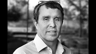
Nobel laureate Eric Betzig traces his path from near-field microscopy to lattice light-sheet microscopy, which images live cells and embryos in 3-D without killing them with light. He explains why super-resolution became a fad, and why the real breakthrough was solving the speed and phototoxicity problem instead.
Chapters
Good afternoon and welcome everyone. My name is Steve Boggs. I'm the chair of the physics department here at Berkeley, and it's a privilege to welcome everybody here to this very special lecture. Our speaker today is Eric Betzig, group leader at the Janela Research Campus of the Howard Hughes Medical Institute and 2014 Nobel laureate in chemistry. This lecture is pretty special. It's co-sponsored by 3 different departments on campus, chemistry, molecular and cell biology, and physics. Eric's unique background and research interests provided the perfect opportunity for students and faculty of all, from all 3 of these departments to appreciate and celebrate his accomplishments. I have a few thank-yous to do before we get started. I wanna ask that people just hold their applause until I get all these names out. So I'd like to thank my fellow chairs for their help with this event. That's David Wimmer in Chemistry and Richard Harlan in Molecular and Cell Biology. I'd also like to thank our deans for working with us on this opportunity. So that's Dean Steve Martin, Frances Hellman, and Douglas Clark. Special thanks to Professor Xavier Darzacq and Hernan Garcia for helping us organize this event. And a very special thank you to Dr. Najee, who is Eric's spouse and Berkeley PhD alum, for presenting her seminar earlier today on imaging the brain and as well joining us here tonight. So let's thank all of them. Okay, now on to our speaker. So Eric Betzig exhibits the best of what it means to look beyond boundaries when exploring scientific frontiers. He was trained as an experimental physicist. He tackled real-world situations in his dad's machine shop in Michigan. He took a turn as inventor at Bell Labs and then as a scientific consultant, and he's the author of 29 patents. He shared the 2014 Nobel Prize for Chemistry for the development of super-resolved fluorescence microscopy. I think we could all congratulate him on that. Congratulations. He's also the recipient of the William L. MacMillan Award and the National Academy of Sciences Award for Initiatives in Research. And now he's tackling even more exciting bioimaging techniques, which I am sure he's going to tell us more about in his talk. Interestingly, instead of dreaming about being a scientist in which field he would like to specialize in when he was a kid, It may surprise you to know that Eric actually grew up wanting to be an astronaut. As a child growing up in the 1960s and '70s, Eric was inspired by the Apollo missions and watching, as many of us did, as humans landed on the moon. His love for space may not have been his full-time career, but still part of his life. And in fact, I'm totally jealous of this. Later this year, he's gonna be meeting with Elon Musk for a private tour of SpaceX. If you can get me on that, that would be awesome. But my understanding is he's not yet signed up for a one-way trip to Mars on Mars One. So we're gonna keep him on Earth here for a little longer. So the title of his lecture today is Imaging Life at High Spatial Temporal Resolution. As our understanding of biological systems has increased, so has the complexity of our questions. Eric and his colleagues have developed powerful microscopy tools for studying biological systems at the highest resolution imaginable. Allowing researchers to track biological process at the molecular level. These tools have excited biologists, chemists, and physicists alike, enabling us all to start addressing some of the most fundamental questions about life from the molecular level and to start addressing them together. Eric will describe some of these tools and challenges for us tonight. Please join me in welcoming Eric Betzig, 2014 Nobel Laureate in Chemistry, to UC Berkeley.
Eric. Well, I'd like to thank Steve and the rest of everybody he mentioned for the hospitality they've shown and setting this up. And I have a big debt to Berkeley because in my career I've only had 10 postdocs, but 3 of them have been from Berkeley, and all of them have been absolutely phenomenal. So everything that I will show you, a large fraction of that can be attributed to the educations that these people got while they were at Berkeley. So I got the Nobel Prize, as Steve said, for something called super-resolved fluorescence microscopy. So what the heck is that? So the problem is that if that's the size of a molecule, then when you look at it in a normal optical microscope, you see a blob like that. Well, this is a problem because if we want to understand, one of the most interesting questions for me is how do you go from these inanimate little specks that are the molecules, and there might be 100 billion molecules in a single cell, how do these things self-assemble, these inanimate objects, and then create something which is animate, that lives, that breathes, that reproduces, and which is robust under perturbation and deterministic in its life cycle? And it's an incredible mystery, but what we need to do in order to answer that question is tie together the fields of molecular biology and cell biology. So a lot of my career has been about the development of imaging tools to make that possible. So you would say still, okay, so a molecule is much smaller than an optical microscope, but many of you know that electron microscopes can see right down to the atomic level. So what still is the big deal? Well, that's where the fluorescence word comes in, is that in an electron microscope you're really limited to seeing just sort of a global contrast of everything and you can't look at living things. But with visible light and fluorescence in particular, where you shine light of one color and then a different color is emitted because there's dyes or other things in the sample, you can buy different types of dyes, you can do multicolor imaging, to identify different organelles and their behavior in the cell. The other nice thing about fluorescence is even the signature of a single molecule in the midst of that 100 billion others is very easy to see. And so you can do single molecule measurements and really tie together these different spatial regimes. Because it uses visible light, and that's what we evolved under, it can be very gentle so that you can study cells noninvasively to understand their physiology for long periods of time. And then finally, the cell's a very dynamic thing, and so you need to be able to image quickly. And so fluorescence allows this fast imaging of dynamic processes in cells. So the idea of getting around that limit of that fuzzy ball and getting close to the molecular size is a problem which has involved me for ever since I entered graduate school. So there I met my eventual advisors, Mike Isaacson and Aaron Lewis. And Mike had just recently— he was an electron microscopist— had done electron beam lithography and figured out a way to, in an opaque film, drill holes as small as 30 nanometers, or about a 20th of the wavelength of light. And so he and Aaron figured that if you took one of these opaque screens and pushed it down near a cell and shined light on one side, the light that came out the other side would be initially confined to the size of that hole.. And then you could drive that around point by point and start building up an image with this sort of nano flashlight where the resolution is equal to the size of the hole instead of the much bigger wavelength of light. That's known today as near-field scanning optical microscopy. The goal back then in '82 was to make an optical microscope that could then study living cells with the resolution of an electron microscope. That sounded to me like something that could be a big deal, and so I wanted to do something that I thought would be a big deal instead of a small deal going into graduate school. So it was the only thing that interested me, and that's what I decided to do. So near-field eventually progressed. I spent 6 years at Bell, sort of did sort of a half-assed job of it, but it was good enough to get me in the door as a PI at Bell Labs, which was my dream job. So I got there in '88. The first 2 years were very hard. The technology was really stalled and going nowhere. But I was about ready to quit, but then had a couple technical breakthroughs. And then sort of '92 to '94 was the golden age of the super-resolution near-field technique. So we were able to do lithography to write different patterns much smaller than the wavelength. At one time, we had the world record for data storage density, writing bits of information much, much smaller than these diffraction-limited bits. 60-nanometer-sized bits, able to look at histological sections. And in '93, I was the first person to actually demonstrate that on living cell— or not a living cell, but a fixed cell by fluorescence, you could substantially beat the diffraction limit, and in this case look at what's known as the cytoskeleton, or this, this structure that gives some of the shape and stiffness to the cell underneath. And so all of that was the golden age of near-field there. But there were two experiments during that era when I was there that were particularly influential for what was to happen later. One was in '93, a very hot topic at that time because W.E. Moerner, who shared the Nobel with me in '89, was able at cryogenic temperatures to see the spectral signature of single molecules. So the idea of single molecule imaging and single molecule detection was a very hot topic at the time. The main problem was not so much how much signal you get from a molecule but how much background there is around that that masks it. But with the near-field probe, because you illuminate such a small area, the background was negligible. So it was easy to image single molecules. And furthermore, we were able to, by knowing the shapes of these fuzzy balls based on their dipole moments, find their positions to about a 50th of the wavelength of light. And do this at room temperature instead of near absolute zero. So this was the beginning of what became a whole field of single-molecule biophysics. Eventually supplanted by much simpler tools than near-field in the end. The other really influential experiment was when I got into Bell and I was having those problems in the early days, I made friends with another guy in my department, Harold Hess. And Harold has been my best friend ever since and is the best physicist I've ever known. Harold was doing low-temperature scanning tunneling microscopy. STM was a hot field in that era. And he was looking at superconductors. But what we did is we took out his STM probe and we put my near-field probe in, and then we started to look at the spectral properties of these quantum well structures. So these are like sandwiches of different semiconductor materials which bind when your light goes in, they create these excitons which are then confined where the meat in the sandwich is, and they diffuse around. And what we were able to show is that, that diffuse— where the light comes out when those when these, when these what are called excitons are created, they can't collapse and emit light anywhere willy-nilly, but only in discrete positions. And furthermore, those positions based on the atomic changes in the roughness of that sandwich would make them glow in different colors. So by that, we were able to actually study a lot about the properties of light generation inside of these structures. So that's all the old condensed matter physics. But what was actually valuable for us later on was the notion that even in the small size of our near-field probe, there might be a dozen or more of these little points of where the light comes out, discrete points. Sort of like consider some semiconductor molecules. And normally that would be unresolvable even with the near-field probe. But because they all glowed in different colors, If you looked at them in a 3-dimensional space instead of just XY but XY and color in this direction, then you could separate them and study them one by one because they were resolved in that bigger space, even though there were a dozen in the same probe. And so by '94, I had been doing near-field microscopy for 12 years, and I was thoroughly sick of it for a number of reasons. The first is that it had fundamental limitations, and the main one is, yes, the light is confined when it comes out of that aperture, but it spreads really, really fast. So even if you're only like 10 molecules away from the sample, you get a significant loss of resolution. The problem is the cell looks like this, and it's way, way rougher than that. So there was no real way that this was gonna be really well applied to looking at living cells, which was my dream when I entered graduate school. So I was very frustrated and disappointed. At the same time, near-field had, because of the successes we had with the single molecule imaging, the semiconductor spectroscopy, the data storage, near-field became a hot fad, a very hot field, and there were dozens if not hundreds of people jumping into the field. And I learned one thing about myself is that if a bunch of people are working in the same field I'm working in, it's time to figure out something new. Because I don't like to do that. But also, you know, anybody who's in science in this room knows that science goes through fads and that, you know, you have these sort of boom-bust oscillations where people jump in and they claim the moon and they say that you can do everything with it at the same time that I'm perfectly aware of what you can't do with it and other people are saying the opposite. And I got to the point where I felt like every good result we had was just providing the justification for 100 crap papers that were published in return. And all I was doing was providing justification for these things, and it was a waste of time, and it was a waste of the taxpayers' money. So I really believed I was a net negative to society by what I had done. Okay. And so all of those things together, and then there was a final thing, which is at Bell you had to work your ass off to succeed, and I did. And I loved it. I mean, my friend Harold and I, we would come in and we would race each other to be the first guy in the building. And so we were at the parking lot, we'd take the two spots closest to the building, we'd come in at 4:30 in the morning, and if Harold had beat me, I'd say, "Shit!" And then I'd put my hand on the hood of his car and by the temperature figure out by how many minutes he had beat me. So, and he would do the same if I beat him. But then, you know, we would work for several hours, we'd play tennis in the morning, we'd eat lunch together, we'd eat dinner together, and it was a great relationship. But you had to work hard. So I got there in '88, and by '94, with all the things I said plus the hard work, I was frankly burnt out. So in '89, Harold and I looked like this. But the other problem was is that in 1984, the government broke up the monopoly of the phone system, and that knocked away the financial underpinnings of Bell Labs. And so 10 years later, by '94, you could see the writing on the wall that it just wasn't going to be able to support and value basic research as they had in the past. So we felt the weight of the world on our shoulders. So by '94, we looked like this. So that was reason enough to get the heck out of there. OK. So that's what I did. And I really had no plan B. I was really fed up with academia because I didn't like the way people were portraying Near Field, and I was frustrated that I had failed with Near Field, and I was just fed up. And so, but one day, about 3 months later, as I was doing my best to flush Near Field out of my head, I was walking my daughter in a stroller around in New Jersey, and it struck me that you could combine that single molecule imaging and finding the positions of those molecule experiment I did with that quantum well experiment that Harold and I did to make a new way of doing super-resolution imaging. So the idea is, is that if you have a bunch of molecules in your biological sample, usually they're so close that those fuzzy balls run together and you can't really do anything with that. But if all of those molecules are different from one another in some way, if, for example, they glow in different colors, they have different polarizations, or they turn on at different times, then just as in that quantum well experiment, you can isolate them in a higher dimensional space. But once they're isolated, you know, then you have a nice isolated fuzzy ball. You can find the center of that fuzzy ball to much better precision than the diameter of the fuzzy ball, and hence find the coordinates of all of the molecules. And so if you do that, in the end, what do you have? You have a molecular resolution image of your sample. And so I was very excited about this idea, but there's a catch. And the catch is that in a normal biological sample, there might be hundreds or thousands of molecules in one focus of your diffraction-limited microscope that would excite all those guys at once. So whatever you're using to separate them out in this original other dimension has to be really, really good to separate them out a lot, to stretch them out that you can see them one by one. And at the time, the only way I knew how to do that would be to do cryogenic near-field microscopy of biological samples. It would be a hero experiment with Harold, and it would be, you know, just not really a viable tool for doing biology. So I almost didn't publish the paper, but it's a damn good thing, even though I was unemployed, that I did, because this was one of the two primary papers the committee cited in giving me the prize. And if I hadn't published that paper, I may not have gotten won the prize. That paper's only been cited about 100 times in the last 20 years. I got to turn this off. This is the advertising. I got to turn that off. Where is it? Okay. My plan B eventually was my dad had started a company in 1985 which was starting to take off to make large machine tools for the auto industry. These machines would be the width of this entire auditorium, cost about $5 million, and make one particular part like an intake manifold or brake caliper or whatever. So I started consulting for him in '94, and by '96 I was convinced that I could do things better. In fact, I was so optimistic about it, I said, well, if one good physicist goes back to the Rust Belt, he can save it. So this was how naive I was. So I went to work for my dad's company, and eventually my baby was this thing, which basically took that big machine like this, collapsed it to something the size of a compact car, and do the same work, and would move 4-ton loads at 8 Gs of acceleration in position of 5-micron precision. It would move anywhere inside a meter-cubed volume in 100 milliseconds. And I was very proud of that machine. I spent 4 years developing it and 3 years trying to sell it. In the end, I sold 2. And so what I learned is that I may not be an academic, but man, am I a horrible businessman. So after burning through about $1 million of my dad's money in about 2002, I said, Dad, you know, I've given it everything I can, but this is just way too out there for everybody and they're not accepting it.. And so I quit again. And so now I've quit twice and now I've failed twice. It's 2002, I'm 42 years old, I have 2 young kids, I'm unemployed and probably unemployable. And so that was the darkest time of my life, as you can see. But I did something smart, which is in '97 Harold had left Bell as Bell continued to implode. And went to work for a company that makes test equipment for disk drives out in San Diego. And he was more successful there than I was, but he was also starting to feel— basically we were going through our midlife crises at the same time. You know, that we said, you know, what is the meaning of life? You know, what value is anything? And we were, you know, so what we did is he was in San Diego, I was in Michigan, we would meet at national parks. About once a month, and we'd just start walking and talking and trying to think about what the hell we wanted to do with our lives before we were dead. And so what we realized in the end is that while neither one of us really had fit in the standard academic environment, we both really pined and missed being able to do science and to do just curiosity-driven research. And we wanted to find a way to do that again. So then I went back and I started reading for the first time in 10 years of scientific literature. And one of the first things that hit me was green fluorescent protein. So in GFP, what happens is you're able to snip a bit of DNA from a glowing jellyfish and splice it to DNA that makes any particular protein you want to look at inside of a cell and coax the cellular machinery to make glowing copies of that protein. Back in the near-field days, like that experiment I showed on the cytoskeleton, that was incredibly difficult because the labeling technology fluorescence wasn't up to being able to densely label with high specificity that structure. It was very difficult to do. And this was magic. To the biologists out there, the standard joke I say is I was the last man on earth to learn about GFP because it came out in '94 right after I left Bell. If I had known about it then, I probably never would have left science because it would have just been too awesome. But I knew, you know, when I was starting to get back into science, I said there are two things I knew I didn't want to do. I didn't want to do machine tools and I didn't want to do microscopy because I had been there and done that. But when I read Chalfie's paper and the other ones about everything GFP had done since, I said, shit, I got to do microscopy again. So I started thinking about ideas about, about how I could make a contribution, and I came up with an idea I was really proud of. It was based on something called optical lattices, which are you bring multiple light beams from different directions that interfere and make intense spots. It's something that's common in the atomic molecular optical field for trapping atoms, but those aren't really the type of lattices that are useful for microscopy. But I found new classes of optical lattices that would allow me to make basically a massively parallel imaging thing in 3D for live imaging. So I started trying to sell that idea and at the same time try to coax Harold to do it with me, because like I said, he's not only my best friend, he is the best physicist I've ever known, and I, I knew I would need his help. So one of the places we went to try to pitch this was Florida State University, where the National High Magnetic Field Lab is. 'cause it was headed by another Bell Labs buddy of ours named Greg Bobinger. And it was there that we met Mike Davidson. So Mike Davidson is an interesting character himself. He's a microscopist who spent his— his day job was to look at the, like, grain boundaries and the materials that would make the magnets and so forth. But what he had done is he had taken polarized light microscope images of cocktail mixes that would make, like, margaritas and stuff. Stuff, imprint those on ties, and then he sold those online. And he made several million dollars doing this. And so he was able to take that money, turn it around, and then he started making the greatest website ever for microscopy about everything you want to know about microscopes but were afraid to ask. Eventually, 3 of the major— of 4 major microscope companies actually had him design these websites for him. He made even more money from that, and then he plowed that into his passion, which was to do live cell imaging. In that era, 2003, that meant you need fluorescent proteins, and like GFP. And so Mike had, by recruiting an army of undergraduates, many of them near dropouts, and turning them around into productive scientists, making a library of 3,500 different fluorescent protein fusions. And so while it was on that trip in 2005 to Florida State that Mike told us about a new type of fluorescent protein. So this is something which is called a photoactivated fluorescent protein. So in this, it isn't normal— a normal fluorescent protein, you shine blue light on it and green light will come out. With a photoactivated protein, you shine blue light on it, nothing happens. But if you shine purple light on it first, it switches to an active state, and then you can shine blue light on it and green light will come out. And so Harold and I were sitting in the airport in Tallahassee to leave, and it just struck us that this was the missing link to make that crazy idea that I had published and abandoned 10 years earlier to work. So the idea is that you, you have your cell and you coax it to produce any protein you want with a photoactivated fluorescent protein. On it, and initially it doesn't glow, but you turn on that violet light so only a few molecules at a time turn on, and then they will be isolated from one another just by chance because they'll be so far apart. And if they're isolated, then you can find their centers. Then you study them until they basically burn out and bleach, and then you turn on a new subset with the violet light again and find their centers. And you do that over and over and over again, as you see here, If you sum the fuzzy balls together, you get the diffraction-limited image. But if you find the centers as you do them like 2 at a time here, you start to build up this high-resolution image. So that's called photoactivated localization microscopy, or PALM. That's why I got the Nobel Prize. Okay. So basically it was taking that idea that I had in '95, but now this discriminating thing isn't color or anything, it's just time. And turning on just a few molecules at a time so we can isolate them one by one. And so there was a main problem though immediately is that Harold and I were unemployed. I had convinced him enough about the lattice that, and he was dissatisfied enough in his job, he hadn't completely decided to throw in with me on the lattice, but he quit his job. And so now we're both unemployed. And so we said, look, it's gonna take way, this idea is way too easy. Anybody could do this. And so we were scared about that, and scared rightly so, because several groups were hot on our heels. And so, so it would take way too long to get VC funding, it would take way too long to write a grant. So we said screw it, let's just do it. And so because Harold doesn't burn his bridges as effectively as I do, when he left Bell he was able to take his equipment with him. So we pulled all of that out of the storage shed and we put about $25,000 each of our own money in it that we didn't have for things that didn't exist 10 years previously. And normally, like, Wozniak and Jobs would do it in the garage, but we were able to do it in the living room because Harold wasn't married. So, so there was nobody standing in the way of doing it there. So, so that's the initial PALM microscope in Harold's living room. We called it La Jolla Labs after because he lived in La Jolla up on top of Mount Soledad, if you know where that is. But we had another limitation, which is we knew zero about biology. And so we needed good biologists to work with us. So part of my frantic effort to find a job to build the lattice microscope involved caging an invitation from a Bell Labs friend who was at NIH. And once we got the palm idea, when I went to actually give that talk, I said, please, please, can I possibly meet Jennifer Lippincott-Schwartz and George Patterson? Because they were the two who invented the first of these photoactivated fluorescent proteins. And so I took them to lunch, I swore them to secrecy, I told them what Harold and I were working on. And most people in Jennifer's shoes would look at this wild-eyed crazy dude who hadn't published a paper in 10 years and said, thank you very much, now get lost. But Jennifer in her typical way said, fantastic, bring it here. And so we did. And this is now that same PALM microscope in the darkroom next to the old EM that she had in a corner. And that's where we did the experiments. And within a month of taking the instrument there, this is a thin section cut through a cell. On the left you're seeing the single molecules pop on one by one as they're photoactivated. If you sum them together, you get the diffraction-limited image. But if instead you find the centers, you start to build something of higher resolution. And after taking 20,000 images of this, you go from something like this to something like this. And to better appreciate it, you go from that to that. So what it is, it's a microscope that can take you from 200-nanometer resolution to 20-nanometer resolution and do it in a microscope that's simple enough to do in your living room. So it was, you know, everybody jumped on this right away, and it was very easy to do, and many groups got involved in it. So in that same year, by a crazy set of circumstances, but again involving networking through my old Bell Labs contacts, I got in touch with an entity I had never heard of before 2005 called the Howard Hughes Medical Institute. Institute. And basically, they're my fairy godmother, okay? So they basically— we went from rags to riches. Harold and I both got jobs there overnight. And so 2006, the doors opened, and from 2006 to 2008, I hired this guy, my second postdoc, Hari, who worked for Jan Liphardt's group here at Berkeley, and we lived and breathed POM. 18 hours a day for those, you know, 3 years from 2006 to 2008. Did a number of things. This is an experiment we actually did with Jan here at Berkeley where he was looking at chemotaxis receptors in E. coli, and we were able to show that you can see there's a bunch of clusters of these chemotaxis things in addition to these big clusters at the poles. And Ned Wingreen, the collaborator from Princeton, was able to show that the sizes of these clusters and their positions along the bacterium are completely predictable in terms of a stochastic model of self-assembly, where the proteins are inserted randomly in the membrane, then diffuse and stick to an existing cluster with a probability equal to the size of the existing cluster. We're able to also do two-color imaging, in this case looking at focal adhesions, which are sort of the feet of the cell where it attaches to the matrix. And there's 90 different proteins in there, but looking two at a time we're able to show the proteins that look like they're co-localized when you look at diffraction resolution are nothing of the kind when you look with high enough resolution by PALM. In another example with Bob Tijan, of course another Berkeley guy who's now head of HHMI, we were able to show that these core promoters that are involved in gene expression, one way of silencing the genes is there's actually a spatial segregation of the core promoters from genes that hug up against the red nuclear membrane, and that acts as a silencing mechanism, this separation. And then finally, on live neurons, we were able to show neurons send out these spines, which are at the end of the spines are where the synapses are to connect to the next one. And the structure of these spines is held in place by the same actin filaments I was describing before, and we were able to show by photoactivating and then tracking the molecules that there's only discrete spots where the actin is formed inside of those, inside of those spines. And so that's all of the good news about PALM, but by 2008, I was as thoroughly sick of PALM as I was of near-field in '94 for many of the same reasons, is that it got to be a hot field, super-resolution. In 2008, Nature Methods calls it method of the year. Everybody and his sister is doing it. And again, every time these fads happen, the signal of the field stays about the same, but the noise goes way, way up. And so— but there's basic intrinsic problems. And the first is that it's— if you can't put your molecules that have the dye or whatever close enough together, you're never going to see the underlying structure. Just as if you blow out 99% of the pixels in your TV, you're not going to see a good image. And so that's getting high enough density labeling is still a major challenge in the super-resolution field. Another is most of the time people would look at fixed cells, chemically fixed, dead, and the fixatives cross-link proteins and they change the ultrastructure. So the 99% of crap that we've learned from super-resolution, most of it has a big fat asterisk next to it. Can we really trust it? And then in order to get to those high densities of label, You have to overexpress the protein, and so now you've changed the whole homeostatic balance of the cell, so you don't even know whether that cell is any, any more of a natural cell. And finally, the intensities to see single molecules are about 10 to the 4 times higher than the light outside that we evolved under. And so oftentimes by any technique, super-resolution technique, that's what starts to happen to your cell. And so you ask yourself, what the hell are you doing? I mean, you can either use these as structural tools, in which case this kind of thing and this kind of thing bites you in the ass, or you can use it as a live tool, in which case this bites you in the ass, right? And so in my opinion, this award was really freaking premature, okay? I mean, super resolution still has a lot to prove. So, and so this kind of summarizes that, is there's 3 major technologies for super-resolution imaging. There's something called STED, which Stefan Hell developed, and which also he shared the prize with W.E. and I on this, and it's a technically elegant way of doing super-resolution. I won't go into the details, but it uses focused light. And then there's localization, which involves single molecules. These two techniques though, they require anywhere from hundreds to thousands of times as much light to come out of the sample to make the image at high resolution as a normal image would if you were looking under a microscope like you would have in a normal laboratory. And if you, if any of you are biologists and have looked at live cells or even fixed cells, you know that you can't get tens, hundreds, or thousands of frames before you bleach the crap out of the sample or kill it. And so that's a problem. The power intensities for STED are anywhere from 10 million to 10 billion times as much as the light outside that you evolved under. And for localization, it's 1,000 to 10,000 times as much. So these methods, these are the two methods that got the prize, okay? But they have major, major challenges for live imaging. There's a third technique though which didn't get the prize, and that's something called structured illumination microscopy. There you put a standing wave of excitation on your sample, and what happens is that, that sort of wave pattern beats against the information in the sample in the same way if you look through, if you look on a porch, screen porch, and you look through two screens at the same time, you'll see these fuzzy sort of, you know, patterns, these wavy patterns that you can see between the two screens. If you look at just one screen, you see nothing. And so that's these beat frequencies called Moiré fringes. That technology can be used to double the resolution beyond the diffraction limit. The reason this technique did not win the Nobel Prize is because they said, ah, it's just a factor of 2. You know, the other ones can do a factor of 10, but they involve way less light that's required from the sample, way lower intensities, and it's way, way faster. So it's much more compatible for live imaging. So the moral of the story is that no matter what method you're going to use to increase spatial resolution, if I want higher and higher resolution, my image has to have more and more pixels. That means it's going to take more and more time, and it means I'm going to— and more and more time means I lose temporal resolution, and it means I'm throwing more and more light at the specimen, generating phototoxicity. So if you're going to be honest, and there hasn't been enough honesty in this field, you're always playing one thing against the other and you're always working somewhere inside of this tetrahedron here. So the guy who understood this limitation before the rest of us, I believe, was Mats Guftason. He was a postdoc at UCSF who then stayed on as an assistant prof and was really one of the pioneers of the structured illumination technique. This is that, again, sort of moiré fringes that you get from the beating of the sample against the excitation. And so Matzett was the real pioneer of this. He was doing great work. In 2008, we were fortunate enough to recruit him to come to Janelia, and he was off on a great trajectory there. And in 2010, he was diagnosed with a glioblastoma. And he died in 2011. And so I managed— I inherited basically his group when he passed and also the technology. And so I've been working to try to extend the SIM technology so that it has its rightful due. As I will describe in a few minutes, Matz was the messiah of the gospel of SIM, and I am his acolyte. And I love localization microscopy. And it has a real role below 50 nanometers. But in my opinion, SIM has not gotten a fair shake yet, and I'm out to try to fix that situation. So the beautiful things about SIM again is that you can really do great live imaging. This is only 100 nanometer res— yeah, only 100 nanometer, but the fact that you're able to get images at sub-second time frames, which is the rates at which dynamics is happening in cells, and you're able to do it for such long periods of time creates a wealth of information that you don't get from a single static STED or PALM image. And so again, if the limitation of SIM and the knock as to why it didn't get the Nobel Prize is that the resolution is limited to 100 nanometers, how can we crack beyond that? Well, the simple stupid thing is to find some microscope objective which can write a finer pattern that allows you to then go to higher resolution. So Olympus sells a specialized objective at very high numerical aperture which allows you to push the resolution down to 80 nanometers. That's pretty damn good, okay? The normal resolution is 200 in a microscope, and you can see tweaking back and forth between those two resolutions. These particular things are called clathrin-coated pits. They're one of the mechanisms by which cells bring different cargoes inside of the cell.. And so they form these little domes on the inside of the plasma membrane, and the cargo sits inside of there. When we do this in what's known as TIRF, we illuminate only the very part closest to the plasma membrane, so these domes look like rings. And now we're resolving those rings with the, with the high-resolution SIM. But again, because it's SIM, we can do it live. And so now you can study clathrin-mediated endocytosis at high spatial temporal resolution, study the entire life history from the formation of these clathrin-coated pits to their internalization of cargoes. And again, for long periods of time and high speed. And so the other nice thing about SIM is STED and PALM require these fancy tricks of photo switching and so forth in the probes, whereas SIM can use any off-the-shelf labels that you have. So it's easy to do multicolor. In this case, you're looking at the actin cytoskeleton again, and at the same time, the clathrin-coated pits. So it's a major controversy to a degree in the endocytosis field about what is the role of actin in clathrin-mediated endocytosis. And admittedly, we have a very small sample set, but in our data, we find that only about half the time does actin actually colocalize with these clathrin pits as they internalize. But when they do, they make a small but statistically significant shortening of the lifetime. There are also these things called plaques, which are large aggregations of clathrin that just look like a fuzzy blob at the diffraction limit. We were able to show in these cells that these plaques are nothing more than aggregations of pits and that they eventually spin off pits. And then finally, we found these crazy rings of actin. Which there's not a lot of record in either the EM literature or anywhere else about these things. One of the models of clathrin-mediated endocytosis said the actin would pinch in a ring to pull off the bud, but these things aren't colocalized with the clathrin at all, so we don't really know what the hell their role is. Then, okay, so 80 nanometers is good, but what if we want even higher? So Matz had developed before he died and demonstrated a couple forms of what's known as nonlinear structured illumination microscopy, where normally the reason it's limited is because the standing wave that you can create is also diffraction limited. But if you can apply either nonlinearities, either by saturating the fluorescence or photo switching like we do in PALM, you can create other higher harmonics. It's kind of like Spinal Tap where you turn the amplifier up to 11, right? You get all the distortion, you know, out of your speakers, and so that creates these higher frequencies that allow you to push beyond in the resolution. And so with that, you can push to 60 nanometers. So now this is the actin cytoskeleton live at 60 nanometers and doing two-color imaging with looking at BRAB5A, which is a protein which is a marker of early endosomes. So after the clathrin-mediated endocytosis happens, These cargoes then become endosomes, and this is part of what's known as the endocytic pathway inside of the cell. And so we can then follow and look at the morphologies of these endosomes, and unlike the clathrin, they have very bizarre shapes like you can see here. By looking at a 20-year-old EM paper and then looking back at our data and cherry-picking and finding ones that look just like the EM ones, you can see, you can see that we actually start to see these shapes and can actually then study the but now we can study the evolutions of these shapes over time, which you can't do by EM. And you can see these dark dots that represent cargo inside of these guys, and you see all sorts of different types of dynamics, from sort of Brownian-type motion to sort of confined motion by the actin cytoskeleton to directed motion along the microtubules. So the moral of the story was the entire field of super-resolution was moving as close to that point as they can. Everybody wanted to say I got higher resolution than you. So what we said is that, and what Motz first said, is that, hey, wait a minute, that's not the only metric that's important. And by backing off, you open a much bigger application space in different directions that the other methods couldn't touch. And so it sort of begs the question, if we backed off to 100 nanometers and learned so much, what would happen if we backed off all the way back to the diffraction limit? So let the entire— remember, I don't like to drive a bandwagon. I don't want to be where other people are. Let them go in that direction. So in 2008, when I was so sick of Palm, I wanted to do something else. I wanted to take that lesson from SIM and say, is there something we can do with the diffraction limit? And in particular, can we make a microscope that improves 3D imaging of living cells by as much in the time dimension as Palm increased the spatial dimension. And so that's what I wanted to make my goal. And so why, why is it so important to have that time dimension? Well, the hallmark of life is that it's animate, and every living thing is a complex thermodynamic pocket of reduced entropy, and matter and energy is flowing through that continuously. So if we really want to understand how inanimate molecules come together to create an animate cell, We really need high resolution across all 4 dimensions of space-time at the same time. And so what are the challenges though if you're gonna do that? Well, the first is that the live cell, unlike the fixed one, is moving. And if you want higher and higher spatial resolution, the velocities of the cell are dictated by Mother Nature. So it means that as your resolution spatially improves, your resolution temporally has to improve at the same rate or else the image will smear just like when you leave your camera shutter open too long. And so you need— and that's a bad trade-off because now you're asking for more information in less time. It's hard to do. The next is that cells are three-dimensional, and much of the imaging that's done in cell biology, even with confocal, which can do 3D imaging, is often done in a single plane because the instruments are too slow and too damaging to collect a whole 3D image. And so here you can see all those chromosomes, but you only see a few because only one plane is happening when the cell is dividing. And then that's all you get is you get partial information, but not everything happens in a plane inside of the cell. And then finally, the normal imaging tools for 3D don't have the same resolution in all directions, and you would— nature doesn't have a preferred direction, so you want the same resolution in all directions. And so the other problem is, okay, what if we do want some gain of spatial resolution? Well, if I'm working in 3 dimensions, I'm not talking pixels, I'm talking voxels. And if I want higher spatial resolution volumetrically, my voxels get smaller. Just to get SIM in 3D means a factor of 2 in each dimension. That means a factor of 8 in volume of those voxels, so you have 1/8 as many molecules, producing 1/8 as much light unless you bang it 8 times as hard, and then you don't want to do that. So that's a problem. And then furthermore, if I'm doing a fixed cell like in Palmerstedt, I can burn out all of those molecules in one image. But if I'm doing live imaging, I gotta get multiple frames, so I can only parse it a little bit in each frame of bleaching. And finally, like I've said already, light— I mean, light is somewhat non-invasive, but trust me, Cells do not like intense light, and so this is often— in this case you can see mitochondria fragment, the cell retract. That's a sad, sad, all too common story to people who do confocal imaging of cells. And so the moral of the story is imaging live cells is much harder than imaging fixed ones, and imaging in 3D is much harder than imaging in 2D. And so the good news is that the standard tools that people use for 3D imaging in biology leave a lot of room for improvement because they're basically the wide-field microscope, which is like what you would use in a high school class and just blast the cell with light and look at the light coming back, or else the confocal microscope where you focus the light to a point, but that point is inside of the specimen. But in these methods, there's only one plane that's in focus, and it's all just fuzzy shit outside of that plane. And so in the confocal, you use a pinhole to strip that out, but you're doing all sorts of damage and bleaching to these other areas while you're just getting information from one plane. In my opinion, one of the most important innovations in microscopy of the last 15 years, perhaps more important than super-resolution, was when, was when about 10 or 15 years ago, Ernst Stelzer at the EMBL reintroduced a 100-year-old concept called plane illumination. So in this technique, instead of bringing the light through the objective and blasting the whole thickness and coming back, You take sort of a cylindrical lens from the side, make a thin sheet of light which is only coincident with the, with the plane that's in focus, and illuminate that whole plane at once. That means you can use a camera to snap the whole plane, boop, all at once. So it's much faster than scanning a point around. It means that you're not bleaching the areas above and below. And so you can go like a bat out of hell and really get very quick imaging. This has been transformative for studying embryogenesis. At single-cell resolution. But it has a problem, and the problem is, is that over the dimensions of even a single cell, and it gets worse as you get even bigger, the light sheet itself, by the laws of physics and diffraction, is on the order of 3 to 5 microns, and a cultured cell might be twice that size at most. And so you're really not gaining a lot of benefit of the technique. And so one thing that immediately hit me as an idea was that, okay, you can use something that came out in the late '80s when I was doing near-field called diffraction-free beams or Bessel beams. In these beams, you illuminate the objective that creates the beam with just a ring instead of fully illuminated, and you create this tiny little thin pencil of light. It's basically the same type of beam that's in your supermarket checkout scanner. And then goes over the barcode in order to read it because it doesn't diffract and diverge. It's a teeny skinny little thing that can be a long spear. And so we figured, well, if we use that, we could then scan that in and out of the screen as you see here and then repeat as we go from plane by plane through the cell and eventually plane by plane build up a 3D image. It sounds slow, but in the modern versions of this microscope, we do that at 1,000 planes a second. Or several volumes per second through the cell. So that worked fine. We were able to do study, for example, the initial development of the C. elegans embryo with that. We were able to study the asymmetric division of some cells with that, which normally are way too light-sensitive to study during their division, but because of this method is much less light-sensitive, it allowed us to do that. But there's a problem with the Bessel beam is that it isn't just that skinny pencil, in cross-section it looks like a bullseye and has these concentric rings around it. And so as you sweep along to create a plane of excitation, those side lobes create out-of-focus excitation. So there's a trick called two-photon microscopy which allows you to create fluorescence which is only where the brightest part of the beam is, and then that creates just the center part contributes, and then you can image as we originally intended in very good resolution in all directions. So this is an example looking at a HeLa cell. So this is like Henrietta Lacks, if you read that book from the New York Times list, cervical cancer. These are very hardy cells that metastasize easily because they have these filopodia that allow them to grab onto things and move along. But now here you can study, you know, for 100 volumes at 6-second intervals for an entire volume, the dynamics of those filopodia. Another method we used to get around this, this problem of the side lobes was instead of sweeping the beam continuously, to step it discreetly and then create a grating pattern. And then we could use Matz's old tricks of structured illumination to actually use the best— the side lobes to our advantage to create a patterned excitation that would allow us to poke beyond the diffraction limit in 3D. And so this is an example of looking at a cell which has been— where what's been introduced is a protein that's a cancer signaling protein that promotes these ruffling mechanisms of the cell, and it folds over the membrane and then creates these bubbles or vacuoles that you can see underneath in this X-ray view here. So one of the problems with that SIM mode though was that it took too long to just step that beam across to create that pattern. So we introduced sort of a fancy diffractive optical element that fanned the beam out into 7 beams. And that certainly fixed the speed problem, but what shocked us was how much less damaging it was to the cell to image with 7 beams instead of 1 beam. And so what I learned from that experience was that while the total dose of light that you throw on the cell is an important metric for the health of the cell, a far more important metric is the instantaneous intensity that you throw at the cell. So what that says is that a line is better than a point and a plane is better than a line. And so you want to spread the energy out as much as you can. It also says the standard technique biologists use for live imaging which is the confocal microscope, may be the absolutely stupidest possible way to do live cell imaging, because not only do you have these cones of light going in and out to create all this damage above and below, at the focus you have this actinic spot of light which is going, "Ergh," and leaving death and destruction in its wake as it drives along. And so you really need to spread the light out. So that got us thinking about why stop at 7? What happens if we start looking with more beams? The problem is, is now you gotta worry about what happens when the side lobes of those beams get close enough together that they start interfering. And so we started modeling what would happen there, and what you find is that there's certain specific separations of these beams where you have a magic thing happen where all of the side lobes destructively interfere with one another, and you get rid of that problem, and all the excitation gets truly confined to the single plane., it spreads the energy out in that plane, and you have great modulation for doing Matz's structured illumination technique. So it's sort of a triple win. Those happen extremely rarely in life, okay? I can say, but it was wonderful. The reason I was kicking myself, though, is because these magic separation periods where this happens were basically— they represent two-dimensional optical lattices, which is that goddamn theory I had developed 10 years before. In 2005 when I was trying to get back in. And so once I realized that these were optical lattices, I was able to dust off all of that old theory I had developed 10 years before and then apply it now to this light sheet microscope. And so this is what we call lattice light sheet microscopy, and you can use it in one of two modes. You can either use it in the structured illumination mode, which is the high-resolution mode you see here, where you see the filopodia a little bit better, Or you can dither it a little bit back and forth to smear it out to just get a plane. And here you have to take 5 images per plane. Here you only take 1 image per plane. So it turns out in the end about 7.5 times faster. It is so noninvasive that anybody who's done live cell fluorescence imaging will tell you there's a very limited amount of time, even in 2D with confocal, that you can look at a cell. But with lattice light sheet, if the protein is expressed heavily enough, We can look at it forever. The natural turnover of the protein is actually faster than the rate at which we lose fluorophores to photobleaching. And so it's really an amazing tool. And so one of the amazing things that it's great for is one of the other skeletons in the closet of the whole single molecule field is that it's really limited to very thin samples because, again, remember you have to see the weak signature of a single molecule, and if there's a bunch of other molecules that are out of focus, they create a whole bunch of background and makes it difficult. So you have to look in thin parts of the cell. You can't look at cells in mitosis. You can't look at zebrafish and things of that sort. But the lattice light sheet is so thin that it's thinner than the depth of focus of the detection objective. So it means that only molecules that are in focus are excited. So you have fantastic signal-to-noise in arbitrarily thick specimens in order to do this. So this has been very useful for my colleagues at Janelia who study transcription, where they're able to look in, in this case, a spheroid of mouse embryonic stem cells and then be able to study the binding kinetics of these transcription factors to the DNA. But the other thing, although I swore I would never do localization microscopy again in 2008, is the other thing it's good for is getting around that nasty problem that I said about getting enough enough dye in your system to be able to get high resolution by PALM. In the same year that we published the PALM paper, Robin Hochstraßer at UPenn published a related idea where you don't use photoactivation to turn on the molecules. Instead, the molecules are whizzing by in solution and they're going so fast that they're just a haze. But if they end up binding and sticking to the, to the part of the sample you're interested in, Then they're fixed and then they look like a glowing spot. So that's called PAINT. And so the beauty about PAINT is that in a normal PALM, STORM, whatever localization experiment, you put your labels in at the beginning and you're done. That's it. Whatever label density you got, that's what you got to work with. With PAINT, the whole media is labeled. So this infinite army of molecules can keep coming and coming and coming. The problem with PAINT is the whole fucking medium is glowing. So it's difficult to be able to use this method at high SNR, but the lattice-like sheet is perfect for it. And so this is an example where you're looking now through a dividing cell that's about 20 microns thick, or about 10 times more than typical localization, and 300 million molecules localized, which is about 100 times more than a normal localization image. So instead of looking like a bunch of dots, which is typical localization, it actually looks like an EM image because there's enough information there. To see what's happening at high resolution. And you can do multicolor imaging with this as well. But I'm tired of looking at dead things, and so the beauty of the lattice light sheet is to study 3D dynamics. So this is an example of looking at a dividing cell where the orange is the chromosomes and the colors, the fireworks, represent tracks of the ends of growing microtubules during the division of these cells. And so from a technical point of view, this is impressive because this is 1,000 3D volumes between the two different colors of the same cell. And cells, when they divide, are incredibly light sensitive. They want to shut down so that they don't create replication errors if anything goes wrong. So they have all these mitotic checkpoints. But this cell went through 1,000 3D volumes, which represented about 300,000 2D image planes, and divided perfectly normally and divided again after that, the daughters divided. And so it's really a non-invasive tool for studying very delicate processes inside of cells. And in this case, we're able to actually show that the velocities of these growing microtubules change as the cell goes through different phases of cell division. Let me start that again. The other thing, if the process isn't super fast, and you have extra time, you can use that time to do multiple colors. So this is 3-color imaging of 3 different organelles inside of the cell. So one is the chromosomes, one is the mitochondria, sort of the power cells, the power packs of the cell, and the other is the endoplasmic reticulum, which is where proteins are sort of processed after they— during their creation and folding. And so what we find is during cell division, this is a whole field of cells. Now we've taken that 3D field and sliced it into 2-micron-thick slabs so you can see what's happening inside. And you can see that what happens is the endoplasmic reticulum, which is that net I showed you in that early SIM image, instead folds up and creates sort of these little pockets or cisternae. And inside of these guys, the mitochondria, which are normally long sausages, fragment and they fit inside of those pockets and they're carried along by those pockets as the cell provides. And so that's the kind of information you can get. So working from single cells now to cell-cell interactions, this is now looking at a T cell, which is the kind of cell that fights infection, but in this case it's sensing an antigen-presenting cell, which makes the T cell decide whether it's the right T cell to fight the infection that's happening. And so now I showed you an early example of that in Sim. Where we were looking right at the boundary of this thing at high resolution. Here it's diffraction-limited resolution, but we're able to see the entirety of the cell. We're able to see this— identify this very fast flow of actin away from the immunological synapse, and once the synapse is established in these guys. Now, one of the motivations for studying cell motility is metastasis in cancer. But normally a lot of cell motility we study is on immortalized cells on coverslip. Which is not exactly the geometry inside of a tumor. So another nice thing about the lattice light sheet is now to finally study cell motility in 3 dimensions in a real matrix. This is a collagen matrix here. This is with Dyke Mullins' group at UCSF across the bay. And so this is a neutrophilic cell, and you can study its motion through the collagen mesh. So the— working up the next length scale, this is now looking at C. elegans, which is a model organism, a a small nematode worm, and looking with our collaborator Josh Bembenek at a protein which is normally thought to be just a remnant that's left over— that's this thing— after cell division, sort of a cytokinetic remnant, and finding that it's actually co-localized to other parts of the cell. And Josh believes these remnants actually have a later role in development of the organism. Some things in the late stages of development of C. elegans, right before hatching, the muscles are going like crazy. And it's just too damn fast to get a 3D volume, but you can park at a single plane and then still image at that plane at fast enough speed to see what's going on. So what I would like to do for the future of my group is frankly to take cell biology away from the coverslip because cells didn't evolve there. They need to be studied at these speeds and at these resolutions inside of the organisms in which they evolved. And there's a problem with that, is that as our light goes in, for example, a lattice light sheet, this is looking at an adult C. elegans, and these are the meiotic chromosomes. When the light sheet starts, it's great and you can see these guys fine, but over here the light sheet has been scrambled by the refractive index inhomogeneities of the specimen itself, just like the water on your windshield scrambles the light coming in to your eyes.. And so the same thing happens on the detection side, that once we create a light sheet, the nuclei that are closest to the detection objective we can see fine, but the ones whose light rays actually have to pass through the organism get scrambled. And so that's a big problem. These aberrations are a big problem. The good news is that we're not the only ones who have had to face this problem, and astronomers have been dealing with it for for 50 years, the military and the ophthalmologists as well. And so the solution that they came up with is something called adaptive optics. So you have an astronomical object that's 10 million light years away. In the last 100 miles when it hits the atmosphere, that flat wavefront is distorted into a distorted wavefront. In the AO system, it bounces off a mirror. You pick a little bit of the light off, you put it into a sensor, that determines the nature of that deformation. But then you put it in a computer and the computer changes the shape of this mirror to exactly cancel those wavefront distortions to create a flat wavefront and get diffraction-limited imaging. So this is what you see of a star here without AO. This is with AO. Without AO, the biggest ground-based telescopes in the world would be no bigger than— no better in seeing capability much better light collection capability, but in angular resolution, no better than a high-quality reflector you could have in your backyard. But with AO, the best ground-based telescopes in the infrared kick the ass of the Hubble Space Telescope. And the next generation scopes like the 30-meter scope and the European Extremely Large Telescope will be fucking amazing with the AO that they have. And so you gain not only resolution back— you don't gain resolution, you recover resolution, and you recover signal, because instead of all the light going everywhere, it comes to a focus, coherent, and interfere. This is showing Neptune without and with AO. So how can we use this in biology? Well, there are two ways that we've done in my group. The first is exactly steal from astronomers, and I give a talk about once a year about the historical connections between astronomy and microscopy, and the moral of the story is Microscopists are the retarded stepchildren of astronomers. And astronomy does everything about 50 years before the microscopists finally catch on and do it. So I just want to be a little bit quicker on the uptake as a thief and try to steal stuff a little fast. So what they do for AO generally is because the light from that distant galaxy is not very bright, and so it's not got enough signal to give you a good measurement of the wavefront error. They shine a laser up into the stratosphere to excite some sodium atoms to create an artificial star by fluorescence up there. And then that one is bright enough, and because it's right next to the thing you care about, then a correction on your guide star, as it's called, will give you a good correction on the, on the object that you wish to look at. And so in 2011, a group in ICFo in Barcelona said, well, what if we used a two-photon laser Because in a two-photon laser, you don't get that cone of light exciting fluorescence everywhere. You only get fluorescence at the focus. So it's exactly analogous to that. So for transparent organisms like the zebrafish, which is a model organism I absolutely love, we can apply that. And so this is an example looking near the spinal cord region of a zebrafish embryo 3 days post-fertilization. This is showing the resolution and the signal without the adaptive optics, and that's when you turn the adaptive optics on. So now this is an example. One of the problems, though, in microscopy, and it's sort of a similar problem in astronomy, is that you can correct in a region, but depending on the application— in the mouse brain, a single correction can cover 100 microns, but in the zebrafish, it's big enough and there's enough variability of the refractive index across the the organism that you have to create many corrections. But this guide star method can do that. In this case, it was 20,000 different AO corrections. We're going deep into the midbrain region, about 250 microns deep. That's with the adaptive optics. This is turning off the adaptive optics. So that's what a normal biologist thinks they're doing diffraction-limited imaging when they use their confocal and two-photon microscopes in developmental biology. But man, they're really far away. And so with the AO, you can actually get back there. And so— and in this case, we were using the two-photon mode of it to then study the development and the motion, the neurite-guided motility of these oligodendrocytes, sort of the glial cells inside of the zebrafish, inside of that. But this is slow and this is damaging and this will bleach. So the thing that's on our plate right now and is about ready to see first light is a lattice light sheet microscope that combines adaptive optics for both the excitation to keep the light sheet good and the detection to see the light that comes out. When we have that instrument, and that can also be combined with the SIM or with the localization, then we will have a tool that will really allow us to be able to study cell biology on its own terms. And I really think we are on the cusp of a true revolution in cell biology between the genome editing tools such as Jennifer Doudna here at Berkeley has developed, coupled with the non-invasiveness and the high speed of the lattice light sheet microscopy, coupled with Matz's SIM microscope, we will be able to see cells as good if not better than we ever have before, but see them in their true physiological state with native levels of expression expression with minimal invasiveness from the light and get everything we want and see all the cell-cell interactions that really are the true reality of the way cells work. And so I think we're really on the cusp of a revolution there. So that's fine and that works for the zebrafish, but the damn problem is that Janelia, 80% of what we do, if not 90%, is systems neuroscience. And while we do finally, thank God, have several people there studying zebrafish, for many years all we were doing was fruit flies and mice. And if you particularly look at the mouse brain, it looks like this. I liken it to trying to image through tofu. So trying to image neurons through tofu, because of all of the scattering that goes on, it would be like trying to image a star when the star is behind a cloud. Well, goddammit, the astronomers haven't figured that one out yet, so we can't steal from them on that. So we have to come up with our own solutions. So I hired a woman crazy enough to come and work on that problem, and that was Naji. And so we had no idea when we got started as to how we would solve that problem. But we came up— I don't have time to tell about the solution, but we came up with some solutions that would allow us outside of a guide star, but doing other means, to be able to figure out how to do an AO correction. So this is looking at structural imaging about 200 microns deep inside of the living brain. So you put a window inside of the skull of the mouse, do your imaging through that window, and then look at the neurons there. And then look at also the functional imaging. So there's calcium indicators that light up when the activity of the neurons are going off. And you can see that you can see much better signal-to-noise, the activity of what's happening inside of these neurons. So that was the initial work that we did up through 2010. And then Na was able to waggle that into getting her own group at Cytogenela. And she's taken this technology light-years beyond what we were able to do at that time. She gave a talk earlier this afternoon where she's applying it to study how the information that comes from first the retina and the algae and then goes to the visual cortex, what layers it goes to and how that's processed to get directional selectivity inside of the mouse. And a lot of that would be impossible to be able to see these weak signatures of all of these things without the adaptive optics to be able to pick them up. So the final thing that I want to say is, you know, Well, first I want to say something to the young people. There are many, many things that disturb me about academia, as you may have guessed over everything I've said. But one of the things is it seems like people get too wound up in titles, and they get too wound up in prizes and things of that sort. I feel like a hypocrite for accepting the Nobel Prize. I really do. I think most prizes are actually— I think the pain and the sting of not being on the winning side far exceeds the pleasure of it. And really in the end it's just fucking subjective, okay? It's a few guys in a room, it's their opinion, there are many opinions, okay? And so the thing that really matters is the accomplishments, being able to do something or really seeing other people being able to use it. And so Nobel Prize or not, I will consider myself when I'm on my deathbed a a failure if these techniques are not able to answer biological questions. And I will never know enough biology to do that myself. So it means I have to get these tools in the hands of a biologist. And that tech transfer problem from something that works in my lab to something that can be turnkey to work for biologists so he can focus on the biology rather than his goddamn microscope not working right. Is it's a big problem. That valley of death is something that everybody in technology faces. And so we're taking a multi-tiered approach to that. So at the simplest level, you know, we've worked with, in my group alone, we've worked with over 35 different labs who've come to work with us to apply our microscopes to biology. And in between 2013, 2014, we were a de facto imaging Center, applying these techniques. But I want to get back to doing what I'm good at, which is making new microscopes. And so we developed, in partnership with the Gordon Moore Foundation, an advanced imaging center at Janelia where we cloned the lattice light sheet microscope. We have a structured illumination microscope, live cell. We have another technology MOTS developed for simultaneous 3D imaging of specimens. And we have Harold Hess's iPalm, which is the most exquisitely sensitive form of localization microscopy ever invented. And so all of those are free to use, and one of my main goals in coming here was to encourage you all to come to Janelia and use these tools. It's a 2-page application. We put you up in a beautiful hotel on site. We feed you great meals in the pub where everything, even the booze, is subsidized. Oxidized, and you'll have a great time and you'll work with great people and you'll get terabytes of data to take home and chew on for the next couple of years. So I highly encourage people to think about the ASC. That's the first step of the problem. Another way we're trying to help out is at least for the Lattice slide sheet, we work very hard to completely document every aspect of the construction of it. So that we will give you drawings, vendors, part lists, down to the penny of what it costs, who to talk to to buy this thing, you know, what code is, you know, we give you the code, we give you wiring diagrams, we give you alignment guides. You can come out and see us operate it to see how we get it, you know, tuned. About 50 people have executed research licenses to do that. How many will build them? I don't know. Xavier would be one, Michael Loy might be another. There's, there's, but you know, we want to disseminate the tech. We don't want to hoard the technology. It's too narrow a bottleneck. We want people to use it. And then the final step would ultimately be commercialization. And so we've exclusively licensed the lattice tech to Zeiss, and they've, but it's again 2 to 3 years to go through the whole product development cycle. To make that bulletproof turnkey instrument. So it's been sub-licensed to a smaller company called 3i in the States, and so you can buy a lattice light sheet microscope now, but which would be basically a clone of what we have in our lab. And so we have all of these mechanisms that we're trying to develop to try to make sure that biologists can use these things and actually answer questions in the 'cause the papers don't matter, it's the insights that matter in the end. And so the prizes don't matter, the papers don't matter, the insights matter, the accomplishments matter. And that's the message I wanna leave for the young people in the room, okay? Don't think about you have to be a professor or you're a failure because that's bullshit, okay? And a lot of you won't succeed at being professors anyway. And there are many, many ways to contribute that are at least as important just as valuable as being a professor. Okay, so with that I'd like to wrap up. I mean, I've been privileged to work with great people. Like I say, Berkeley people, this is my new guy, David Hoffman, who came from Rich Mathey's group. He's been in my group for a little less than a year and he's equally fantastic to Na and Hari. My group has never been bigger than 5 people and it's averaged 3, so everything you've seen has been done with a group of that size. And I think that's one of our strengths is that we do keep it small and a family and tight. Worked with many great collaborators, but the one guy I have to single out is Harold, because he was my mentor when I was at Bell and he was— I would not have had the courage to do Palm on my own, but with Harold by my side, I knew there was no way we could fail. And the bittersweet thing about winning the prize is is that Harold didn't share it with me. But I fully view the prize as his as much as mine. And he is the greatest physicist I've ever known. And among many people I've been privileged to work with, he's number one. But with that, I'd like to thank you for your time.
Dr. Betzig, if you don't mind, we'll take a few questions. I'm coming to the back because the students usually hide back here, so they're all— start with questions from students in the back.
Any questions? Raise your hand. Come on up.
Oh, all scared.
Is that you? Ah, come over. There we go. Thank you. So with GFP and photoactive fluorescent protein, since you're basically tacking on an extra protein complex to a protein that's already existing, how do you ensure that that doesn't interfere with, well, the way the proteins work or how they move? It absolutely can interfere. So basically you just have to do as much functional imaging at the diffraction limit as you can to see that the physiology as close as it can. But there are— there's all sorts of tricks in the trade about linker links and so forth of the FP to the target protein to make it incorporate more noninvasively, but it's a whole black magic art of different fluorescent proteins have different levels of invasiveness to different other target proteins and how they assemble. But yeah, it's a big problem. I mean, one of the major goals of the field, which nobody has been able to address at really high resolution yet, is label-free imaging of not having to use fluorescence. I have a love-hate relationship with fluorescence, in part for the reason you said, but also because of bleaching and so forth. So if somebody could come up with the perfect label-free technique for super resolution, that would be a big advance. More questions?
Oh yeah, one of the images— hold that away from your mouth— one of the images you showed looked like a cell attached to a substrate with some sort of fibers, and I'd never seen an image like that before, and I wondered what the fibers were.
So I'm not sure which one it is, but the paint one? Oh, OK. Yeah, yeah. Those are called retraction fibers. OK. So when the cell divides and it starts— normally it's flattened out like a fried egg. OK. But when it divides, it humps up on itself and rounds up. But it still leaves these actin cytoskeleton protein-rich cables that bind it like a tent, right, to the surface as they go apart. And so those are called retraction fibers. They're seen in the EM images as well when you look at them like that in a dividing cell. But they're not typical in a cell that's in its normal non-dividing interphase state.
Yeah. Pass it down. You showed that wonderful image of imaging the mouse brain, but you had to drill a hole in the skull.
So I was wondering, would it be possible to get to fraction-limited imaging without having to do that? There are people who actually try to work on that problem, so there are different that bind it like a tent right to the surface as they go apart. And so those are called retraction fibers. They're seen in the EM images as well when you look at them like that in a dividing cell. But they're not typical in a cell that's in its normal non dividing interphase state. Yeah. Question,. And then it normally becomes very diffuse. But what they do is if they can measure that, they can actually use something called a spatial light modulator to create a very fine set of beams which exactly boom, boom, boom, boom, boom in a way that all those pinballs come together at one point. It's been demonstrated. The problem is that, A, it takes a long time to correct to get to one point like that. And then once you do it, when you move even half of a wavelength a part, then they will pinball in a completely different way through the thing. So you would need to do the whole damn process over again. And it took so long and took so much light to do one, it's just not practical to do many. It's far easier to actually then take the goddamn drill and drill a hole and put a window in.
So yeah. If everybody can join me in thanking Dr.
Eric Betzig once again.
More from Joshua







Scientist Stories: Hans Waterham, The value of phenotypic assays in peroxisomal disorders




Scientist Stories: Joel Zlotogora, Genetic carrier screening programs for reproductive purposes