Scientist Stories: Eva Nogales, Visualizing Dynamics of Complex Macromolecular Assemblies
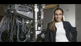
Eva Nogales describes how cryo-electron microscopy evolved into a near-atomic resolution technique for visualizing massive protein complexes. Her lab used it to map microtubules, gene-silencing machinery, and the transcription pre-initiation complex, revealing mechanisms that inform drug design for cancer.
Questions this video answers
In principle the observed cryo‑EM states correspond to points on the free‑energy landscape, but the relative populations are distorted by sample‑preparation biases (air‑water interface, cross‑linking, surfactants) and classification artifacts, so quantitative PMFs cannot be reliably derived at present.
Chapters
Good morning, good afternoon, good evening, good night, wherever you are. Thank you for attending the 2021 Biophysical Society Awards presentation and the Biophysical Society lecture. I'm going to start right in with the awards. So please, if I could have the next slide. I'd like to first acknowledge the program committee chairs, Patricia Bas Ho and Bertrand Garcia Moreno, who are co-chairs of this meeting, and all the other members of the program committee for 2021. I am particularly grateful for all the work that they've put in. They'd already done almost a year's worth of work when they learned that they had to throw everything into turmoil and make this an online virtual meeting. And they did a great job. And so thank you all very much. It's, it's, it's been wonderful. I've attended many sessions. So I'm going to move on now to recognize the 2021 Ambassadors of the Biophysical Society. So the Ambassadors program is a relatively new program. You may not have heard about it. So Ambassadors are Society members chosen to serve as local resources in their countries or regions to develop biophysics as an inclusive, interdisciplinary, and international field to promote biophysics. And so this year's 2021 ambassadors are Canan Atiklan from Sabancı University in Turkey, Irep Gozen from the University of Oslo in Norway, Gonzalo de Prat-Gay in Instituto Le Loire-Conicet in Argentina, and Siti Ngalim from the Universiti Sains in Malaysia. So thank you all for being Biophysical Society ambassadors. I'm sure that your work will bring us many, many more new members from around the world. Next, I'd like to move on to undergraduate poster award competition. This competition recognizes high-quality undergraduate research and presentation thereof. The 2021 SICS awardees are Yuna J. Choi from Amherst College, Sean H. Lin from Wesleyan University, Lydia Good from Georgetown University, Daria Wunderlich from the University of Oregon, David Tang, University of Chicago, Adith Srivatsa from the University of California, Davis. Thank you all for attending and sharing your work with us. We're very, very proud of you. Next, I'd like to move on to the Student Research Achievement Awards, the SRAA Awards, which are given basically in all the, by the sub- groups of the society. So in bioenergetics, mitochondria, and metabolism, Jiemin Shen from Baylor College of Medicine won the award. In bioengineering, Akash Sahai from the University of California Riverside and Trishit Banerjee from Tohoku University are the awardees. Biological fluorescence awardees are Hari Krishnan Balasubramanian from National University of Singapore and David Schott from the University of New Mexico. Biopolymers and VIVO are Fatima Dhoul of the University of Arizona. Channels, Receptors, and Transporters recognize Williams Miranda from the University of Calgary, Dheeraj Prakash from the University of Leeds, and Matthew Brook from the University of Rochester Medical Center. Cryo-EM recognizes Hamidreza Rahmani from Florida State University. Intrinsically Disordered Proteins recognize Lavi S. Bigman from the Weizmann Institute, Malina Theoni Geparaki from the University of Pennsylvania, and Anindita Mahapatra from the Indian Institute of Chemical Biology. Macromolecular Machines and Assemblies recognized Sebastian Kenny from Purdue University, Inés Martínez-Martin from the Spanish Center for Cardiovascular Research, and Upasama L. Malimadugula from Washington University in St. Louis. In Mechanobiology, we recognized Susan Masoy from the University of Cambridge and Amruta Patkunarajah from the University of New South Wales. Membrane structure and function recognizes Hammad Ali Faizi from Northwestern University, Valeria Zoni from the University of Freiburg, and Chantal Leveille from the University of Washington. Membrane transport recognizes Mark McRae from New York University Grossman School of Medicine, Nandan Haloi from the University of Illinois at Urbana-Champaign, Victoria C. Young from Texas Tech University Health Sciences. Motility and Cytoskeleton recognized Lena Nering, University of California San Francisco. Multi-scale Genome Organization recognizes Fatima Zahra Rashid from Leiden University. Nanoscale Approaches recognized— recognizes Giulia Armigliore from University of North Texas, Jugal Saharia from South University, and Weiqing Xu from Arizona State. Physical Cell Biology recognizes Mali Malikah from the University of Washington and Yanitza Trozel from Memorial University of Newfoundland. Thank you all for participating in this competition. Again, we are super proud of you all and really happy to have you among us. So we're going to move on to the Biophysical Society Fellows for 2021. The Fellows Program honors the Society's distinguished members who have demonstrated excellence in science, contributed to the expansion of the field of biophysics, and supported the Biophysical Society. So I will name this year's awardees in alphabetical order. So Hashim Alhashimi from Duke University for developing and deploying NMR methods that reveal the dynamic nature of nucleic acids and their functional complexes. Hermann Gaub from the University of Munich for his breakthrough introduction of single molecule force spectroscopy to directly quantify biomolecular interactions. Vincent J. Hilsor from Johns Hopkins University for his outstanding contributions to protein biophysics by combining creative and rigorous ensemble-based theories with state-of-the-art experimental strategies to understand how proteins function and evolve. Limor Joshua-Tor from Cold Spring Harbor Laboratory for her contributions to our understanding of the molecular basis of nucleic acid regulatory processes, in particular RNA interference and DNA replication. Sarah L. Keller from the University of Washington for her pioneering fundamental experimental contributions to the understanding of misability phase transitions in membrane systems. Carol B. Post from Purdue University for her fundamental work spanning molecular simulation, NMR, and theory in the areas of virus infectivity, kinase activity, and protein thermodynamics. Lucas K. Tom from the University of Virginia for his impactful contributions to our understanding of the structural biology and functional biophysics of cell membranes and membrane proteins. And Ada Yonath from the Weizmann Institute in Israel for her seminal contributions to the structural understanding of the ribosome and its functions. The Biophysical Society is incredibly lucky to have such fantastic scientists among our midst, and we congratulate you all and hope to see you soon. So moving on to the Society Awards, we have quite a few. First, the Anatrace Membrane Protein Award is funded by Anatrace Incorporated and was established to recognize an outstanding investigator who has made significant contributions to the field of membrane protein research. This year's award goes to Nian Yan from Princeton University for her seminal contributions to our structural understanding of the molecular mechanisms of membrane protein function and modulation. Next, the Avanti Award in Lipids, established by Avanti Polar Lipids Incorporated, is awarded to an investigator for outstanding contributions to our for our understanding of lipid biophysics. This year's award goes to Richard Pastor of the NIH for his pioneering molecular dynamic simulations of lipids and development of widely used lipid force fields. The Michael and Kate Barany Award, established by an endowment from Michael and Kate Barany in 1988, recognizes outstanding contributions to biophysics from a young investigator who has not yet achieved the rank of full professor at the time of nomination. This year's award goes to Tanya Mittag from St. Jude's Children's Research Hospital for her rigorous and foundational contributions to the field of macromolecular condensates and their biological relevance. The Margaret Dayhoff Oakley Award honors the memory of Dr. Margaret Dayhoff, former president of the society, professor of biophysics at Georgetown University, and director of research at the National Biomedical Research Foundation. The award goes to a woman who holds very high promise or has achieved prominence while, prominence while developing the early stages of a career in biophysical research. This year's award goes to Randi Stockbridge from the University of Michigan for her pioneering and innovative contributions to our understanding of fundamental paradigms in ion channel and transport Transporter Biology. The Founders Award was established by the Society and is given to scientists for outstanding achievement in any area of biophysics, often reflected by acceptance of and use by others in the field. This year's award goes to Angela M. Gronenberg, born from the University of Pittsburgh, for her many contributions to the field of NMR protein structure determination with a particular emphasis on their functional dynamics. The Emily M. Gray Award is given for significant contributions to education in biophysics, including classroom instruction, mentoring, development of novel educational materials, scientific outreach, or a track record in attracting new students to biophysics. This year's award goes to Doug Barak from Johns Hopkins University for his masterpiece textbook on biomolecular thermodynamics, which is being widely adopted for teaching undergraduate biophysical chemistry. The Biophysical Award in Health— Biophysics of Health and Disease honors a Biophysical Society member who has made significant contribution to understanding the fundamental cause or pathogenesis of disease or enabling treatment or prevention. This year's award goes to Andrea Meredith for her biophysical discovery of BK channel behavior that has resulted in translational medicine advances in the treatment of epilepsy. The Innovation Award of the Biophysical Society recognizes a BPS member who advances fundamental understanding of biological systems through the development of novel theory, models, concepts, techniques, or applications. This year's award goes to Gregory A. Voth of the University of Chicago for his theoretical and computational mesh— methodologies that enable understanding of the behavior of complex systems, including membrane protein interactions, transport, and self-assembly. The Kazuhiko Kinoshita Award in Single Molecule Biophysics recognizes outstanding researchers for exceptional contributions in advancing single molecule biophysics. The award honors the memory of Professor Kinoshita, who helped establish the field and carried out many elegant experiments, including proving conclusively the rotation of individual molecules of F1 ATPase. This year's award goes to Carlos Bustamante from the University of California, Berkeley, for his leadership in the field of single molecule studies of DNA elasticity and molecular motors, and for his generosity in outreach and collaboration. And the final award is the Inacio Tinoco Award of the Biophysical Society, It honors the life and work of Inacio Tinoco, whose wide-ranging contributions deepened our understanding of fundamental biophysical principles. In keeping with the generous and inclusive environment that Nacho promoted, the award recognizes fundamental contributions to the physical chemistry of biomolecules and promoting a collaborative, inclusive, and engaging research environment This year's award goes to Peter von Hippel from the University of Oregon for his longstanding contributions to our understanding of nucleic acids and their interactions. I'd like to thank all— to congratulate, sorry, all of the award winners of the society and tell you how proud we are of you and how happy we are to have you among us. So thank you very much. Now to move on, to the 2021 Biophysical Society Lecturer. So one of the best things about being president of the Biophysical Society is the honor of choosing the annual Society Lecturer. I'm exceedingly pleased to announce this year's Society Lecturer, Dr. Eva Nogales, one of our Biophysical Society's most prestigious, productive, and enthusiastic members. Eva is a professor of biochemistry, biophysics, and structural biology at the University of California, Berkeley, She's also co-director of the Cal Cryo Facility there. And I imagine to her great relief, she recently stepped down in 2020 after 5 years as head of Biochemistry, Biophysics, and Structural Biology Division of the Molecular and Cellular Biology Department. In 2000, Eva was named and remains a Howard Hughes Investigator, and in 2015, she was elected to the National Academy of Sciences. Last year, Eva was elected fellow of the Biophysical Society. She's been named fellow of several other societies and is the current president of the American Society of Cell Biology. She's been named keynote or plenary lecturer for literally dozens of conferences. Of course, the Biophysical Society lecture is the most exciting. It is particularly fitting that among her many awards and honors, is Distinguished Role Model in the Life Sciences, conferred by Northwestern University in 2015. Indeed, when I chose her as this year's Biophysical Society lecturer, in addition to all the fantastic science she will tell you about in a few minutes, it is also because I believe that she will serve as an inspiring role model for all the young biophysicists among us. How we trace our paths in biophysics is the result of a myriad of seemingly small choices and our ability and desire to seize upon opportunities when they present themselves. So I will tell you a bit about Eva. She was born and grew up near Madrid, Spain. She obtained her undergraduate degree in physics at the Autonomous University of Madrid in 1988. According to her bio, she had thought about entering the medical profession, but after reading some medical textbooks, decided that physics would be much less gory. That was fateful decision number 1. After her undergraduate training in Madrid and having met a scientist from the synchrotron at Darlington in the UK, she made a second fateful decision, she moved to the UK to carry out her thesis work at the University of Kiel and the synchrotron radiation source in Daresbury under the direction of Dr. John Bordas. Her thesis work centered on the structural dynamics of the tubulin assembly using X-ray solution scattering. This is also when she began using cryo-electron microscopy as well, well before the explosion of cryo-EM as a premier technique in structural biology. It's safe to say that Eva's decision to enter the cryo-EM field has definitely panned out. Having obtained her PhD in the UK and spoken with Dr. Robert Glaser, a pioneer in cryo-EM, Eva decided to pursue her studies of tubulin with Kenneth Downing at the Lawrence Livermore Berkeley Laboratory. Fateful decision number 4. So in 1993, she moved from Europe to the US for a postdoc at LLBL, where she solved the first tubulin alpha-beta tubulin dimer structure by cryo-electron crystallography on 2D crystals, and that was published in Nature in 1995. Her postdoctoral work yielded a total of about a dozen publications about the structure of tubulin, including a highly notable paper in Cell in 1988 reporting a high-resolution model of the microtubule. Then in 1999, she was offered and accepted a position as assistant professor of biochemistry and molecular biology in the Molecular and Cell Biology Department at the University of California, Berkeley, where she still is today and has risen through the ranks to full professor and, as I noted, division head recently. At Berkeley, she met Robert Tjian, whose work on transcription factors inspired her to begin some studies on these complexes using cryo-EM, fateful decision number 5. Cryo-EM would not necessitate crystallization of these unstable, dynamic, and rather large complexes. Indeed, they are uniquely suited to structural analysis by cryo-EM. And in 1999, she and Tijen published the cryo-EM structure of the transcription factor TF2D. Today, about half the work in Eva's lab involves the structural biology of transcription and other aspects of genome function, including a long-time collaboration with Jennifer Doudna, also a 2018 BPS lecturer on CRISPR-Cas9. She's branched out as well to consider the structural biology of proteostasis, bacterial pathogenesis. With all these new interests, though, she never abandoned tubule in her first love. Recently, she published an eLife report on the effects of post-translational modification on microtubule structure and stability. With over 200 amazingly impactful publications, Eva is, as I said, one of the most productive members of our society. Her excitement for science and evident joy in building strong and long-lasting collaborations is contagious. At every stage in her career, Eva seized upon the opportunity to delve into fascinating structural aspects of biological function. Her brilliance and perseverance together have allowed her to be amazingly successful in these efforts. I've really enjoyed hearing her present her work on several occasions over the years, so I'm sure you will be inspired as well by her 2021 Biophysical Society lecture entitled Visualizing Structure Dynamics and Interactions of Complex Macromolecular Assemblies.
Eva. had made me look good and, and have really enriched my scientific life and made it very enjoyable and exciting and always keeping me on my toes. So I, you know, the title of my talk is kind of the summary of the theme of my lab. We are interested in molecular mechanisms and understanding them through the visualization of structure dynamics and interaction of macromolecular complexes. So we do these using cryo-electron microscopy where our pure purified assemblies in conditions where they are active are quickly frozen and in this frozen hydrated state imaged in the electron microscope. Cryo-EM has really gained a lot of attention in recent years. In 2017, the Nobel Prize in Chemistry went to three pioneers in this field, Dubochet, Frank, and Henderson, especially for their work in the '80s. And it has taken all this time to get the Nobel Prize, I think, because it has only been in the last 7 or 8 years that, that cryo-EM has become a mainstream structural biology technique capable of atomic resolution models. So I also would like to thank two of my favorite pioneers that have really influenced me in Berkeley. One is Bob Glaser, who was the first one to demonstrate that cryogenic temperatures allow reduce radiation damage to allow high-resolution information to be obtained from biological samples. And that he has been a constant inspiration to me and members of my lab with his unstoppable vision, energy, ideas, and optimism. I also would like to thank Ken Downing, my postdoctoral advisor, another pioneer and expert in transmission electron microscopy and electron crystallography. That, you know, is the single one person that was important for my scientific career. And if I'm here today, it's because of him. And very sadly, he passed away a few years ago. So in, in cryo-EM, molecules are imaged with very low doses to minimize radiation damage. So the projection images are very noisy, and we use a lot of computation to align the images classify them, average them to improve the signal, and then combine them to generate three-dimensional structures. And the big advantage is crystallization is not needed, so we can study fully assembled complexes without the need to break down to crystallizable units in near physiological conditions where they are biochemically active. And we can do it with very small amounts, so whether you do it by choice or by need, you can even rely on endogenous materials, even for complexes that are very scarce in the cell. So this has been true always, but has become a reality in the last few years, thanks to the development of new detector technology, direct electron detectors, and new software that have taken full advantage of the improved resolution, and contrast in the image to be able to separate in very often heterogeneous samples, different conformations or different compositional states. And this in turn has contributed to increasing the resolution in our analysis. So today most cryo-EM studies reach resolutions between 3 and 7 angstroms that ultimately ultimately leads to some atomic modelling. The best behaved samples with state-of-the-art methodology hardware can now get down to 1.2 angstrom resolution. So I've never worked on well-behaved samples. We work on a number of areas that Kathy already introduced to you, but most importantly, the three stems in our lab are microtubules, the gene silencer PRC2, and transcription initiation. I will tell you a little bit about the first two very quickly and then really concentrate on telling your biological story on the last one. So I'm extending beyond what would be a normal, more compacted story because I want to thank as many people as possible. And I already apologize to those that I'm not going to mention their work because I just don't have time. Also, apologies for not mentioning explicitly all our collaborators that contributed to the richness of these studies, or those that brought to our attention all the biological, beautiful biological systems. Also won't be able to thank all of those that make technical developments that really push our studies forward, or all of those doing biological studies ahead of us that really led and inspire our experiments. This is— science is a collective effort, and we all stand on each other's shoulders, and that's the beauty of it. But I apologize for not mentioning everybody in my acknowledgments. So as Kathy said, I've been working on tuning of microtubules all my career. In my lab, it was only when Greg and Gabe came together to to optimize our image pipeline for microtubule data that we were able to reach the resolution that I did during my postdoc working with Ken Downing and with Sharon Waltz doing electron crystallography. And their studies really were amazing to push barriers that existed for the study of microtubules concerning resolution. And they did this with film before direct detectors. It was an amazing effort. Many others in my lab that are shown here have used this methodology to gain biological information on microtubules, in many cases improving the technique. And this is especially the case for Rui that really did, allowed us to deal very beautifully with the problem of the seam that breaks helical symmetry in microtubules. So, we've worked on many aspects. I'm just gonna highlight a few studies that are summarised in this lovely animation that Liz made for me thank you so much, Liz. And that shows how now we can systematically obtain microtubule structures around 3.5 angstrom resolution. This has allowed us to study, for example, the role of nucleotide states in dynamic instability, which is at the heart of microtubule function. And in the past, we use GTP analogues to visualise how alpha-tubulin changes conformation with GTP hydrolysis. So it's both the GAP of the GTP-inhibitor tubulin and the reader of that change. And actually, that conformational change then propagates into a change in the lattice that both destabilises the microtubules and also can be detected by different factors that regulate their function. Most recently, and I'm not going to show you the data we've used GTP hydrolysis mutants in collaboration with Thomas Serre to get to similar conclusions and to enrich our understanding of the process. We have also studied antimitotic agents that work by binding to microtubule and actually blocking depolymerization. Some of them, like Taxol, are very broadly used anti-cancer agents. And this has allowed us to see not just the binding site, but also what effects it has on the microtubule structure. But in many cases, we've been studying how microtubules interact with different things. We started with the kinesin motor domain, actually using it just as a fiducial to facilitate our studies, but we've studied many others like the EB3 plastic tracking protein that binds between four tubulin dimers, allowing it to sense the conformational changes that we visualise during the GTP hydrolysis cycle. And then we've looked at ever smaller and smaller microtubule-associated proteins, which makes the data analysis actually more difficult, like the microtubule binding domain of PRC1, which is a microtubule binding protein that organises the spindle. And these are examples of three well-folded domains, a motor domain, calponin homology domain, a spectrum domain of the variety that combine microtubules that have a defined structure that had been characterized previously by X-ray crystallography. But what we would be able— we were able to see was how they bind to microtubules, how in almost basically all cases to some extent change conformation about binding to microtubules, and then they themselves either read or affect the microtubule structure. And then we will study others, other microtubule-binding proteins that that do not have a structure, the microtubule binding domain is unstructured until they bind to the microtubule and they have very small footprint. And these have been particularly challenging. Among them is the mitotic microtubule assembly factor, TPEX2, or the neuronal MAP tau that is so important in tauopathies like Alzheimer's. We have to obtain ab initio. In some cases like TPEX2, the density and the nature of the interactions was such that we could trace and establish the sequence that binds and test it with single point mutants. But in other cases like tau, for reasons that I don't have time to explain, this was a lot more challenging. And we, in that case, complemented our studies with Rosetta modelling that used the constraints of the density map to then obtain the lowest energy state structure that fitted the density. And then we could test these with different tau repeats to get a consistent answer. Very recently we've done this also for MAP7, which binds as an extended alpha helix, and the Rosetta modelling has also been great to complement the cryo-EM study. So that's just a vignette on microtubules. Just a few words about the gene silencer polychrome repressive complex 2, which is essential to establish cell identity during embryogenesis and to maintain it through the life of the individual. This was work carried out by 3 sequential spectacular postdocs. The complex acts by trimethylating lysine 27 in histone H3, a modification that ultimately leads ultimately leads to chromatin compaction and gene silencing. In addition to the protein that carries out the methylation, the complex contained a reader of that modification that then acts allosterically to activate ECH2, and that is to contribute to the spreading of the modification and the silenced state. When we started, Claudia started studying this complex, which is about 300 kilodaltons, before direct detectors. It was too small and too flexible for cryo-EM. So he did negative stain, and he was able to interpret his 20 angstrom structure by very cleverly and painstakingly adding GFP and Mbp through the different domains in the different subunits and come up with a map of where the different domains would be in the complex that was then complemented with cross-linking mass spectroscopy, which is something that we do a lot, both for PRC2 or for complexes like TF2D that I'll tell you about in a second. Eventually, with direct detectors and with a lot of resourcefulness, Vignesh was able to obtain an atomic model for the whole complex. And then we, meaning Simon, did the great job of trying to visualise the complex bound to chromatin using a beautiful dinucleosome that contained both a modified allosteric nucleosome and a substrate nucleosome. And yet he had to face the problem that PSC2 is one of those samples that really gets disrupted at the air-water interface, something that we cope with by cross-linking, but the cross-linking was incompatible with the interaction with the nucleosomal DNA. So, he had to deal with some sample disruption, but otherwise this was beautiful work to show how PRC2 engages with chromatin. Now we've improved the methodology for sample preparation, and in work that Vignesh presented in this conference on Monday, we've been able to obtain— we can obtain beautiful structures, really improve the modelling of PRC2, but also in this case study how it engages with with monoubiquitinated nucleosomes that are deposited there by Policon repressive 1 and contribute to its recruitment. And the details of how the ubiquitin is recognised are very beautiful. But I really would like to dedicate the rest of the time to our studies on transcription initiation. We work on TF2D. I started with Patricia doing cryo-EM showing that the complex was very flexible, but it was Mike that showed that that flexibility was extreme really stream and link to DNA binding that then Robert described in great detail and Abby ultimately led to a model of the whole complex. All the work on transcription would be impossible without Jay's purification from huge growth of HeLa cells of these very large transcription complexes. Yuan carried out all the studies on the assembly of the Pol II transcription pre-initiation complex, and I will end with that of TF2H, a member of that PIC, that were carried out by Basil in the lab. The Pol II pre-initiation complex is a large assembly that has to assemble at core promoter DNA of each one of our protein coding genes, includes the TF2D complex, very large, that contains the TATA binding protein as well as 13 TBP-associated factors or TAFs. Binding ultimately leads through a number of protein interactions to the recruitment of the also very large RNA polymerase and ends with the recruitment of TF2H, which contains both ATPase activity to open the duplex DNA and kinase activity to phosphorylate polymerase and allow it to transition from the initiation to the elongation phase. So we started working on TF2D with Tijan in the early days. This is a very important factor because recognizes all Pol II core promoter sequences. It is involved in the recruitment of the rest of the PIC and itself is important for gene regulation, gene expression regulation, by interacting directly with gene-specific activators, repressors, as well as chromatin marks. But it is very challenging, is very large, very difficult to reconstitute. We've relied on endogenous purification, but it is very scarce. We have very limited It has very poor stability. The biochemists can get along with it by using glycerol or sugars, but these are incompatible with getting contrast in cryo-EM. This was challenging. Most importantly, it is dramatically flexible, and I mean dramatically. So Mike was able to show how of the 3 main lobes of this 1.3 megadalton complex, one of them, lobe A, moves across the complex by more than 150 angstroms. He quantitated the position of lobe A with respect to the more rigid AB core, and he was able to show that there is a bimodal distribution between what we call the canonical and rearranged states. Then most interestingly, when he added DNA and TF2A, which others have shown is important for binding to the DNA, he saw the shift to the rearranged state, and more importantly, It is that rearranged state that actually binds to DNA. And this has a lot of implications for the role of TF2D in controlling binding of DNA only at core promoters, as I shall show you, and it gives lots of opportunity for regulating. Now, this work was done way before direct detectors, and it would have been impossible to deal with this heterogeneity if it wasn't for the Avinishu method that Andres Lecina developed when he was in my lab working on chromatin remodelers and that he called the orthogonal TIL reconstruction. So, thanks to him. So, then Robert wanted to improve using the direct detectors and a simplified sample that was steadily bound to DNA, and he used a methodology developed for Yuan for the study of the PIC. In which we can work with tiny volumes, you know, a few microliters with nanomolar concentrations, where DNA is bound to a substance-coated bead. We can assemble the complexes there, wash the beads, and then just release the DNA, say, using a restriction enzyme. And he was able to visualize that now a stably bound complex. And what he saw is that of the yellow lobe that moves, only one part remains there, is stably bound to the DNA. The rest is flexible. We have to play with the threshold of the density to see it, and it's contributing to misalignment. So we masked it, masked it out, did focus refinement, and Robert was able to improve the resolution to the point that he could dock the crystal structure of TPP2A and TATA DNA into the density and then see where the rest of the core promoter sequences were that were present in our DNA construct. And then he was able to go farther and really concentrate on the most stable part of the complex, getting to 7 or 8 angstrom resolution where it was then possible to dock in the crystal structure that existed for TAF1 and TAF7 that is binding the downstream DNA called promoter elements, and then as well as generate homology models for the rest of this lobe C. And then Avi decided to pursue the structure of the whole complex in the Apo estate. And he did this using a huge data set where he did massive sorting, massive, you know, then masking and focus classification to get the structure in pieces. He was able to get low B and part of low C at about 4 angstrom resolution, low A around 9. The TAV1/7 module is completely flexible in the absence of DNA, which is very interesting. And then he was able to really assign the densities to the different subunits and ultimately can now come out putting it all together with a model of the whole complex. And this was greatly facilitated by the fact that there are two copies of some of these TAFs. This we knew from previous work by others. And it turns out that these correspond to lobe A and an extended copy of lobe B, and that repeats in lobe A with some variation. So let me show you that. In lobe B, it's made up of TAF5, which is this beautiful WD40 domain and this N-terminal. Sorry, I just cannot see my cursor, but hopefully this is all very clear. WD40 beta propeller domain and an alpha helical N-terminal domain. And that buttresses against 3 pairs of 6 histone fold-containing TAFs where TAF9 pairs pairs with TAF6, TAF4 pairs with TAF12, and TAF8 pairs with TAF10. This repeats in lobe A, instead that TAF8 is substituted by TAF3. And this now is a platform for the binding of a fourth pair of TAFs that contains histone folds, TAF13 and 11, which then bind TBP. And TBP TBP in the context of TF3 is further anchored to the complex through the interaction with peptides at the N-terminal end of TAF1 that actually block other functional surfaces. So, oops. So, we know that TBP is in this mobile node bay and that the movement is linked to DNA binding. So, what we did was to go back to a mixed dataset where we put together TF2D, TF2A, and the core promoter DNA and did sorting to find out the different stages that are involved in this process of DNA binding. So, we see the Apo complex, you know, unbound to anything in this mixture where LobeA carrying TPP is bouncing off between interacting with one end of the complex or the other.. In this state, TPP is completely inhibited by binding to the TAF11/13 pair and these TAND1 and TAND2 regions from TAF1. Now, we also see some complexes that are in this intermediate state where the downstream DNA has been bound by the TAF1/TAF7 module. Now it is engaged, is being held, and this facilitates what we call a scanning state with low base and TPP attaching on the DNA and trying to compete away the TAN1 peptide. And they are able to do that because of the geometry of this arrangement, how the TF2D is holding the DNA. But in this case, TPP is just touching and letting go of the DNA without binding very strongly and without bending the DNA. We see another state that is very similar, but it contains TF2A. TF2A interacts with Lobe A and then is able to again start competing for those TAN, for the TAN2 peptide, given the geometry, really allowing now through its interaction with TPP to, for it to finally engage the DNA in its high affinity state where the DNA is bent, and in the process pop off the, TAF11/13 that is already hindering this interaction. And at this stage, when TPP has been freed from the rest of LOBAIN, it exposes the surface that now allows for the interaction of TF2B. And this is very important because TF2B is the component that now binds and recruits the polymerase. So I'm going to tell you the rest of the assembly from that point on, but I'm going to take a little prank emphasis to tell you about something very recent that I'm very excited about, which has to do with the fact that there are two, not one, but two transcriptional coactivators that contain TAFs and carry TBP. And one is TF2B and the other one is SAGA. So we have studied this complex, which in addition to this yellow region that contains the TAFs, it also ubiquitinase and histone acetyltransferase activity. It contains a splicing module and a TRAP subunit that binds to gene-specific activators. So this is the work of Dominic in my lab in collaboration with Megan in the DARSAC lab. Megan has been able to CRISPR attack in one of the unique subunits of SAGA and purified from human cells, and Dominic has done done this beautiful structural analysis. What I'm showing you is a hybrid model. The part that concerns in gray, in orange, that concern the splicing module and its interaction with the heat repeat of the TAF6-like protein within TAGA are very flexible. So we only see at low resolution. The core that you see in the different colors and the top one in gray, he's been able to obtain at 2.9 angstrom resolution.. And the DAVN modules are really flexible. They are at the end of long tethers. We think this is important actually for function, and we don't see. So there's a lot that I could tell you. I'm just going to concentrate on this snippet. So this is just showing you the structure. I'm just going to concentrate on something that has to do with the core lobe that contains the elements, some elements that are shared with the low base of TF2D. So that's the structure. And this is the core module. It contains, in addition to a TAF5-like protein that has again the beautiful WD40 domain that buttresses against 4 pairs of histone fold-containing subunits, and then an N-terminal domain. And the WD40 domain binding against this histone folds distorts what otherwise will look similar to a nucleosome. And we can establish otherwise this relationship between the histones and the TAF subunits. And in context, in comparison with TF2D, you know, the TAF6 and TAF9-like proteins, paralogs, form a pair. The TAF12 and TAF10 that are shared, literally shared with TF2D, bind to equivalents to the TAF1 or TAF8 subunits. And then there is a structurally equivalent to the TAF11/13, which is the SAPT3H subunit in orange that actually contains two of these histone folds. And these are the elements that are supposed to interact with TBP. So interesting, the WD40 domain is shared in the way it interacts with between SAGA and TF2D, but the N-terminal domain of the TAP5 or TAP5-like protein and the heat repeat of TAP6 are in completely different position, and this gives rise to completely different architecture. And this is true between TF2D and SAGA, but even between human and yeast SAGA, these elements can move and give rise to an organisation of the complex that are completely completely different. So taking you back, the, you know, I told you about this LoA and LoB that build in this scaffold, but otherwise they're doing completely different roles in the process of binding and deploying TBP that ultimately leads to this situation in which TBP is free to engage and start with the rest of the PIC and start building the active complex. And this was pursued by Yuan, studied at a time when there were no direct detectors. So we have to be very careful with the limited resolution of how we assign the different components of this very large assembly. So what we did was to build biochemical data on the assembly pathway to use a bootstrap strategy where in this DNA-bound, these beads-bound DNA, he built increasingly large complexes, starting with the smallest ones that contain the polymerase bound to DNA, and for which there were crystal structures for all the pieces. And then adding components one at a time, seeing both conformational changes that occur, but also extra density that we could then assign to that particular factor. In some cases, like TF2F, there were crystal structures for the pieces that we could dock in, but then building again up and out and assigning the position of all these to every component. Eventually we had direct detectors, so we concentrated on the fully assembled complex, and it was initially disappointing that our resolution was not getting much better, but then we could analyse where mobility was, determine that TF2H is moving, you know, is hinging with respect to the rest of the complex in, you know, impairing proper alignment and building the resolution. So again, we mask TF2H concentrated on the core and improve the resolution to the point that we could generate atomic models. And then we did this for different DNA substrates that mimic the different stages during the transcription initiation process, the opening of the bubble, of the transcription bubble, the synthesis of the first nucleotides of mRNA. Just to give you an idea, this is the initially transcribing complex, and we are looking into the active site. And this is the kind of detail that we can see of, you know, the active sites, the template DNA, the RNA is being built. And in these structures, we saw the full length of the transcription bubble and how it is established. So in the process, we dropped TF2H. It was very hard to do the masking in this context and improve its resolution. So Basil studied it by itself. Ultimately, he was able to get 3.7 angstrom resolution and determine the structure of the 7 components that form the core of TF2H, which is important both in transcription and in DNA repair, as well as part of one of the subunits MAP7 that is part of the CAC subdomain that includes the kinase CDK7 that phosphorylates Pol II. So he was able to get this structure, see how subunit serves as a scaffold for the two ATPases Xpd and Xpb, of which Xpb is the one that is active during transcription, and the position of MAP7 this subunit that then tethers it to the rest of the CAC. So, we could now take that structure, put it in the context of the full PIC. We saw that there is a conformational change that has to occur that we think activates Xpb and allows it to bind to the DNA. And then putting it all together back to the different states, we could see how Xpb can serve as a DNA translocates to open the duplex DNA, something that had been proposed based on biochemical data, but now the structure really showed how the translocating and turning the DNA generates supercoiling that is relaxed by unwinding, and then how the polymerase and TFIIIB stabilize that bubble. And the very last thing that was left was the CAP domain itself. So in this case, because again, it's at the end of a long tether, so Basil expressed it and studied it by cryo-EM. This is now really small. The structural part is only 85 kilodalton, and he was able to get better than 3 angstrom resolution, generate a model that shows how the T-loop in the CDK7 is in an active conformation by interacting with the rest of the components. And that now gave us the full structure of TF2H that we could pull put in the full structure of the PIC. And most recently, in work that I'm very happy made the COVID of, of the last issue of Biophysical Journal, he's been able to push the resolution in the study of an anti-cancer agent that is in clinical trials and been able to describe at 2.5 angstroms the detail of that active site, allowing us to explain affinity and specificity. And I think this is very exciting moving forward for drug development and improvement. So just finish with this animation that basically summarizes all that I told you and that we've done over the years. It has some interpolation between the structures, some extrapolation of certain things, but it shows how elements in TF2D are recruited to the to specific chromatin marks in the plasma nucleosome, then allowing for the specific recognition of sequences that then hold the DNA and with the aid of 2A allow TPP to be ultimately freed from TF2D, allowing the interaction with 2B that then recruits the polymerase that comes to the site bound actually to TF2F. This now is the platform for the binding of the next component. So the biochemistry makes sense with respect to the structure. TF2E now binds and interacts with the polymerase and TF2F, and this itself is the platform for binding TF2H, which binds to the polymerase and TF2E. As I told you, TF2H changes conformation upon engaging the PIC and the DNA, and this conformation allows for the activity of XPB, which is placed at the right place to work on the DNA, pushing it while it is being held at the other end so that it has— can do nothing but open up and then be stabilized by the polymerase and the rest of the peers. So this is the story that I wanted to tell you. All that is left for me is to thank people again, everybody that has worked in the lab and all those that have helped our studies, our funders, HHMI and NIH, Biophysical Society, for this sonar and for all the work that it does for us. I want to thank Janet Iwasa for generating the movie and this image that you've been seeing till, you know, to death during the conference. And to thank you all for your attention.
So, Eva, the chat has amazing, jaw-dropping science. I think, I think you wowed them as much as I knew you would. Yeah, it just keeps coming in. There is a question that I have here. Others can add theirs to the chat. Inspiring work. Can free energy profiles, PMFs, be calculated directly from histograms of cryo-EM structural states?
Excellent. So, you know, in principle, the, the states that we see through the analysis of many, many proteins that were frozen in time statistically correspond to the landscape of states that one molecule molecule follows through time. Instead, we've completely lost the temporal, you know, no ratio— the ratios are lost. But otherwise, that will correspond to that landscape. However, there is a caveat. And if you have looked at— the caveat is that during the image processing, We do classification into different states, but the most important thing is that the states are— the images that go into one state are consistent with each other. It's a consistency that is defined in an objective way in these software packages. So there are different states. Some of them are better represented than others. And there are also what we call bad particles. And bad particles can be a few percentage or could be a huge percentage. It depends on the protein and how badly it behaves with respect to the air-water interface. So there's one step during the sample preparation on where we have to reduce the volume of buffer containing the protein complex to something that is just about the size of the molecule and then very quickly freeze it. And during that time where the surface-to-volume ratio is just huge, okay, the molecule have many opportunity before it's frozen to diffuse, touch the air and touch the air-water interface. The air-water interface is a huge hydrophobic sink. So it can do deleterious things to some samples. It can open up complexes, and it can even slightly unfold proteins, it can aggregate them. You know, there are ways in which we cope with this. We can use surfactants, we can use a substrate so the molecule binds and doesn't diffuse. There are many, you can cross, lightly cross-link, many different ways. But because of these, if we do any of these things, the proportion of molecules that get disrupted at the air-water interface is not the same at the different stages, at that point our stages no longer quantitatively reflect the equilibrium in solution. This is a long answer, something that may get me into a lot of trouble, but it is— the truth is, until we have methods that are completely that do not affect the equilibrium in any perceptible way, we can describe it, states that exist in there, but we cannot be absolutely sure that the ratios between those states are truly accurate.
Well, thank you, Eva. I think we've run over time, but I think that We're collecting all the comments and we'll make sure that you get to see them. I hope you've all been completely blown away by what Eva showed us. I certainly was. Thank you all for coming. Thank you for attending the BPS meeting this year and have a good time for the rest of the day. All right. See you in San Francisco.
More from Joshua







Scientist Stories: Hans Waterham, The value of phenotypic assays in peroxisomal disorders




Scientist Stories: Joel Zlotogora, Genetic carrier screening programs for reproductive purposes