Scientist Stories: Hans Clevers, Organoids to Model Human Diseases
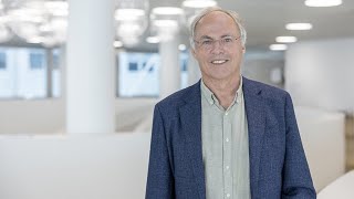
In this comprehensive lecture, Hans Clevers outlines his pioneering work on adult stem cells, Wnt signaling, and the development of 3D organoid cultures that recapitulate the architecture and function of various human epithelia. He describes the discovery of Lgr5 as a marker of intestinal stem cells, the molecular mechanism by which R‑spondin amplifies Wnt signaling through Lgr5, and the establishment of intestinal organoids using a defined cocktail of growth factors. The talk then expands to organoids of the liver, salivary gland, and other tissues, highlighting their use for modeling cancer, infectious diseases (HBV, SARS‑CoV‑2), metabolic disorders (steatosis), and for high‑throughput CRISPR‑based genetic screens. Clevers presents data on engineering organoids to mimic the stepwise mutational progression of colorectal cancer (the Vogelgram), measuring somatic mutation rates in single‑cell derived organoids, and uncovering a distinctive mutational signature caused by colibactin‑producing E. coli. He also discusses organoid‑based SARS‑CoV‑2 infection studies, identifying ACE2 and TMPRSS2 as essential host factors while questioning the relevance of chloroquine in primary epithelial models. Finally, he reviews ongoing clinical transplantation trials (inflammatory bowel disease, salivary gland restoration), explains why organoids cannot yet replace whole organs (lack of vasculature, innervation, immune components), and outlines future challenges such as vascularization, immune integration, and scaling for therapeutic use.
Chapters
Good. So, um, hi everybody. Um, it is my absolute pleasure to be able to welcome Hans Clevers today. He's a group leader at the Hubrecht Institute in the Netherlands. So thank you for joining us today to give the Caesar Milstein lecture this year. I have to say that this might be the most daunting introduction I've ever had to do. I've known Hans for a few years now, and he's, you know, a giant in my field, but I don't think I really appreciated all the accolades that he's collected over the years until I started reading up a bit more on him in preparation for this. And I can't tell you everything about what he's done, all of his achievements, because it would take the entire hour just to tell you. So I'll try to instead focus on some of the most sort of key discoveries and accomplishments. So many of our audience members probably will have heard of him for his work in organoids— I mean, really his invention of them, specifically intestinal organoids, which really beautifully model the regenerating intestinal epithelium— but he's actually received most of his awards for his earlier work and his continued work, really, on adult stem cells and The Role of Wnt Signalling in Regeneration and Cancer. So Hans characterised key factors downstream in the Wnt pathway, specifically TCF transcription factors, and their roles in various contexts, but then sort of more extensively in the gut. His focus on the gut then revealed many key mechanisms of how Wnt signalling controls intestinal regeneration, how it's abnormal activation leads to colon cancer. And the realization that Wnt signaling was driving intestinal growth and regeneration also led to the identification of the Wnt target Lgr5 as a marker of adult stem cells. And that really set up— really set the stage for, you know, establishing our current understanding of adult stem cells. And he used this really elegant Lgr5 labeling and lineage tracing experiments, which I'm sure he's going to talk about a little bit or introduce. And this allowed him to identify for the first time the adult stem cells of the intestinal crypt. And just to stress how important these cells are, I mean, they are responsible for essentially remaking the entire intestine every 5 days. So really beautiful regenerative tissue to study in this context. And so based on this knowledge, he was then able to tease apart how Lgr5 is activated by R-spondin and used this knowledge to establish the first 3D self-organizing in vitro models, the intestinal organoids. So his career, I feel, is a really beautiful example of how the scientific method should be carried out. I mean, he's made all these beautiful observations and then taken them, followed them, you know, followed the science from one hypothesis to the next to really carefully and elegantly reveal the cellular and tissue mechanisms. And so For this work, he was awarded the several prizes as I, as I mentioned, but I'll just highlight a few. For example, the Ernst Jünger Prize, the Breakthrough Prize, the ISSCR McEwen Award. He was elected a Foreign Associate of the U.S. National Academy of Sciences. He's a Foreign Member of the Royal Society, and he was recently awarded the Kao Medical Science Prize. So I'll stop there. I, in terms of Q&A, if you have any questions, feel free to type them in the chat, type them in the Q&A, or raise your hand, however you want. And if you want me to call on you and we can try to unmute you, then we'll see if Mark can help us with that. But otherwise, just type it in and I'll ask it on your behalf. So with that, I will hand it over to Hans Clevers. Thank you.
Okay, thanks very much, Madeline, for kind introduction and also for the invitation to speak today. I'll try to share my screen. There we go.
Looks good.
I think this works. So yeah, so yeah, I started as an immunologist, actually. And that's how we found these T cell factors, as we call them, TCFs. And it took a while to figure out that they were actually the endpoint of the Wnt pathway. Bart Bijlmer also made the same discoveries. And then when we started knocking them out, we actually hit upon the gut, where we found that Wnt was driving driving the process that you see here. This is one villus. So, mouse small intestine has about a million of these, we about a billion. And every villus is surrounded by a number of these crypts of Lieberkühn, that you might remember from your histology lessons. And it was known when we started reading about this process that there were stem cells at the base of crypts that were not really identified. There were some, some cell types were proposed to be the stem cells. But we found it most interesting because this was by far the most rapidly self-renewing tissue that we know of over the mammalian body. So cells indeed are born at the base of the crypt, where the stem cells must reside. They divide for about 2 days while they move up, these progenitors. After about 2 days, they pick up one of about 10 different cell phenotypes. They then keep on moving up, with the exception of paneth cells that actually turn around and move down and settle at the base of the crypts. All other cells will move up, and when they're 5 to 6 days old in the mouse, in human a little bit longer, they reach the tips of the villi and they undergo apoptosis. Now this process entirely halts when you block the Wnt pathway. So this set us on the track of exploiting the Wnt pathway to try to find the stem cells. And simultaneously, together with Bert Vogelstein in the collaboration, we found that colon cancer is also driven by Wnts, but then not by Wnt factors, but by activating mutations in the Wnt pathway. And I'll show that a little bit later. So Nick Barker in the lab, who's now independent in Singapore, worked with Pat Brown, who used microarraying, precursor to RNA-seq, but this allowed us to determine what the gene expression program is that is activated by Wnt in a colon cancer cell line. It's about 50 or 60 genes, we then mapped them back to the normal gut. And indeed, we found that all of those genes are crypt genes. So they are associated with proliferation and possibly with stemness. And one of them, when Nick worked his way through this list, stood out because it revealed the presence of cells that we had entirely missed. And I guess everybody in the field had forgotten about them. They were actually seen earlier by LeBlanc and even by Joseph Panette a century and a half ago. They're tiny. They're not much more than a nucleus. This is a knock-in mouse where Nick created a mouse with GFP in the ALGER5 locus and also CreER, an activatable version of the Cre enzyme. And you can see these, so every little stripe that you see here represents one of these cells. So there's about 15 at the base of the crypt. We thought were the stem cells, but it didn't really fit the bill very well. Mostly because they were quite abundant, and people thought that stem cells have to be rare. Most importantly, they don't— they divide constantly. In a mouse, they divide every 23 hours. And there was a strong belief that stem cells should not do that. Stem cells should be mostly quiescent. And actually, people use DNA label retention as a marker for stem cell quiescence as a way of identifying stem cells. So these cells would never be found by DNA label retention because they basically divide each day. Now then, using this CreER, he could now permanently mark some of these cells in blue using the ROSA lacZ reporter. I won't go into too much detail here. This allows us in a lineage tracing experiment to mark one of these cells in blue in a crypt, and then watch what it produces. So you see blue daughters over the first 2 days dividing. They leave the crypts indeed, as predicted, after 2 days. They then start forming all of the cell types of the gut. The most abundant ones, the enterocyte and the goblet cells, you saw here. Day 3, 4, and 5 after initiating the label in an adult mouse, they now work hard. They're on the flanks of the villi, they keep on moving upwards. And then by day 5 or 6, you see the first blue cells arriving at the tips of the villi, and they undergo apoptosis. Now, if you don't terminate the experiment then, but we leave the mice in their cage for 2 years, you'll still see these blue ribbons implying that the cell that we turned blue is long-lived. So it just doesn't live 5 days, it lives years, as long as the mouse. And second, we could observe in these ribbons that if we do the experiment correctly, are clonal by labeling a small number of these cells rather than all of them. It turned out that in one of these clonal ribbons, you would see all of the other cell types of the gut. So they're long-lived, multipotent, and by our definition, they were the stem cells of the gut. There's a lot of discussion in the field because they didn't really fit the bill, as I already said, but I think now people accept that these indeed are the stem cells. Now, While we were doing these experiments, we also found that LTR5 was expressed in many other tissues, particularly in tissues when they were actively dividing, like the stomach epithelium or hair follicles. But also when you damage other organs where normally there's not much proliferation, like the liver or the pancreas or the lungs, you'll see LTR5 coming up. And LTR5 is essentially a Wnt target gene, an indication that Wnt activates these stem cells. And I think we would now feel safe to say that every epithelium in a mammalian organism will use Wnt as its major driver of stem cells. And when the stem cells are activated, they will actually express Lgr5. Now, what does Lgr5 do? On the left here, you see Lgr5, a 7-transmembrane receptor that we don't believe to be coupled to G proteins. On the right, you see Frizzled, another 7-transmembrane receptor family, also not coupled to G proteins. And you see LRP 5 or 6. And these are the very well-defined co-receptors for, for Wnt factors throughout the animal kingdom. Now, during development, this is what happens a lot, is Wnts get secreted, they're lipid modified, they're very difficult to work with. They can maybe only travel from one cell to the direct neighbour. When you engage their co-receptors, signalling through beta-catenin and TCF leads directly to activating of target genes. Amongst those target genes are these E3 ligases that you see here. And what they do, they directly move back to the activated complex, and they put ubiquitins on. And this actually breaks the signalling cycle that you see here. I'll stop the movie here. So this is great during development, this would only take 2 to 3 hours. And now the cell is ready for the next signal. And you might realise that, for instance, zebrafish fully develop, vertebrate zebrafish, in a matter of 6 days. So they have to make thousands and thousands of decisions while they're developing these embryos with not Hedgehog, BMP, these types of pathways. And so you don't want the signal to be too strong. And particularly, you don't want it to be long lasting. Now, that's great during development, but not good enough for a stem cell. So stem cells tend to have Lgr5. We found, and actually simultaneously some other labs also reported the same thing, that a molecule, a secreted molecule called R-spondin, known to be a Wnt amplifier, by unknown mechanisms. But so what it does, if there is a Wnt signal, these Wnt signals are usually weak. But when you now add R-spondin, they become very strong. And so we found that R-spondin binds to Lgr5. It's actually Lgr5 is a receptor for R-spondin. And this is the mechanism that Feng Kong's lab and our lab resolved. So what happens is when a cell undergoes active Wnt signaling, and Lgr5 is expressed on the surface of these cells, because they're stem cells, R-spondin will bind with high affinity to Lgr5. It will also bind with good affinity affinity to these E3 ligases, remove them from the surface. And now the active Wnt signaling complex stays on the surface of the cell and isn't gone in a few hours, stays for days. And if you read out the strength of the signal, it goes up from maybe 5-fold to something in the order of 200 to 500-fold. This would paralyze embryonic development totally. So that would not work. But this is exactly what adult stem cells like. So they like these robust, prolonged Wnt signals. So you need to have an engaged Wnt receptor complex. And in addition to that, you need to have an ALGA5 family member and an ARSPOTIN family member secreted in a niche of these stem cells. And then you get these very potent Wnt signals that, that adult stem cells need. Now, when Toshi Sato joined my lab, maybe 14 years ago or so, we didn't know that ALGA5 was the receptor for this respondent is Wnt amplifier. But we sat down and we were thinking about ways to culture these stem cells. Because they were constantly dividing, we thought it should be possible to create conditions that mimic the crypt environment, and to take one stem cell out of one of these mice and then just make many, many stem cells. Now, what Toshi did is we actually came up with three recombinant proteins based on old work from our lab. Instead of Wnt, like Wnt3a or so, hard to produce lipid modified, very sticky. We were lucky to to choose our spondin as a Wnt amplifier, not knowing then that it actually is the ligand of Lgr5 and allows this very potent amplification that I just showed you. Anyway, so when we put these three, these three proteins together and advised by Mirna Bissell, we did this not in 2D, but we did it in Matrigel in 3D. And what we get is we get fantastic growth, but rather than a lump of stem cells, like what you would get with iPS cells or so, we get these structures. And here you see movies, they run over about 3 days or so. The structures essentially contain a central lumen that are lined by all of the mature cell types that you'd see in a normal epithelium. So, enterocytes, goblet cells, tuft cells, enteroendocrine cells, etc., etc. And these buds that stick out are crypt equivalents. They have multiple stem cells at the tip of these buds, so at the base of the crypts, interspersed with the cells I already mentioned, these paneth cells. And paneth cells essentially are daughter cells of the stem cells. They also secrete a number of the niche factors. So they're also part of the niche of these stem cells. These structures have grown for many, many years, and actually they outlast the original owner. They keep their telomeres long, so they have good TERT expression, etc., etc. And the question was, are these now still normal cells, or have they transformed into cancer cells? Particularly because, as you can see, they remain extremely vital. Now, by sequencing, we never found oncogenic mutations. So I don't— sorry, I don't show this here, but sort of implying that they are not— they've not undergone malignant transformation. But when we transplanted them, I don't show this here, and this was done in collaboration with Mamoru Watanabe in Tokyo. So Toshi grew up a single stem cell to about 100 million cells, sent them to Tokyo, and there he could transplant— Mamoru could transplant dozens of mice with inflammatory bowel disease and essentially create tissue from our single stem cell in each of these mice. That cured the mice and actually stayed fine, so didn't turn into cancers, also wasn't exhausted, so didn't disappear for the rest of the lifetime of those mice. So really remarkable. We never intended to do this, but to take a single stem cell in a rather simple culture condition, 3 recombinant proteins, no serum, matrigel, will create structures that essentially recapitulate many of the primary structures and cell types and also functions we now know of the gut epithelium. Now, since then, we, but then also other labs have joined in, have come up with variations on the theme. So, the basic cocktail is a strong amplification of Wnt signals. So, Wnts are spawned in. CHIR can be used, small molecule, we don't really think it's too good. Then we need to block BMP and TGF-beta. That's done with, in our case, with Noggin, which binds BMP factors. And we need to activate a tyrosine kinase, receptor that depends a bit which one it is on the tissue that you're trying to grow. And often EGF or IGF or HGF or FGF will allow you to do that. And the combination of these three signaling pathways will actually allow you some growth. And then for particular tissues, you might add additional goodies, memory gland like estrogen and progesterone, prostate, not yet here, like testosterone, etc. And some of these cocktails will have up to 10 different defined components, but there is never serum in these media. It's highly defined. The only problem so far is still is, is Matrigel, which is collagen laminin, but might have other undefined proteins as well. But it's magical for these organoid cultures. Now we can use these to do all sorts of interesting experiments, but I'll say quite a bit about modeling cancer and modeling infectious disease in these primary tissue, primary epithelial tissue organoids. One example of a non-gut organoid, the liver essentially has these two liver-specific cell types, so endodermal cell types. Cholangiocytes build bile ducts. Bile canaliculi here are formed between, in between two hepatocytes. They merge their apical domains to create a channel that's called the bile canaliculus. This allows them to secrete bile into this canaliculus that then goes on into the cholangiocytes. The cholangiocytes form bile ducts. And these are hepatocytes that, keep an eye on the clock, that are the major chemical factory of our body, but also a source, a major source of, for the serum proteins. Now, the liver is probably also the most regenerative organ. Chronic infections will destroy the hepatocytes, and then the cholangiocytes will dedifferentiate into oval cells, and they can repair the liver. This would be viral infections or intoxications that last over longer time periods. The other way by which the liver can repair is rather spectacular. So, if a surgeon removes two-thirds of a liver, for instance, because of metastasis from colon cancer, the liver can grow back in a matter of weeks. And that's done not by stem cells, but essentially hepatocytes that go into cell cycle. They don't dedifferentiate much. They just, if all of them divide a few times, you rebuild the capacity of the original liver. Other cells in the liver are not endodermal, so they migrate in, like Kupffer cells are really macrophages from the bone marrow. Stellate cells are from the mesenchyme. Blood vessels are, of course, and lymph vessels, sorry. Nerves are also part of the liver. But we try to model liver organoids. So Miri Huch, who's worked in Cambridge for quite a while, is now in Dresden, came up with protocols to grow cholangiocytes. And this is remarkable. Almost every cholangiocyte will give rise to a cholangiocyte organoid. But the hepatocytes, the dominant— these are these cells here— the dominant cell population in the liver, we could never grow. And this has been a major issue in the liver field. There are companies selling human primary hepatocytes for all sorts of metabolic studies, but also for safety studies in the drug development process. But these, the moment you put them in culture, will slowly deteriorate, and they will— after a week, they're rather useless. People have tried to make hepatocytes from iPS cells, but they really don't get much beyond a very fetal-like immature state. And also, the hepatocytes we make from these cholangiocyte organoids they remain rather immature, they never look like hepatocytes, so large, big nuclei, 4N or 8N chromosomes, etc. So we've tried hard to get this going. That was actually solved by Hui Li Hu, a Chinese postdoc who is now back in China. This is a mouse still. So we sort a single hepatocyte, and she defined a cocktail of about 10 different components. We mark hepatocytes here with albumin-Cre rosa tomato. So we know for sure this large cell is a full-blown hepatocytes. As you can see, it goes into mitosis, divides, and within a matter of 3 weeks— they're not the fastest growing organoids— but we get these structures. And then with some adaptations, we could do this for human organoids. And here I should state that unlike most other tissues, the older the donor is, the shorter the number of months, the briefer the time period is in which we can culture these exponentially. So an old donor will allow us growth of maybe 2 to 3 months, a younger donor maybe 6 months. So we have now referred to fetal human, fetal liver. In culture, they become, the hepatocytes become adults. So if you compare them to adult hepatocytes, they are virtually the same. Yet these will often grow forever. So we have a number of fetal hepatocyte lines, organoid lines that we can now grow forever. Here you can see production of one of these abundant serum proteins, alpha-1 antitrypsin in red. You can see how abundant the expression is. You can also appreciate how large the cells are, 40 micrometers, the very prominent nucleoli, the expression of a mature transcription factor of the hepatocyte lineage, HNF4 alpha. So they really are mature hepatocytes. But you might also appreciate there is a mitosis here, so they keep on dividing. They expand maybe 3-fold per week, 2 to 3-fold, rather than the 10-fold that we see with gut organoids. But they remain in cycle exponentially for at least a year. They not only just create hepatocytes, but maybe better seen here, They also— so these are these large hepatocytes with the nucleoli. They also create a bile canaliculi. So I just told you these are the structures formed by fusion of the apical membranes of two adjacent hepatocytes. The MRP2, the marker that we stain here, secretes— is the channel that allows secretion or the transporter, sorry, of bile acids. So it marks these bile canaliculi. And as you can see, they from anywhere in these organoids, they feed into a central space. Eventually, this will burst. But this is where we think that normally the, the bile ducts formed by the other cells of the cholangiocytes should sit, and will take the bile out of the liver into the gut, into the gallbladder. So really remarkable how well these hepatocytes on their own behave as if they're stem cells, but at the same time, they rebuild structures that are very close to the real liver. Now, these can be transplanted, much like what I just told you earlier. This was done by Ipid Yong in New York, our collaborator, and postdoc in the lab is Helmut Geijert, who did it. It's now in Zurich. And you can see even 2 or 3 months after transplantation, so this is the islet of human albumin expressing human hepatocytes in a mouse background that has a defect, has a genetic change in which, on normal Ciao, leads to the destruction of its own hepatocytes. And in yellow, you see Ki-67 positive nuclei. So there are even 2 or 3 months after transplantation, these hepatocytes are still dividing, and they slowly fill up the liver of these mice. Now, this is now a model that turns out to be very, very easily modified by CRISPR. We can do, you know, all sorts of knock-ins, knockouts, single base changes with base editors. And we have two, just two slides on ongoing projects in the lab. One is, both are models that have been lacking from the liver field. HBV, hepatitis B virus, is a huge problem worldwide. But there are no good models to study HBV infection long term. That's where treatment really should be aimed at, try to remove latent hepatitis B in hepatocytes in patients. And as you can see here, this is now a month, but we think we can do it much longer. What we do here is we grow these organoids, we then plate them out in 2D, we take the most important mitogenic factors out, so they essentially make mature human hepatocytes. And when we now infect with HBV, we see HBC antigen, the core antigen from the virus abundantly expressed in these hepatocytes after about a month in culture. So this MBC-OLBC integration of HBV virus in the host genome. And by all means, we think this is now a good model to study chronic HBV infections. And we hope that we can use this to identify host genes that are key for this. We actually already found a few by CRISPR, we knocked them out, and we can no longer expand the virus. And also maybe for drug development, this could be a very useful platform. And another disease, just one slide, is steatosis. So this is lipid accumulation, an epidemic in the Western world, also in India, for instance, and then coming up in China as well. It's essentially people overload themselves with calories, and the calories can go to your fat tissue, but they can also go to the liver. Here you see normal human organoids under normal culture conditions stained for lipids. There is no lipid, as you can see here. But we can either genetically modify the organoids in ApoB G mutant will not allow secretion of lipid. So you see an enormous buildup of triglyceride droplets here in these organoids. They actually keep on growing if you do this, which is very surprising. And we can do the same thing by giving high doses of fatty acids. They get incorporated in these organoids, and they will actually create a steatosis-like syndrome. And we've actually been testing about 25 or so compounds currently in clinical development. There's no registered drugs for steatosis and for NASH, and we find that only 2 or 3 will block block the buildup of lipid in these organoids. They do this very well. But all the other drugs, we cannot find any effect in this system. So, we also think this is a very useful model to help understand steatosis and also maybe help develop therapies for steatosis and for NASH, or NAFLD, as it's called here, nonalcoholic fatty liver disease. We've done a lot of work on cancers. Because we are growing epithelial cells, we definitely should be able to grow grow carcinomas. Carcinoma by definition are derived from epithelial tissues. It's about 90% of all adult cancers are carcinomas. So what you see here is we can grow side by side— this is done by us but also by many other labs— healthy and cancer tissue from the same patient. You can see the different types of carcinomas that we have been growing in the lab, but you can find in the literature many other approaches to also other rare forms forms of carcinomas. You can sequence these organoids very well. I should stress that they only contain cancer cells. They will not have any immune cells or blood vessels, obviously, or stromal cells. And most importantly, we can perform drug screens on these cells. So this would, in principle, have the promise of using this technology for personalized medicine approaches. And that indeed works quite well, as I'll show you in the next slide. So this is one of those pairs normal and tumor tissue from the same patient. We have, we now have about 300 or 400 in Utrecht. Many other living biobanks, as they're called, are being developed elsewhere. You can perform these drug screens, as already said, and while we were trying to show that these drug screens on organoids predict very well the response of an individual patient from which the organoids were derived, it's a beautiful paper from the UK appeared in 2018. Claiming that organoids have a very high predictive value for the behavior of drugs in the patients from which the organoids were derived in the order of 90-95% correct predictions if a treatment will or will not work in the patient. We were then involved in a number of other studies, but these were all done primarily not by us, coming up with similar numbers, 80, 70, 80, 85, 90% correct predictions of drug sensitivity or resistance. You have to realize that the average cancer patient probably overall only benefits from first-line treatment in 40% of the cases. So even if you're classified very well in a group of patients for which that particular treatment works the best, you're never sure whether it will work for you. And actually, the majority of patients will not benefit from cancer treatment. Now, if this all holds up. And it has to be much, much cheaper, has to be much faster. Probably we need machines to do this rather than highly skilled lab personnel. But then possibly this could be a very nice addition to the standard diagnosis that's performed on cancer patients, of course, which includes imaging, pathology, and DNA sequencing. But this is ongoing. And we have high hopes that this might be an addition to the armamentarium of the diagnostics in. You can also use organoids to model cancers. And this is a paper of a number of years ago done in my lab by Jarno Drost, but totally independently done by Toshi Sato. We both had the same ideas simultaneously, and luckily we published simultaneously. And we both realized that the most common mutations in colon cancer, these 4 genes, actually reflect— at least the top 3— reflect exactly what we had empirically found to be the key signaling pathways for normal stem cells of the colon. And here you see, so here you see what I want to say. So we need Wnt, I told you Wnt are spondin. We need EGF for colon organoids, and we need Noggin, the BMP inhibitor, a recombinant protein here. Cancer, colon cancer tends to first lose APC, or sometimes undergoes other activating pathways and activating mutation in the Wnt pathway. And the prediction is if a cell loses APC, it no longer needs Wnt, because you take this Wnt inhibitor out of the cells. KRAS, similar relationship with EGF. If you activate KRAS or any, any other mutation in the signaling pathway of the EGF receptor, you would no longer need EGF. SMAD4, a transcription factor in the BMP and TGF-beta pathways. If you knock out SMAD4, you would no longer need the BMP inhibitor Noggin. And by losing these, or by undergoing these three mutations, cells would become independent of the exogenous factors that we that we can't see applied to the cultures, but that we believe are present in normal crypts, create a normal crypt environment. And we also modeled p53. This was all done by CRISPR. And originally we did this because we wanted to recreate what's called in the field the Vogelgram. So Bert Vogelstein and Jörg Fehren in 1990 proposed that there was an ordered array of mutations that would take a normal colon cell into, you know, through these different adenoma stages into a malignant in invasive and metastasizing colon cancer, colon carcinoma. This would first be APC, as you see here, then KRAS, then this turns out to be SMAD4, and then eventually p53. And that would then let the cell go from here to here. Now, to put this in a movie, again, you'll first see the vogelgram. So starting from normal epithelium, an ordered series of mutations would take the normal from normal, presumably stem cells of the colon epithelium through these early, late adenoma stages to carcinoma stages. And we were going to model this in organoids. And here you see a normal human colon organoid. It requires the growth factors you see on the right. We target APC with CRISPR, we leave Wnt and R-spondin out, every cell will die in the culture. But the APC mutants that no longer require Wnt, predictably, will actually grow out, and that worked. So we had one mutation. Now we target CRISPR, two alleles, we can kill p53 wild-type cells in NUTLIN. That works very well, and only the mutants will grow out. So now they have two mutations. There is now KRAS. We now have to knock in G12D. This is done with a long oligo of about 100 bases. We leave EGF out of the medium. Now they have three mutations, and they only require the BMP inhibitor knock-in. And finally, SMAD4, the BMP transcription factor, we target the gene, we leave the BMP inhibitor out of the medium, everything dies, only this MET4 mutant will grow out. This worked actually beautifully. And we had a little library of all of the different combinations, you can actually do this in any order, you can do it at the same time, as I think I'll show you in the next few slides. They can be transplanted orthotopically into the colons of mice. And only the one with the 4 mutations will actually give us invasive and metastasizing growth. So this one rapidly metastasizes to the liver, as one will see in humans. And all these, these other combinations of fewer than 4 mutations will give us something that would be scored as an adenoma, or might even not grow at all, as you can see here. So this essentially confirmed the flowgram, also made this very nice link between the empirically defined set of growth factors in this for organoids and the mutations that cancer apparently selects from the very same organ. We since then have used this a lot, this approach. So it's the same as people have done with mouse engineering, for cancer models in the mouse, it's much, much faster, because we can essentially mutate genes in a matter of 2 or 3 weeks, and then come up with the next, but we can even combine. And nowadays, we don't use the crude approaches with CRISPR/Cas9 in its basic form, but we use these base editors, and also prime editing, if base editors don't work. Base editors, the ones that we use can do 2 things, they can either create, turn a C into a T or a T into a C, when you— this would be 2 of the 6 different base changes that one can think of. So that would be 40%. But it turns out that you can create about 50% of all the SNPs that are seen, or all the single base changes that are typically seen, as a result of somatic mutations. And they work by linking Cas9, so blocking its nuclease activity, and linking it to an enzyme, ApoBec2. For the C to T, I believe, and TAD-A for the T to C mutation, as you can see here. Now, they can actually— so here, Martin Hürth in the lab loads one of these base editors with— so it's one base editor with three different guide RNAs. And at the same time, he creates a stop code on APC, a stop code on p53. And he activates— he puts an activating mutation, a single base change in PIK3CA, as you can see here. And by sequencing, that all works. Now we can play this trick again with leaving growth factors out of the medium to select for APC mutants or for PI3 kinase mutants. And that's— for that we use a MEK inhibitor. And we can kill the— we can select the p53 mutants by adding Nutelin again. And you can see, if we select with all three at the same time, we still get clones that grow out. And when you sequence these, they will have all the mutations you want. And when you select with two, you typically find only the the organoids that have the pathways for which you are selecting. So very rapidly, you can actually create a large collection of organoids with all the desired genotypes and then perform any functional assay that you want to do. This is a paper we hope to write up soon. This again first refers to an approach that we published with several collaborators a few years ago. So whole genome sequence of single cells is still not feasible at the high— to generate a sequence of high high quality. That's probably getting closer. But what one can do is expand single cells in the form of organoids briefly, and then when there's enough cells, maybe 1,000 or so, then sequence that organoid. And this gives you a readout for the genome of the single cell. Now, we originally used it with collaborators that you see here to determine what the mutational somatic mutational rate is in 3 different organs. And we did this for a reason, because Vogelstein and Tomasetti had published that the rate of cancer frequencies in human organs correlated directly with the stem cell activity in those organs. So stem cells that divide a lot would have a lot of mutations, stem cells that don't divide would have no mutations. Now, the liver would be an organ with almost no proliferation. Paul stated that the colon has the most active stem cells and the small intestine would be less. We don't find that that is true, actually, they have sort of equally active stem cells. Anyway, we, from primary tissue from patients of very different ages, we selected single cells, we briefly grew them up, and then we sequenced them, we compared them to the germline sequence. And this gave us a mutation rate that's actually linear. We now have many, many more of these. And what essentially happens is that independent of age, every year in your colon or in your small intestine, you add about 40 single base changes to the genome of every single stem cell. And this is linear, doesn't increase this rate of the addition of mutations. This goes from 0 to 80, as you can see here. Very surprisingly is the liver. So the liver doesn't divide much. If you never, if you don't have liver disease, it divides very rarely. Yet the rate of somatic mutation was still the same. We now have younger and older, and again stays on the same rate. So 40 single base changes per year. A group in Voxel, a collaborator, has since done this for bone marrow, and there it turns out to be a little bit lower. It's more in the order of 20 single base changes per per stem cell. So this approach is actually used for another collaboration that we did with Ruben's lab in Utrecht. And in my lab, Cayetano and Jens worked together with Axel, they actually came up with this project without Ruben and me knowing. They had read about a particular strain of E. coli that is quite common. It's carried by— well, it's published to be carried by 10 to 30% of people with colon diseases. We've since gone back and looked in healthy populations. And we find the same rate. So about maybe 1 in 5 individuals carry this particular E. coli in their microbiome at any given time. And how this is different from a normal E. coli is it has an extra 60 kilobases of DNA in the genome encoding about 10 genes. And these genes together constitute a synthesis pathway for a substance that's called colibactin. I'll show it in the next slide. And when E. coli have this particular extra piece of DNA in their single chromosome, chromosome, and you culture them on top of human cells like HeLa cells, you'll see the rapid appearance of double strand breaks. And that's why these PKS E. coli are called genotoxic. That is old literature. I had never heard of this, but our PhD students found out about this. Surprisingly, particularly after I finished this little story, PKS E. coli, a particular strain, is very popular as a probiotic. So it's given to people with chronic colon problems and other chronic diseases. And it's actually still as we speak, it's in clinical trials for psoriasis, inflammatory bowel disease, irritable bowel disease, etc., etc. But we don't think it's a very smart idea. This is colibactin. It's never been synthesized in its entirety. It's a very labile substance, it probably needs to be injected by Colibacter into neighboring cells. And it's assumed that they use this to fight with other bacteria around them in their constant ongoing competition. Competitive battle with other species in the microbiome. This paper proposes, and we think we would agree with this, that there are two warheads on colibactin, and they can covalently bind to A residues in DNA, bacterial DNA, but also in host DNA. And if that would happen on two opposite strands, you would have an interstrand crosslink that would not be compatible with life of the host cell. E. coli itself is actually protected against colibactin. One of the genes is an enzyme that destroys colibactin. If you knock that out, actually, that's not compatible with life of a pks-positive E. coli. So this is the first experiment that they did. So we got a potent pks E. coli strain and an isogenic mutant in one of those genes, the q gene, you just saw that here on the right. So left is the bad one, right is the good one. The bad one makes a lot of colibactin, the good one cannot produce colibactin. They can be grown quite easily on agar under normal conditions, as we normally grow E. coli in the lab. You can pick them up with a glass needle and then inject them in organoids. We tend to do this in 3D, but you can also plate out organoids in 2D after they've been expanded, and then you just put them on top. So this is— they're injected luminally. This is where they would normally be in a real gut. So we leave them in for maybe 1 or 2 days, and then see if we can reproduce what was in the literature. And indeed, if we inject E. coli, we find that if we inject the bad guys, or the one with the complete pks island, we get lots of double strand breaks. If we inject the negative control, there was a single double strand break in the entire culture. So every dot here is a single organoid that was injected with ColiBac. You can see the breaks are everywhere, and we visualize the breaks by a stain for γ-H2AX. So they then did this heroic experiment, they injected multiple times on Mondays, let them live inside the human organoids, and then on Fridays cure the organoid cultures with antibiotics, because they have to be trans— they have to be passaged, so broken open, broken open. And if we would do that, the E. coli would escape and would overgrow the culture. So So every Friday they treated antibiotics, the next Monday they again inject E. coli. They did this for about 3 months. And then if we would now sequence these and compare them to these on the left, you would expect to see many more mutations if all was true that we were hypothesizing. However, if we just sequence the entire organoid, we would probably not see the mutations because every single cell will have its own unique makeup of somatic mutations induced by bacterium. So they then, the trick that I just showed you, they subclone single cells. So each of these cells has been exposed for a long time to colibactin, and the ones on the right are the negative control. We then sequence these whole genome. We could then basically look for single base changes, but also for small deletions and insertions, and classify them. This is done according to Mike Stratton's system originally, that you probably are aware. And what you then can see quite easily is that indeed, we get more mutations in the PKS-exposed organoids. But in particular, we get a particular class of mutations, T2Cs, the green ones here, and one here, this pink one. And the way Mike classifies is he not only looks at the T2C change, but he also looks at the direct base upstream and downstream. And that actually gives him a way of classifying mutations. I don't think I think I want to explain this. But what we find, that actually, we see very specific single base changes. So the E. coli bacterium always targets a TA pair. And it's just not any TA pair, but it's a TA pair in an AT-rich context. And in particular, there's an A at -3. And what happens is either this TA pair changes into any other base, or this TA pair gets deleted. So there is a a single base change signature and as a deletion signature in the same context. And if we model this now, taking into account that the colibactin can covalently bind to As, what we think is happening is that colibactin will bind covalently to this A here, it will bind covalently to the A opposite the T that we have in our little scheme here, and now you have a double strand break, an interstrand crossing crosslink. The cell has to resolve this. In the process of resolving this, it apparently creates mutations that are very recognizable. It's a very unusual mutational signature, where you change this base pair into any other base, or you delete this base pair. And sometimes you also delete one or two more base pairs upstream from this TA here. If you now look where they, if they really occur in real life, because this is of course extremely artificial, and we went, we turned to the heart the Grachtwig Medical Foundation, who have more than 3,500 whole genome sequenced metastases of a large variety of different cancers. We essentially only find the mutational signature in colon cancer, fitting very nicely with the idea that the signature is caused by E. coli. We see occasionally a case of head and neck cancer where E. coli can also live, and bladder cancers, and we have a few more now. And we then turn to another large cohort owned only of colorectal cancers from Genomics England. And the same observations were made. And actually, I think there is a higher rate of these types of cancers with this type of mutational signature on the left side of colon compared to the right side of colon. And again, the frequency was in the order of— we now think it's maybe 12 to 14% of colon cancer patients show clear signatures of the presence of Colibectin and PKSI E. coli in their cancers. Now, these are just random mutations, it can be anywhere in the genome, but they also target oncogenes, cancer genes, and particularly the APC gene is a very good target for this mutational process. And we find that many APC mutations in these cohorts bear the signature of colibactin, so are likely caused by the presence of Pseudomonas aeruginosa, sometimes before this cancer developed in the patient's colon microbiome. Yeah, this I think I already said. So we think it would be a wise idea to see where PKS E. coli is. And if this all holds up in other cohorts, maybe eradicate PKS E. coli. It's very easily done with antibiotics. And with a fecal transplant, you could give back a healthy, healthy colon microbiome. And I guess the use of PKS-positive E. coli as probiotics should really not be not be advertised. And finally, a few more minutes. When we went into a lockdown, we actually were allowed to do corona research, SARS-CoV-2 research. We realized that ACE2 is probably expressed to the highest levels in the small intestine, not in the lungs. And what I show you here in the small intestine also works in lung organoids. We also realize that many COVID patients present with GI symptoms. They will never die of these symptoms, but diarrhea, gas, nausea, stomachaches. And actually, there are quite a few documented cases where there was never respiratory symptoms, there were only GI symptoms. And this was March last year, people had already detected viral RNA in the stool. So this might be a way of transmitting the virus. And also it might be a way of tracking the virus. And it's actually being used now by taking samples from the sewers of cities and see if RNAs for the virus go up or down. And this is a very generic measure for the state of the pandemic. And it's certain population in the city. Now what we did is we asked, can we use organoids to score if the virus can infect particular organs? That worked very well. You see SARS in blue, so the original virus, SARS-CoV-2 in red. So on gut organoids, there is a dramatic increase in viral particles, live particles here, and in viral RNA, and much, much more than you would get in airway organoids. We've also done it in airway organoids, and airway organoids do better than alveolar organoids, for people who are interested in the lungs. And when we stain, so when we manage to infect one cell in an organoid, if you then go back 2 days later, so this is only 60 hours after infection, you can see that almost every cell in organoids are now infected. And by EM, we see that the viruses are secreted from the apical side, so on the luminal side, and the virus can therefore propagate very readily inside organoids but cannot jump easily from organoid to organoid, as you can see here. Now, we did a lot of RNA-seq, etc., etc., but in essence, this, this was actually confirmed that the lung is not the only only target organ. We now know there are many target organs, several other groups published at the same time that we published this. We've gone on, this is now a paper that's out, to look at host genes, because with CRISPR in the gut in particular, we can create libraries of mutants targeting gene by gene. We started with ACE2, when we knock it out as our positive control for the approach. When we knock it out, SARS-CoV-2 in green affects the wild type but not the ACE2 mutants, as SARS itself, you know, from 20 years ago also requires ACE2 as its receptor that was known. MERS is a family member that is very lethal, but hasn't really jumped much from human to human yet. Jumps from camels to humans in Arab countries, doesn't use ACE2 as its receptor, and indeed it doesn't care about us knocking out ACE2. MERS is proposed to use DPP4, another surface receptor. Also, by the way, SARS-CoV-2 has been proposed to use this as an alternative receptor. When we knock DPP4 out, MERS no longer infects, but now SARS-CoV-2 infects. So essentially, this shows that this system works. And because we can do CRISPR much more easily in gut than in lung, we have concentrated mostly on the gut. We then went through a long list of genes. And there were several papers, several papers in Cell, actually, I think 3 genome-wide CRISPR screens that came up with 30, 40, 50 host genes. We knocked out almost all of them. We cannot confirm any. We can confirm ACE2, I already showed you that. And the only other gene from those lists we can confirm by creating single knockouts, and we have multiple clones from multiple donors for each of the genes, is TMPRSS2. And TMPRSS2 is a surface protein known to activate spike, so it cleaves spike, it activates it, and then allows the virus to fuse the virus envelope with the host cell membrane. None of these other genes actually affect infectivity of the virus at all if we knock knocked them out. There's many other TMPRSSs. We've knocked the ones that were expressed out, they also don't, are not involved. So TMPRSS2 appears to be a very nice additional target. If you want to, TMPRSS2 small molecule inhibitor would probably block infection of the virus entirely. Most importantly here, cathepsin L was one of the strongest hits in the genome-wide screens. In our case, it only enhances. So this is the knockout that only enhances infectivity. Cathepsin L is a cathepsin that is involved in the endocytotic pathway. So keep that in mind. Chloroquine is an inhibitor of endocytosis. In our hands, as published last year by a number of labs, works fantastically well to block viral entry into Vero E6 cells. And Vero E6 cells and similar cells are widely used by virology labs. They have to be, I believe they have to be transfected with ACE2 because they don't carry the receptor. Receptor, which is typically epithelial. Vero E6 cells are not epithelial cells. Chloroquine works fantastically well. Hydroxychloroquine works well. At 10 micromolar, you totally block infection by SARS-CoV-2. As you know, many patients have been treated with hydroxychloroquine and chloroquine, and we now have to conclude that it never benefited them. They got the side effects, but no, no beneficial effect. When we knock out or when we add chloroquine in to our cultures at 10 micromolar where we get full blocking in our hands also in Vero cells, we have no effect whatsoever here in blue when we do this in organoids. How do we put all of this into a scheme? And this is my last slide. So what we think happens in these Vero E6 cells, the virus binds ACE2, it is then endocytosed, and then finally it's released through the action amongst others of cathepsins. The viral RNA is then released from the endocytic vesicle and can then essentially perform its whatever bad functions in the cytoplasm of the cell. This is a step that's blocked if you knock out the cathepsins, but also is blocked when you add chloroquine. What happens in primary epithelial cells on the apical side where ACE2 is expressed, and where we know the virus will first encounter host cells in the respiratory tract or in the intestinal tract. So here the virus doesn't undergo endocytosis, it essentially binds ACE2. TMPRSS2 is expressed on the surface of epithelial cells on the apical surface. Actually, TMPRSS2 has not appeared in these genome-wide screens. We know it's essential. So TMPRSS2 will activate spike, and now the viral RNA is directly injected into the host cell cytoplasm. There is no endocytosis, and therefore there is no predicted sensitivity to hydroxychloroquine, as we find in primary epithelial epithelial cells. So we would argue that for a next pandemic, probably it's great to use these, these workhorse cell lines like the RE6. But maybe it would be good to also add an additional step where you take whatever you find into primary epithelial cultures, like organoid cultures, like I showed you here, and only then start treating patients with those drugs. And with that, I'm a little late, I reached the end of my seminar, I'll stop and I'll give the floor back to Madeline.
Thank you very much. Thank you, Hans. That was wonderful. That was a real tour de force, I think, on multiple fronts. So we already have actually several questions. So I think I'll— there's a couple actually that are kind of going in similar directions. There's one from Aurelius Roskofen, and there's another one from Dina Ashour, both of which are sort of asking about about whether organoids are reaching the point for transplantation. Can we, can they become large enough that they can be actually transplanted into human patients with conditions and we can start thinking about organ replacement therapies?
Yeah, yeah. So first of all, these organoids are not complete organs. They are only the epithelial parts of the organs. Now, if you think about, you know, the functions of your internal organs, the essential functions are performed by the epithelial part of that organ. Then there's, of course, the blood vessels and all the stroma, et cetera, et cetera, that you'd also need. So, having said that, there are several trials ongoing. One by our collaborators in Tokyo, Mamoru Watanabe's lab. I showed, and I just discussed, that you can treat mice with mouse organoids. He currently is now transplanting human human patients with inflammatory bowel disease, refractory inflammatory bowel disease, with their own organoids. So it takes about a month to take a small biopsy and to amplify it 10,000-fold under GMP conditions. And as far as I know, as we speak, delayed by the way by COVID, of course, he is now transplanting the first patients and hoping to break this chronic inflammatory cycle by healing the lesions with with healthy, happily growing organoids. And another trial that has started or is starting in Groningen, Holland, is salivary glands. Corpus is doing this. So essentially, head and neck cancer patients often get radiated, as other cancer patients do, to the head, and then the salivary gland tends to be very sensitive and you lose salivary production. This leads to dry mouth disease, terrible terrible condition. Difficult to speak, extremely painful, your teeth fall out, and very difficult to treat also. And there, so mouse to mouse works very well, human to mouse also. He's now going to do human to human, essentially injecting suspensions of salivary gland organoids. The organoids are never bigger than maybe a millimeter at most. Just injects them, so in the mouse it works very well, into the parotid gland or the submaxillary gland, and they basically will fill up the organ much like what I showed you or liver, where if you leave them long enough, nowadays, sort of, you replace the missing tissue by healthy normal tissue. So what he's seen in mice, he hopes to see in humans, and that's, that's started. So those are the only two trials I'm currently aware of. So we will never build entire organs with this technology, I think, but we can use the infrastructure of an organ and then replace the epithelial parts of the organ by cultured epithelial cells.
Endothelium. So can I follow up then and ask you, what do you think is the limiting— what is limiting them from becoming full organs? Why can't we do that?
Yeah, I think that was very obvious. There's no circulation. That is the big thing. You would need blood vessels, but not only endothelium, no forming nice channels through your organoids, but then you need, of course, circulation. There's no innervation, there's no immune system. So the immune system, I know many labs are trying to add, you know, to both to the iPS-based, what Madeleine, would you do, but also to these adult organoids. What's remarkable, what we've seen, but many other labs have seen, is the self-organizing capacity of— so these cells know very well, the stem cells know what to produce and where to put it, and other cells you add know exactly where to make the connections or where to go. So it's really surprising, but I guess a billion years of evolution we shouldn't have taken care of that.
So we have a question from David Baker, actually, who's asking, you mentioned that you block TGF-beta signaling to create the organoids. Due to the high levels of TGF-beta in the tumor microenvironment in certain cancers, do you think that this will hamper efforts to model the tumor microenvironment? And can we get around this?
Yeah, so here I would have to refer to work from my old postdoc Edu Baci in Barcelona. So the cancers that have high levels of TGF-beta typically have the cancer cells themselves have loss of function mutations in the TGF-beta receptor pathway. So they basically have made themselves immune to TGF-beta, and then they fire TGF-beta at the environment around them. This activates fibroblasts, you get this malignant stroma in the tumors. So that is, yeah, so indeed, yeah, so we can model this very well if we knock out TGF-beta receptor, or you want to knock out some smat4, then you can add, you know, whatever you know, how much TGF-beta you want to add, the cancer cells no longer are harmed by that. A typical epithelial cell will terminally differentiate, and you'll lose it. So that's, I think, how the cancer cells get away from TGF-beta and then can use it to create an environment that they like. So I think that can be modeled. We haven't done it, but Edu does this.
So there's another couple of questions that are kind of related. So there's one from Vaithish Belajahan and one from to Sutcliffe. They're both sort of asking about the speed of growth of the hepatocyte organoids. You said that they were slower compared with, for example, the gut organoids, and also you also talked about generating hepatic organoids from different age donors. So the question is sort of, why is it slower, and is it different depending on the age of the donor? So for example, are more like even fetal stage age, you know, tissues that you then make organoids from, are those fast? You know, are they, do they go the pace of development that would be happening?
I can make a few remarks. So generally, I think the younger the donor, the more vital the tissue is and the better they grow. Having said that, so far, so on the one extreme is the gut, the gut tissue grows fastest, and there's actually, we have things that grow even slower than the hepatocyte organoids. I think that's sort of tissue intrinsic. The gut organoids never age, so they never go into senescence. They have high levels of TERT. TERT tends to be a Wnt target, as published a while back. There's maybe one other remark. We think that the hepatocyte organs get in trouble because they do undergo senescence. So, when you take it from an adult, they will will not keep their telomeres at length. So they actually shorten the telomeres. And this actually, I think, is the only culture where we see this. So there they behave as people would predict, that they will, every cell division, they will lose a little bit of the telomeres. And interestingly, liver cancers often have activating TERT promoter mutations. So there is something in the biology of the liver that also cancer needs to overcome. So it needs to do something about the limiting activity of TERT, which in other tissues we don't see. So there the stem cells typically have high TERT and the organoids have high TERT. The fetal organoids, so there seems to be a black and white difference between some of the fetal lines that we can grow forever and other little bit later maybe fetal lines or early neonatal lines where they can maybe grow for a year, but they eventually will undergo senescence and they will have lost their telomeres. We can overcome it by, by pressing through it. But that's sort of an oncogenic event, and we don't want to do that.
So I was actually really curious— nobody asked this, but I'll ask it— about the ploidy of the cells. And I was wondering, when you're making these hepatocyte organoids, do you know what the ploidy is you're starting with? And when they're 4N or 8N or something, are they actually going back to 2N before dividing, are they staying 4N and just, you know, what's going on there?
So what I know is we can grow them from 4N epidermocytes. We cannot grow them from 8N epidermocytes. We actually have movies where we have cells with 4 spindles or 8 spindles that actually manage to separate, you know, to segregate the chromosomes very well in separate nuclei and don't have lagging chromosomes and things like that. We haven't really would be not so easy. We haven't really followed a single tetraploid cell into its first division. But the field would believe that 4N epidermocytes cannot divide. But we can start totally healthy organoids from 4N epidermocytes. The overwhelming majority in those organs will be 2N, by the way, so they revert to a 2N stage.
Okay. So yeah, that's really interesting. Okay. So then there's a Going back to the gut, and in particular, the E. coli infections that you've done. So this question is from Dina Asher. Do you know whether there are microorganisms present in the human gut that can metabolize colibactin and therefore neutralize the negative effect of the PKS-positive E. coli?
No, so E. coli itself can do it. It has this enzyme. The PKS islander also reports that it's not only E. coli, but other gram negatives can have this this piece of DNA. So it might jump between species, horizontal transfer, I think it's called. The problem with colibactin, that's so labile that nobody has ever seen colibactin in its native form. That's actually what I didn't say, because I felt I didn't have the time. But when we published the signature, Mike Stratton, who's of course, you know, the hero of these signatures, had been sequencing single crypts from healthy individuals and found the same signature. Sometimes in a few crypts where many other crypts wouldn't have the signature, and then a few crypts would have it. And he also showed you could actually do a lineage because crypts can undergo fission. And he found one case where 4 crypts had the same signature. So this crypt had divided twice, but after this division, it no longer added the signature. Implying that there was— so in his paper, which came out almost simultaneously with ours, he says there is an exogenous factor that induces this type of mutations that can transiently be present and then disappear again. So what we think is happening, that when people are colonized, and as long as E. coli is in the lumen of the gut, you're safe. But when it managed to actually penetrate the crypt and colonize a crypt, then it's close to the stem cells, can probably directly inject colibactin into stem cells. And that's when then you really are in trouble.
So I will ask, I think, probably just one more. It's a bit of a broader question, I think, because I want to make sure you have at least 5 minutes to take a short break before you start with your meetings. So this is a question from Fiona Gribble, and it's kind of asking a general question about how well do you think these different organoids are really modeling the phenotype of their derivative, you know. So, you know, are they really good avatars for people, and how can you convince her otherwise?
Because she's— Yeah, so simple analyses like single-cell sequencing comparisons, like you do in the brain. So for the gut in particular, we probably make the most complete organoids. It is really remarkable how, you know, they are identical. So, when you keep the organoids growing, you know, under high mitotic conditions, then you tend to see the cell division signature that normally is not present to that extent in the gut. But when you titrate this well, essentially, well, the enteroendocrine cells of interest for this question, but all of the other cells are essentially indistinguishable by RNA expression from their, from the in vivo counterpart, which— so we don't see what you get from iPS typically is the sort of late fetal, somewhat immature stages, but we start from mature, fully specified stage. Having said that, I think if you now want to do more complex features like, you know, drug resistance or drug sensitivity, it gets to be more complex. And if you want to know about sensitivity to immune oncology drugs, for instance, then a single organoid will not tell you. You then need to add immune elements, and it depends on how you do how well it predicts. I think it depends on how well you build your model. It'll never be equivalent to a real in vivo situation. But as long as you know what you're looking at and that you model it well, I think they are great tools. But there's never— it's never the real thing. It's always reductionist.
Yeah, it's always a model. Good. Thank you. I think on that note, that's a perfect note to end on. And I just want to say a really big thank you again, Hans, for giving this thought-provoking lecture. And I'm sure that the audience enjoyed it as much as I did. You have 5 minutes, and then I think you have your link for the next— the first meeting that you have.
So it's a different link?
Yeah, it's a different link. So we're going to go off this link, and it should be in your schedule. But if you have any problems, just email me.
Okay, well, thanks very much again.
Thank you, everyone. Bye.
Bye-bye.
More from Joshua







Scientist Stories: Hans Waterham, The value of phenotypic assays in peroxisomal disorders




Scientist Stories: Joel Zlotogora, Genetic carrier screening programs for reproductive purposes