Scientist Stories: Hans Clevers, Stem Cell based Organoids in Human Disease
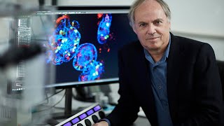
Hans Clevers traces his discovery of Lgr5 stem cell markers and organoid culture, which lets single cells self-organize into mini-guts. He shows how organoids model diseases like cystic fibrosis and colon cancer evolution, and enable CRISPR gene correction for precision medicine.
Chapters
Well, thanks very much, Halnd, for a kind introduction. I think we failed to get together last year, but it worked this year. And it's an honor to, I understand, to give the second Jim Lopski lecture. I'm not a geneticist. I actually used to be an immunologist, and I'll give you some background of how this ended up in studying the organ that you see here. This is a small intestine of a mammal. This could be a human or a mouse. And when we first saw this about 20 years ago, we realized that this is probably the organ that looks like it's designed to study stem cell biology. It is the fastest self-renewing organ in our body, at least the epithelium. It was known that these structures that are called crypts of Lieberkühn, that they harbor stem cells. They had not been identified. They generate daughters every day. These daughter cells proliferate with a life— with a cell cycle of about 12 hours. For 2 days while they move up. So at the age of 2 days, they exit the crypts, they mature out into one of a number of different cell types that we need to digest food and take up nutrients. They move up for another 2 days, and 4 days after they are born, they die at the tips of the villi. So every 4 days as you sit here, you totally replace the lining of your intestinal tract, and this amounts to about 100 to 200 grams of cells that you produce per day. And then die 4 days later. So it's the speed of the system, far faster than skin or bone marrow or any other self-renewing tissue. But it's also the design. A mouse has about a million of these crypt villus units. We have about a billion in our small intestine. The colon doesn't have villi. It's flat. But other than that, it's really molecularly very similar. But also this design, it's so stereotypical that it makes life of a scientist here very easy because you can just look at a small image, microscopic image, you know this cell is 3 days old, this cell is 4 days old. By H&E, you can recognize all the cell types. Very simple. You don't need any markers to do so. Yeah, and then as I said, small intestine and colon is very similar. There was a lineage. This is what developmental biologists or hematologists like to draw. So a rare stem cell that was presumed to divide asymmetrically. You probably remember these images from your textbooks. Biology textbooks, transit amplifying daughter cells that would then generate all of these different cell types. So these are the abundant enterocytes, 90% or so of the cells that sit here. But there's a large number of different hormone-secreting cells that really have not been characterized at all. Goblet cells are important. Paneth cells are important. And there's some other cells. Our entry into this had to do with TCF. So as an immunologist, we hoped to clone transcription factors a long time ago that were T lymphocyte-specific. TCF1 was the first to be cloned, T-cell factor 1. It took us a number of years to figure out sort of what DNA-binding proteins on their own, they did not control transcription, but they required beta-catenin or armadillo to activate transcription. And Groucho that you see here, or is it to repress transcription. And in essence, this put TCF at the endpoint of what's called the Wnt signal transduction cascade. I won't spend too much time on it. But in essence, beta-catenin is the switching molecules in a cell that does not receive Wnt signals on the outside. Wnt being lipid modified, difficult to work with. Secreted proteins, we have about 20 in our genome. So when the— when Wnts don't engage their receptors, the APC complex, very well known to gastroenterologists, degrades beta-catenin, and TCF is a repressor. When Wnt activates its receptors through the APC complex, which no longer works, it now leads to the stability of beta-catenin. It goes— half-life goes from minutes to days. Β-catenin rapidly builds up, binds TCFs, and then activates transcription. And while we were finding this out, we also learned together with Bert Vogelstein that colon cancer cells that almost invariably have lost the APC gene, tumor suppressor, both alleles are not functional anymore. In those cells, you always find high levels of β-catenin in the absence of Wnt. And we proposed that this would transform cells by creating constitutive complexes in the nucleus that would constitutively activate some TCF-driven transcription program. Not so different from what you would see in a cell that normally is activated. And when we knocked out TCF4, one of the 4 members— this is the starting point of what I'll tell you— we had mice that would be born, would be happy, but then die about a day later. And we found out they had no crypts in their gut. Took us a long time as immunologists look in the gut. And this is the first link between Wnt and adult stem cell activities. And we now know that almost all adult stem cells use Wnt as the primary driving force when they become active. And it was not so difficult to link the colon cancer situation where the pathway is overactivated by mutations. There's various mutations that can do this alongside APC with observation that normal Wnt signaling drives normal controlled stem cell activity. We then had to wait for Pat Brown to invent microarraying. This was in 2000. We collaborated with him, and we solved the TCF target gene program. About 200 genes are under the control of beta-catenin and TCF in colon cancer. The very same genes are active in crypts, so that links colon cancer biology and crypt biology. We went one by one through all these genes, made many knock-ins and knockouts, and expression analysis, and then we found this gene that was really not studied, Lgr5. I'll show you later what it does. Lit up cells at the base of the crypt. Here you see this in brown that we had entirely missed. Also, the textbooks still don't show them, but they had been seen by LeBlanc in the '60s, and I actually found a very old paper by Joseph Panette who discovered the Panette cells a century and a half ago. And in his images, he also shows these slender cells that are slightly bigger than a nucleus. Which is essentially what you want to see for an active stem cell. The stem cells have only one role, is to produce daughter cells, so they only have to faithfully copy their genomes. That's the only thing they're good at. Nick Barker, who first saw this gene, then made a number of knock-in mice, and then the best one is here of the first generation of Lgr5 knock-in mice. He knocked in GFP, so, and this works, the cells emit a green light, and then separated by an internal ribosome entry site,, which allows us to express two proteins from one messenger RNA. He inserted a tamoxifen-inducible version of the Cre recombinase, which allows us at will in an adult mouse to induce recombination of LoxP sites. We crossed this mouse to a ROSA26 lacZ Cre reporter, and in essence what this mouse allows us to do, if we— so it has no blue color. If you inject tamoxifen, you activate the Cre transiently. Cre will then activate its reporter. Cells turn blue in a permanent way, and if they divide, also their daughter cells will turn blue. We gave a low dose of tamoxifen. There's about 15 of these cells at the base of the crypt. We hit 1 of these cells in every 10 crypts, and this summarizes a lot of experiments. Over the next few days, Nick indeed saw that this blue cell here, when you mark it, makes blue daughter cells. That after about 2 days, while they also proliferate, will exit crypts, will take up one of these various cell types, phenotypes. Over the next 2 or 3 days, will move further up the flanks of the villi, as you can see here. So now they are exposed to the gut lumen. They can help digest and then pump the nutrients into the blood and lymph vessels here. And by day 5 or so, Nick saw the first blue daughter cells arriving at the tips of the villi and undergoing apoptosis. If we wait, you can see this here, this is a real experiment. So one day after injection of tamoxifen, there's about 10 crypts here. In 2, we hit one of these cells already divided. One day later, there was a surprise because we had believed that stem cells are usually quiescent, and I'll say more about that. By day 5, you can see you have these ribbons. By day 60, the ribbons are still there, although the the epithelium has already turned over 12 to 15 times. 6 months later, they're still there. And you might note that here the crypts are still mixed, so there's a blue stem cell, but there are also non-marked stem cells. But invariably, by 6 months, our blue stem cell has won, and I'll get back to that. And then, so it's long-lived. You can follow the mice for 3 years. The blue ribbon is still there. It lives as long as the mouse. It is multipotent because in a ribbon that's produced by one cell, you see all of the cell types of the tissue. So by our definition, this was a stem cell. Many of our colleagues in the stem cell field argue this is not a stem cell for a number of reasons, and I'll show them to you later. We then designed a fancier version of this experiment. We created a Rosa confetti locus brain locus set we obtained from Jeff Lichtman at Harvard. These are two inverted pairs of fluorescence proteins flanked by inverted LOX-P sites. When LOX-P When the two P sites point in the same direction, Cre will create a deletion, but when they point in opposite directions, Cre will just flip them around back and forth. We gave it a roadblock sequence for transcription, strong promoter, and we knocked it in a ubiquitously active locus in the mouse, ROSA26. This mouse has no color, but when we activate Cre in any of its cells, the cells can— that cell can randomly pick one of the four colors, and in the homozygous situation, it will pick one of 10 false colors. So this allows us actually to follow 10 stem cells independently because they're marked in different colors. Now this would be the prediction by Jeroen Heijboer who makes these movies. So we mark all of the stem cells at the base of the crypt in different colors. And as you can see, they're active, they divide every day. Eventually one of them wins. We now know that this is not a result of the fact that it's a better stem cell. It just turns out that stem cells divide constantly. They compete neutrally for space, and eventually one of them wins. But this is not, as I say, because it's a stronger stem cell. It's just it was lucky. It was luckier. So we don't see any asymmetric divisions. We just see divisions. Every day you double numbers of cells, and half of them get kicked out. And if you play this game with 10 colors, you lose one after the other color, and eventually you're left with a single color. We think actually that probably all Most, if not all, stem cell systems in adult tissues use this strategy. So they fix the space of the niche. Stem cells just divide, and as soon as too many, they are kicked out. If not enough, they will fill up the niche again. So much more robust system. This is what the images look like in these mice. Muscle here, the lumen here, and on a blowup, you can indeed see if you wait long enough, individual crypts will be the offspring of a single stem cell marked in one color.. And the cells— so around the villus are about 8 to 10 crypts. And they produce these parallel bands of cells that move from the base of the crypt to the tips of the villi. And these mice will actually retain this pattern for the rest of their lifetime. What I didn't tell you yet is that when we made these mice, we realized that Lgr5 not only marked small cells in the gut, but it also marked small cells in many other proliferative tissues— stomach, the skin, inner ear, taste buds. And if you damage organs like liver or pancreas or lungs where there's normally no proliferative activity, as soon as a damage response is initiated, you'll see ALGER5-positive cells. So apparently there are ALGER5-negative cells that do nothing, but as soon as they become active, they start expressing our marker. Now we now know what ALGER5 does. It is one of the more recent findings. It explains a lot of what I'll show you afterwards, but that was really, we actually had some lucky guesses there. LGR5 encodes a 7-transmembrane receptor. The Wnt receptors that you see here, Frizzled and LRP, are well known for 20 years. They have to dimerize, have to be dimerized by Wnts to initiate a Wnt signal that you'll see here. Wnts, as I said, are lipid modified. They don't really travel. They probably are just handed over from one cell to the next. Next, you see Wnt. It engages its receptors, as you can see here. It leads to phosphorylation of the tail of LRP. A signal goes into the nucleus. Target genes are transcribed, TCF target genes. Amongst those TCF target genes are genes that we now know are involved in genetic cancer syndromes, but also tumor suppressors. These are E3 ligases that specifically target the active Frizzled receptors. And remove them from the surface. And this actually leads to a situation where a wind signal will never last longer than maybe an hour or 2 hours. Now in development, this is fantastic because you want to rapidly move— zebrafish develops from the fertilized oocyte into something that swims and eats things in 6 days. But not good for a stem cell. A stem cell needs a robust signal. This is where LGR5 comes in. It binds our spondin that you see here. Then these will absorb away the negative E3 ligases, and the Wnt receptors that normally are removed within hours now stay on the surface of these cells for days. And if you read out the strength of the signal, it goes from 4 to 5 fold up to 200 to 500 fold. So incredible amplifiers. So what Lgr5 is, it sits on stem cells, and it can, if our spondulin is active, it can amplify weak wind signals into extremely strong signals. These are the signals that cancer cells use, but also the signals that probably all stem cells use. But we didn't know this when we were doing the experiments that I'll show now. To summarize what we saw in these mice is everything that we could note in these stem cells was wrong, because stem cells, adult stem cells, were believed to be quiescent. The belief is that cells divide, they will create mutations. And if you're a stem cell and you divide lifelong, you're very likely to become a cancer cell. And the Hayflick limit says that cells can only be— they can only divide 50 times, somatic cells. Now we find that these cells divide in the mouse about 1,000 times, in our guts 15,000 to 20,000 times, and apparently they can do this. So the logistics must be enormous, but that's what we measure. So they're not quiescent. They're not rare. That was another thing— stem cells are magical, very difficult to find, extremely rare. They're up to 10% of crypt cells are these stem cells. In the skin, we think it's the very same situation. The basal layer of the skin, I think, should be considered all stem cells. There's many of these cells. Some of them win for a lifetime, others don't. But in principle, they're all stem cells. So they don't divide asymmetrically as we read in the textbooks. And what I didn't show, but what we often find, is that There is always a hierarchy with active stem cells, daughter cells that differentiate out. But there's fairly free movement within this hierarchy. So if you kill the stem cells, cells further down in the hierarchy can actually move back and become a stem cell. So a stem cell is not really a fate. It is a state in which a cell can live. And it's really programmed by the niche, by the niche signals, wind being a crucial one, whether a cell will behave like a stem cell.. It cannot flip identity, so a liver stem cell will never— liver cell will never become a pancreas cell. But within the liver hierarchy, cells can move freely. Bile duct cells can become hepatocytes through an intermediate state and vice versa. I'll give you examples. So because we saw that these cells are constantly dividing, we thought it should be possible to actually culture them. We had hopes that we could take a few of these stem cells out of our mice and then expose them to the niche environment that you have in a crypt and have them create a ball of stem cells. We got much more. So here we showed that we can actually sort GFP-positive ALGF5 stem cells out of a mouse. It takes a few hours. And then in a fairly simple cocktail of growth factors, R-spondin-1, we then knew was a Wnt amplifier, Wnt agonist. We did not know what was the ligand of our marker.— we're just very lucky. Epithelial growth factor we knew was important, and from an old transgenic experiment we knew that we had to block BMP. There was actually reverse BMP gradient, so Wnt is high at the base, low at the tip of the villus. BMP is high at the tip of the villus, low at the base. 3 recombinant proteins in matrigel. There's now a paper in Nature where it's shown you can do it in a totally synthetic gel, so it's a fully defined system. A single stem cell. Will grow out in a mass of cells. We thought, well, these might probably be a ball of stem cells and progenitors. This is what they look like when they grow. We have them now in culture for 6 or 7 years. They've not stopped growing. And actually, they are much more than just stem cells. They rebuild the original structure of a gut, at least of the epithelium. There's a central lumen. There's something wrong with my movies. But there's a central lumen that's lined by all of the cell types you'll normally see in the lumen of the gut. So the hormone-secreting cells, same ratios as you see in vivo. There are multiple crypt-like structures with multiple stem cells at the base of the crypt, multiple transdifferentiating cells. So you see all of the cell types and even the kinetics of being born from a stem cell, proliferate for 2 days, differentiate out, live for another 2 days, and die. Is retained between the in vivo situation and this situation. I'll quickly exit my presentation. Because I think something with my movies. So it was really a surprise that these structures will grow forever and will they will never change over time. So morphologically, even after 3 or 4 years, they still make the exact right cell types in the right numbers. And where was I here? And genetically, they also are exceedingly stable. So they have 2N chromosomes. Telomeres stay long over years in culture. We see essentially no structural aberrations in the chromosomes, and we get a very low number of single base changes.. And typically these avoid the expressed genes. So there appears to be an even more powerful transcription-coupled repair active in these cells. You can fuse them together. So if you have one cell, you can make a billion cells in culture. And you can then push them together into a tube. And as you can see here, they'll make a 2-centimeter-long tube that essentially looks like a normal gut. So a central lumen, multiple crypts, and again, all of the cell types are there. We've transplanted them. So again, the offspring of a single cell I don't understand this. Sorry, sorry for this. I just updated my PowerPoint, and apparently it doesn't see everything. So what we did was we took a single cell, grew it up to about a billion cells in Utrecht from an RFP-positive mouse, sent it to Tokyo to our collaborators where they had about 50 mice with colitis. They infused— that was what the movie showed— infused these rat organoids into the anuses of these 50 mice. They will actually find the lesions. Basal side of the epithelium is on the outside. They attach to the matrix, open up, and they form large lesions, large patches of essentially Dutch red tissue in a black Japanese background. And this tissue will actually live with the mouse for another 2 or 3 years. It never forms cancers. It doesn't exhaust. That would be another prediction, because the cells have gone through so many cell divisions that they should be exhausted. They just live fine, and they live as long as the mice. And you can actually, from these, you can establish secondary organoid cultures, and you can do the very same thing again and again. And this again shows that the only way to find the transplanted patches, again, the offspring of one cell transplanted to 40 mice. Fully functional, and you only can find them by looking with a confocal for the RFP expression. Now we've since modified the protocol of growing mouse organ— mouse small intestinal organoids to a large series of human tissues. First one was human colon and human small intestine. And just because this is a rare disease symposium, we've got a few examples. This was a study we did with Alain Fischer in Paris who does immune deficiencies, as you probably know. And he had several families that had mutations in the same gene. Some families had no mutations, and they essentially had no continuous lumen in the gut. So these kids at birth are fully dependent on intravenous nutrition. A more minor mutation in other families would cause a very severe colitis at birth, probably because the epithelium was leaky. There's also an immune defect. In these patients, and they have no hair apparently. This is what the histology looks like. So it's really the epithelium appears to be heavily affected. We established mini guts, so it was really unclear what this gene does. It sits in the cytoplasm, the protein, but has no functional role. And what we found here, this is Hannah Fahren in my lab, it's difficult to grow them. They grow inside out, and the ones that do grow inside in will actually try to create multiple lumina, as you can see. The lumen here, the apical side is marked with Avectin. These are the villi, the microvilli, and the basal side is stained in green with Integrin. So this would be an organoid of a patient that grows with the right side out but actually has these multiple lumina, probably explaining why these patients have multiple lumina but never create a functional lumen in their gut. And this is one that actually grows inside out, as you can see. The actin is on the outside and the integrin is on the inside. And these organoids will actually— we'll lose them. They will never grow. And then somehow, Hanna read an old paper, and he thought that rho kinase might be involved. And if you add rho kinase, they actually flip back. And the only— so they become normally polarized, and the only way to expand them is to keep them in rho kinase. You can grow them forever. But as soon as you take the rho kinase inhibitor out, they flip inside out again, and they start making— trying to create multiple lumina, and then they will die. So this is one demonstration of how you can actually use organoid technology on patients directly from the affected tissue. You can grow a small biopsy. It's always epithelium that we grow. We grow nothing else. So lymph vessels, blood vessels, immune cells, fibroblasts, all are lost. But if it's a disease that affects the epithelium, This is a very rapid way. You can do it in a week or so to establish a patient-specific model of the disease in the lab. And then these we can culture for years, and genetically we think they really are— they remain very stable. Now we since have developed technologies for a large series of different tissues. Other labs have now added additional ones. For instance, taste buds have LGR5 cells. You can actually grow taste bud organoids. It's not done by us, and they taste. You can inject bitter or sweet, and you can see calcium signaling. There was very recently a paper of Lgr5 cells from the inner ear that make inner ear organoids when you grow them, and these inner ear organoids produce massive amounts of hair cells. These are the cells that get lost often in many syndromes of deafness. So creating opportunities, I guess, for research, but possibly also for regenerative medicine. So for the cases where we've looked, they are remarkably genetically stable, both at the structural level but also at the sequence level. And for human liver, for human colon, we've also demonstrated that we can grow a single cell into large numbers of cells, transplant them to immunodeficient mice, to the liver, to the colon, and show that they actually will live. If you transplant these cells to non-orthotopically. So if you transplant colon organoids to liver, they will die. They can only live— so it's very different from embryonic stem cells. They usually will form teratoma when you transplant them in the wrong place. These cells will live and be normal, or they will die. Another example is the stomach. Again, this breaks a dogma in the stem cell field. So here we find that these structures that we now zoom into, these are called the glands. They're very similar to crypts in the small intestine and the colon. Again, it's self-renewing tissue. Stomach is a little bit slower, maybe for some cell types 2 weeks, other cell types half a year. But all proliferation is observed in the isthmus that you see here. And I hope you can see them divide. Cells will move in 2 directions. The cell types that are produced are very different from the small intestine. It's the cells that produce the perihedral cells that lower the pH to 1 or 2, produce protons. Chief cells that you see here make pepsinogen that help under these low pH conditions to digest proteins. And we found actually that chief cells that are fully differentiated and are beautiful cells, very large ER and Golgi, they have stem cell markers. They divide with a very, very slow rate, once per 3 months or so.. But if you mark them again by a lineage tracing experiment that I just demonstrated, they will divide slowly. And in about a year, they will— so they start at the far end of the hierarchy. They will apparently divide. And once they hit the isthmus, they dedifferentiate, and they can then make everything that you see in a stomach. So I think a very strong illustration that these hierarchies really are extremely plastic. So this cell will never become a small intestinal cell, but it can become any of the cell types. Of the stomach. And this is a summary of a lot of tracing experiments. Now we can sort chief cells from mouse or human stomachs. Asparagus organoids, they grow well. We have them growing for years again. Again, they're genetically very stable. They have, in this case, not all cell types, but most cell types you'll see in the stomach are present in these organoids. And you see that Bartfeld showed that you can actually use them for micro- bacterial modeling, and she injected Helicobacter pylori, which is the causative agent of stomach ulcers and most stomach cancers. They don't grow very well on cell lines, but they colonize these stomach organoids. They have— so stomach organoids have low pH, the thick mucus layers, and what you see is that the Helicobacter bacterium actually drills through the mucus and then touches and establishes these direct physical contacts with the surface of the epithelial cells. And these images you get exactly— you see exactly also when you get biopsies from Helicobacter-infected patients. So it looks like a very good model to study Helicobacter biology. There's another paper last year in Science. Norovirus, probably well-known to you, can wipe out cruise ships and homes of the elderly, causes a massive, very infectious diarrhea. That virus has never been cultured. You always had to isolate it from patients. It turns out it proliferates on differentiated cells, and our human colon organoids are extremely well suited to infect, and then they'll produce lots of norovirus. So it's actually now— new norovirus drugs are currently being developed by infecting norovirus, infecting these organoids. Just one slide here. Franz Schützkes is a PhD student in the lab. We think he spun down his own urine. There's always some live cells floating around, as you know, and then he plated it out under organoid conditions, and he gets a zoo of organoids. And some of them we know what they are. So this is bladder, P63 positive. This is kidney, but there's probably also urethra and ureter, and who knows what else is in, in this organoid. Just starting to sort them out. We find this interesting in our pediatric oncology hospital because we can also now grow Wilms tumor. Straight from urine. So live, and kids in Holland with Wilms tumor are diagnosed by imaging. There's never a biopsy, so there's never a DNA sample. And only after chemotherapy are they operated on, and then usually the tumor is basically melted away. So now we actually have tumors from kids before they get the chemotherapy, and we can follow them quite easily over time to see what happens during treatment and during recurrence. This is liver. Here again, we first try to understand where the stem cells are in the liver. We again think it's a case where the differentiated cells moonlight as stem cells, so there's no professional stem cell, we think, in the liver. One type of differentiated cells that can do this is illustrated here. So if you kill the central hepatocytes around the central vein, this would be a liver lobule here, the triads are here. What we find is that over the next few days, first there's a lot of destruction, then there's the wind signals go up enormously. Cells exit from the bile ducts. Pathologists call these cells probably oval cells for people who are interested in this process. They become LTR5 positive, LTR5 being a wind target gene but also being this involved in this amplification of the wind signals. And when we now mark them again with a lineage tracing trick, we see that they divide divide as small cells. So this is sort of a damage response that emanates from a fully differentiated cell. And then at the end of this process, by 2 weeks, they become bigger and are now hepatocytes. So here, a bile duct cell goes back, proliferates, and makes hepatocytes. It also makes some bile duct. We can do this for humans as well on small liver biopsies. We can sort hepatocytes under these conditions. Hepatocytes do not grow, but pure bile duct cells, cholangiocytes, when we sort 3,000 cells into a Petri dish, we get more than 1,500 organoids. So it looks like almost every, maybe every bile duct cell can make a human liver organoid. And again, these will grow for many, many months. So what they look like under the confocal microscope. And again, here we've used them for disease modeling, alpha-1 antitrypsin deficiency. I'm sure it's very well known here. When you stain— this is from a donor, and actually the patient who received this liver. So this is a piece of the donor liver. You stain for alpha-1 antitrypsin. It's a protein produced in the liver. It inhibits proteases. And if you lack this protease inhibitor, mostly your lungs get destroyed. You get lung fibrosis and eventually need a lung transplant. That's one problem that these patients have. Some of the mutations— lead to misfolding of the alpha-1 antitrypsin protein in the liver, precipitation of the protein in the liver, and then destruction of the liver as well. So these unlucky patients lose their lungs, but they also, which is the target organ they try to protect, but also lose their liver because the protein that you can see here, these precipitates, destroy the liver of these patients as well. Liver organoids we expand under high growth factor conditions, and we have to differentiate out into hepatocytes or bile duct cells by taking the growth factors out, and then Notch controls the fate switch. This is what they look like when you normally grow them. So in the growth phase, they're fine. But when you drive them to become hepatocytes, they will start producing large amounts of L-1-antitrypsin and albumin and other proteins. But this one is mutant. Precipitates, and actually it kills the hepatocytes. So the only thing we can eventually grow here are cholangiocytes. So again, straight from a patient, we take a small liver biopsy, and about 2 or 3 weeks later, we have a molecular model in the lab showing what actually the functional problem is of this particular mutation. Human lung, this is by confocal. You'll see that if we can grow these out of sputum, or lavages from the lung. So it's quite easy to get material. We don't need biopsies. You see that we have ciliated cells here in green. There's the basal cells, all of the other cell types of the airway epithelium. Human lung organoids look like other organoids, but when Norman Sachs first made these, he realized that the mucus is spinning around. It's not spinning at the moment. There we go. So they produce mucus, but there's some movement. And when you zoom in, you can actually see that there are— there's not— let me go back— there it goes well, yeah— that there are actually ciliary movements and somehow they are connected and they all beat in the same direction and in a way they make waves. And here, unfortunately, we don't have the movies yet, but we have a big collaboration with Kiram Eitan in Jerusalem who has a large series of patients with ciliopathies. Some by EM have no cilia, but other by EM have cilia. We actually find you can establish organoids quite easily from these patients. You'll see they have cilia, but the cilia don't move. They don't beat. So there's something wrong. And actually we have one where they beat, but they all beat in different directions. So the coupling seems to be gone. Again, straight from a patient with a mutation, you can actually grow the tissue. Here we have a few patients with candidate genes. We're currently transfecting back in the candidate gene, and so if it's really the cause of that particular disease, we should be able to restore cilia movement. So we don't have to go through knockout mice and things like that to do so. Another very well-known disease, cystic fibrosis, as you all know, CFTR gene is mutant. It's a chloride channel that's essential to keep equilibrium of liquids over various mucosa in the lung, in the small intestine, in the colon. Colon, and the liver, and the pancreas. When I did medical school a long time ago, patients would succumb to pancreas fibrosis, pancreatitis, when they were 12, 13, 14. Now the care is much better with antibiotics, and they live to 30 or 40, and then they are usually dependent on a lung transplant. Vertex has developed miraculous drugs. So many of these patients have missense mutations, no stop codons. And it's believed that the cholera toxin targets this channel. And when you get a cholera infection, the fact that you develop this massive diarrhea, which might kill you, stems from the fact that the toxin, the cholera toxin, opens the channel. It raises cyclic AMP, channel opens, and then you produce about 20 liters of diarrhea per day. It's believed that this is the most common hereditary disease in the Western world because being heterozygous protects against against cholera complications, much like sickle cell anemia would protect against malaria. So it's always the same gene, yet of course there are many different alleles. The most common allele in Caucasians is the delta 508 deletion, so a single amino acid is gone. About 40% of the patients in Holland, probably the same in the UK. There are many other mutations as you can see here. Some hit maybe 5% of the population of CF patients. Some are unique to a village or an ethnic group or even a single family, as you probably are well aware in genetic diseases. What Vertex has done, they have developed two types of molecules, small molecules, and they're quite amazing. So one of them in a mutation-specific way works as a chaperone to help the correct folding of the protein. So the protein is produced, it doesn't fold correctly. Gets stuck in the ER, and the cells have no functional CFTR channel. But this small molecule somehow attaches itself in a pocket near this missense mutation, pushes it in the right shape, and then brings it out to the surface of the cell. That's one group of— one class of drugs. The other class of drugs helps correct gating of the channel. So some of these mutants are always open or always closed, which doesn't work either. There are, however, extremely mutation-specific. So Vertex was smart. They've developed it for the common mutations, particularly for delta-508. In that patient group, if it works, it works fantastically well, but it only helps maybe half of those patients. There are other background genetic effects that are apparently also important for these drugs. Most of these other mutations are too rare to ever be tested in trials, and in our healthcare system in the West, This will lead to a situation where the patients will never get refunded for their drugs. And this is not unimportant because Vertex charges $500,000 for the combinations of these drugs per year. So if it works, you cost $500,000 per year. You probably live 80 years as a patient. So times 80 is $40 million. It's a good business model. However, yeah, so many patients probably might benefit from these drugs, but but you can never test them because you cannot do trials because there's only one family. And I'm sure there are many diseases that you all discuss, investigate here that are in the same situation. So together with Jeff Beekman, a co-founder in Utrecht, we— this one also doesn't work. It's always dangerous to— well, I can— so the assay is very simple. We take a rectal biopsy. It's painless. Kids really don't care very much. In about a week to two weeks, we have enough mini guts to do the test. The test is that we raise cyclic AMP by forskolin or by cholera toxin because sort of the cholera diarrhea, in this case, the minigut version. So liquid enters these miniguts and they swell very rapidly. It takes about 2 hours and they swell. And if you take 100 normal individuals, we have software now that scores this. It's about the same rate. So they're all within the same range. If you do this for a CF patient, this one will also not work. But first of all, you can see that the organoids, they grow well, but they're collapsed. There's hardly a lumen. So already in the ground state, apparently CFTR is needed to have some liquid in the lumen of these organoids. If you now challenge them with cholera toxin, they don't budge because there is no channel to open. And this is probably why these patients will survive any cholera infection. And then with the Vertex drugs, this particular patient— and again, the movie doesn't work, but Combination of the two, so the half a million dollar per year combination for this particular patient worked very well in culture. And we've now done— in our biobank we have about 200 patients. Probably half of them have been clinically followed as well, and there's a one-to-one correlation between the mini-gut response and the clinical response of these patients. This is Fabian. He was in the— probably the most popular Dutch talk show, explaining his situation. So he was one of these unlucky patients that shares his mutation only with his aunt. And his aunt is about 30 years older, was fine. He was very, very sick when he was 16, and his mother believed that he would not be much longer with her. His mutation mapped quite close to delta-508, and there was a reason for Coris van der End to contact us to see if we we can actually set up this assay and test him. And it turned out that he responded very, very well to the first Vertex drug, totally unexpectedly. They had some of these pills left from a small trial. They gave it to him, and he cleared his lungs in about 2 hours. Had pills for about a month, so was on the pills, was fantastic, back on the soccer fields in 2 or 3 weeks. Pills stopped, and then he appeared on television telling his story because we wanted to convince the Ministry Ministry of Health that actually cases like him that have never been in a trial, but that based on a single assay on one person should be given the drug. And that's now the situation. We have now identified about 10 of these kids, I think, that have an unusual mutation but that respond in the assay, and the insurance companies have been told by our Minister of Health that these patients should be refunded for their drugs. Now the insurance companies now have asked us to screen another 500 in Holland of the patients. 1,500, and they also hope to find cases where the drugs don't work, although the patients are within a group of— have the right mutation to actually be statistically responsive to the drug. And that's, as I said, about half of the patients that have the right to the drug do not respond at all, but there is nothing else, and very few side effects, so they probably would like to take these drugs. And that's now a legal situation, should they be given the drugs, although we know that they don't do anything. We can promise these patients that there's a biobank. 3 or 4 more drugs are currently in development. We've already seen that some of these drugs will treat an entirely different spectrum of patients than the currently available drugs. So we think that we can keep on reusing the biobank whenever there's a new drug to find patients that will benefit from this new drug. Also, about 4 years ago, we've used this miraculous CRISPR-Cas9. Bongkyoung Koh, who is in Cambridge, Joshua Elkington had heard about it. I hadn't. And he had actually secretly ordered the kit or got the Cas9 and the guide RNA recipe from somewhere else. And we realized— so this is the assay that you show here— realized that because we can clone these organoid cells at will at any time of culture, that like making knockout mice, we should be able to correct the CFTR mutation in in our patient group. And this would be the idea. So probably quite obvious to you. So we'll find— we'll design a guide RNA that's very close to the mutation that we try to correct. And actually, we did Fabian and one other person cause a double-strand break with CRISPR-Cas9, provide a longer piece of DNA that bridges the gap, but will replace also by homologous recombination, will replace the the mutation by the correct sequence. And then we do this in multiple cells. We grow up clones. Whenever we transfect and we grow organoids up, the organoids tend to be clonal. We just sequence, or we can do a functional test, and then hope to find organoids in which we replace the gene. And actually, the very first try— so this is in the first year that CRISPR-Cas9 got published— we actually managed to take Fabian's cells and a few other patients and correct the gene. And we did whole genome sequence. We find no off-target problems, at least with the guide RNAs we've used so far. This worked well. And as you can see, after we went through this whole procedure, the CFTR is indeed functionally now expressed on the surface of these cells, and we can open it. Next step would be that we give these organoids back to the patients. CF is not the best disease because it's a multi-organ problem. Also, the lungs, the primary problem now, are very difficult to transplant. But we're now pursuing a strategy where we aim to to treat genetic liver diseases like alpha-1 antitrypsin, where we know we can transplant, and there is a single organ that's really the target of the disease. Other applications— this is a paper we did together with Edwin Koeppe, a geneticist in Utrecht. It's actually not known— as far as it was not known what the mutation, the somatic mutation rate is of cells as we sit here, because if you take a patch of skin or muscle or there is a mix of cells and only the earliest mutations that happen during development will be present in enough cells to actually find them, but mutations in individual cells or individual stem cells, for instance, you can never find by— or you have to sequence extremely deeply. So it's really not known what the mutation rates are. And there are all sorts of assumptions. It's assumed that dividing cells have a much higher chance of creating mutations than non-dividing cells. Now, what we did is we have from 3 organs, small intestine— sorry, small intestine, colon, and liver. We generated clonal organoids, so straight out of the patient or out of the volunteer, we cloned single cells, multiple single cells, grew them up as organoids to create enough DNA for a high-quality whole genome sequence. Assuming that we have so relatively so few mutations in culture that it gives us a snapshot of the genetic state of the cell that we took out of the patient. And if you do that out of— for a number of cells from the same organ, we should be able to see all the somatic mutations and say something about mutation rates. Okay, very surprising results. So if you do this for small intestine starting from age 0 to age 80 or so, you can see that the mutations we measure are mutations that happened in vivo, because otherwise this would not go to zero. They all have been expanded in culture for maybe a month or 6 weeks or so. So we add very few mutations in culture that we pick up in a— we don't sequence very deeply because we only want to see the sequences that are present in all the cells in these organoids. And the rate of mutation is about, on a whole genome sequence, is about 1 per week.— so 40, 50 per year. In small intestine, we see this. In colon, we see the same rate. Not very surprising because the stem cells in colon and small intestine are very similar. Yet for cancer biology, it was strange because humans get colon cancer and you never get small intestinal cancer. And people have always assumed that the risk of cancer is directly related to the risk of mutation. So that is not the case. So something else afterwards should take care of the fact that we get colon cancers and we get no small intestinal cancers. Whereas in mice where you find the same curves, although mice get 10-fold more mutations per cell division than humans, we measure, but mice get their cancers in the small intestine and never in the colon. So there's probably something that happens after your mutations that allow a mutant cell to grow out as an adenoma or as a cancer. These are cells that divide every day, as I told you, so at age 60 they've gone through whatever, about 15,000 cell divisions or so. Liver cells hardly divide. It is very rare to see division in the liver, and only after damage or chronic inflammation you'll see this. Yet we find— and we now have more ages— we find the very same line. So it's again about 40 mutations per year, 1 per week, despite the fact that these cells never divide. So apparently division, at least when we compare liver to colon, cell division is not a risk at all. It doesn't look like it. Maybe it's even more dangerous to not define it. What we do see, and this is following Mike Stratton, director here, that the mutation signatures tend to be different. Small intestine and colon have a lot of his signature 1, which I believe is a methylated C that becomes a T. And we see less of that in the liver. So that— and we are now hoping to do this for all organs that we can culture. There are always epithelial epithelia. But the first few tries, so this would be a warm biopsy. The first few tries, the very first try, we only grew Candida. Turns out that the patient had a Candida sepsis. And actually one of my postdocs then developed a Candida project in the lab based on— but anyway, we hope to be able to make a statement about mutation rates. We would assume, for instance, that in skin you'll see different types of mutation, but also many more mutations than you would see in other tissues. We now have a paper under review with Mike Stratton's people that you see named here. What we did in Utrecht is we repeated the wild-type stem cell experiment on a piece of surgically resected gut where there is colon cancer here, but normal tissue here. So again, we picked normal stem cells. From a tumor, we took different parts of the tumor, and from every part, we again created 4 or 5 different clonal cells, clonal organoids. And then we grew them up and we sequenced them. And 3 cases so far, it's a lot of work. This is a mismatch repair deficient tumor. So these are probably 20% of all colon cancers are MSI positive. They, as you can see, they have this particular case, 60,000 to 80,000 mutations in individual stem cells taken out of probably a 60-year-old patient. The wild-type stem cells are normal. Apparently, in this case, actually, we know there was hypermethylation of the MLH1 locus, silencing of MLH1, and this then led to an explosion of mutations. But also, this excites Mike a lot. There's a lot of signatures, mutational signatures that appear. You see these different colors that you would never see in normal cells. So the assumption has always been that cancers have more mutations than wild-type cells, but this has never been measured. And also that they would speed up mutation by activating different mutation processes. And that's exactly what you see here. These are the two more normal, more common colon cancers, loss of APC, p53, activation of KRAS. They have fewer mutations, but again, you see compared to their own stem cells, so this cell essentially used to be a normal stem cell, but then became a cancer cell. It has probably 4 or 5-fold, 10-fold more mutations. Mutations. So there was an acceleration of the mutation rate. It's a different way of looking at these patients, and I'm sure that Mike can tell these stories much better, but this is the same patient with 4 of its stem cells now whole genome sequenced. You can almost calculate when the final common hit occurred, because you can see that the cell developed with the normal signature that's pink here, pink-orange.. Then MLH1 hypermethylation happened. The other common drivers are also in all of the branches. But then you, for instance, see that this cell here and its relatives that are not in the image probably have about 80 to 90% of the mutations are unique. They don't have them in common with other cells from other parts of the tumor. When they come from one region, they tend to be much more similar. So these come from one region, these come from a second region, these come from a third region. So there they share maybe 70 or 80% 80% of their mutations, but they still will have their own unique mutation. And you can also see that the types of signatures indicated by these colors, it becomes much more colorful, so that many more mutation types are arising. And for many of these, Mike doesn't really know what the mechanism is behind it. This would be one of the more common ones, but again, the same thing. For normal stem cells, this would be the stem cell that would be at the the trunk of the cancer. There was a whole genome duplication very early on, so the cells became tetraploid, then started continuing their mutations. And then afterwards, you see this purple signature coming up that was never seen before in normal stem cells. And this is another unusual one where APC and KRAS were hit here, but then in this branch, p53 was hit. These cells can actually be killed with a small molecule called NUTLIN. These cannot. So if you would treat this patient with Nutlin, you'll kill part of the tumor but not the whole tumor. And we have various other examples that I won't discuss here. But also this yellow signature appears in the branch of the tumor, not here. So apparently there's yet something else that happens in this branch that now causes yet another mutational signature to add on to what's already going wrong. When you take these individual cells— so we have about 50 colonial organoids from the same tumor and you run them against 20 or so different cancer drugs, all the drugs that you can give to a colon cancer patient, for all 3 cases we have clones that are fully resistant to these drugs. And patients have actually never seen these drugs. They were selected to be primary surgery, surgery patients. So apparently resistance to any colon cancer drug, at least in these 3 patients, is already built in in in the primary tumor before you ever start treating this patient. And we're now hoping to use the genetics to actually find out what indicates and what is in the genes of these cancer cells, what's responsible for the resistance that we see developing. I have another 5 minutes or so, yeah. So you might probably all heard of Bert Vogelstein, who in the colon cancer field has been pioneering essentially viewing colon cancer as a genetic sort of experiment, if you might say that, of a patient where the addition of mutations and the selection of mutations that would make the cell ever more malignant eventually leads to an invasive and metastasizing colon cancer. And he actually in '89 or '90 proposed that it was a very regular order to this process. We now know that it's not really regular and the mutations can occur occur in different order. Also, often there are multiple different genes in the pathway where you can hit either one or the other. There are multiple pathways that can replace for each other. Anyway, sort of Vogelgramm, as it's called, is probably still a good model of how normal cells progress to become colon cells. What we realized, Jarno Drozd in my lab and then independently Toshi, who invented this organoid culture method in my lab, it has been back in in Tokyo for a long time. They actually had identical papers together at the same time, and we didn't know this last year, that the mutations you see in colon cancer one-to-one reflect the pathways that we activate with our growth factors. And what I mean, and I'll show it here, so what we do here, we start from normal colon organoids from a healthy individual, no mutations in any of the cancer genes. We need to add Wnt or spondin to them EGF or any other tyrosine kinase receptor ligand, and we need to block BMP and TGF-beta. So the three pathways are crucial. If we knock out APC, you might recall from one of my first slides that APC is a negative regulator of the Wnt pathway. You knock out APC, they no longer need Wnt. And you see this here. Here they grow under the complete medium. If we leave Wnt in our spondin out, so the Wnt signal is now gone, they die. Within a day. However, if you hit APC with any of 4 different guide RNAs, we see organoids emerging in a few days, and when you sequence them, they are clonal events, and you see that both APC alleles have frame shifts at the targeted site. Now these organoids are probably comparable to early adenomas. They are usually APC mutant in humans, and they no longer need Wnt, but they still need EGF and they need noggin. The BMP inhibitor. KRAS is one of the very commonly mutated genes. It's in the EGF pathway. If you leave EGF out of the medium, one of the two remaining proteins, our organoids die. But if we, by homologous recombination and CRISPR-Cas9, create the G12D mutation, for instance, an oncogenic mutation in the KRAS allele, we get clonal organoids. And if you sequence them, at least one, sometimes 2 alleles will have G12D mutation. Now they only need Noggin, the BMP inhibitor. The third most common gene mutated in colon cancer is p53. So here we play a little trick. I already mentioned this molecule, Nutlin. It's a small molecule that inhibits, for the insiders, the interaction of MDM2 and p53. So this kills cells that are wild type for p53. P53, and indeed you see that happening here. But if we target with guide RNAs, this works extremely well, the P53 locus, and we now add NUTLIN, the organoids happily grow, and if you sequence them, P53 protein is gone, and there's frameshifts in both alleles. So now they are— they have 3 mutations. And you can do this in any order. You can do it all at the same time. And then the 4th mutation, SMAD4, is the 4th most common mutation in colon cancer. We only have Noggin in the medium, so EGF and Wnt was already out. If we leave Noggin out, the BMP inhibitor, organoids die. But if we take SMAD4 out by guide RNAs, SMAD4 being a transcription factor downstream in the BMP, TGF-beta pathway, they happily grow in nothing. So now they need no growth factors whatsoever. They still need the gel, but that's it. And then RPMI or something like that. So they're now close to being a real cell line, because cell lines, usually you don't add any growth factors. We start transplanting these. We can only, as I told you, the ones with 1, 2, or 3 mutations will only grow in the gut. So if you transplant them in the liver or under the skin or in the kidney capsule, they will die. We just cannot find them back. But if you transplant them in the gut, you can find them. But typically, they try to integrate and be normal. One combination of 3 mutations will give us a slowly growing adenoma. Adenoma, and only when we have all 4 mutations will they form an invasive and metastasizing cancer. And actually, this one is also the one that we can inject anywhere in the mouse and it will grow. So a very, very simple view of metastasis essentially is as soon as you, as a tumor creates a cell that needs no local signals anymore, that's what we did here. As soon as you have a cell that knows, it can actually go anywhere in the body and it can grow there.. And that's what we at least in this simple model show. It's a subsequent paper that I don't have slides for here. But these cells will grow anywhere in the mouse. You can inject them in their bloodstream, and they invade the lungs, and the brain, and the bone, et cetera, et cetera. And again, I'd like to repeat the fact that we empirically came up with a culture method for stem cells with 3 growth factors, cancer-select. So we had tried hundreds. But cancer selects exactly the three that we have selected to support normal stem cells. Again, underscoring the link of cancer with normal stem cell biology. And I'm sure that in other cancers, other types of cancer, you'll find very similar relationships. And then finally, just a few more slides. We're using this not only for rare genetic diseases where we can take samples and see if we can build a molecular model or a cellular model for the disease directly from a patient. We also build large biobanks of cancers. We have about 200 colon cancer cases. We have about 150 breast cancers, 100 pancreas cancers, prostate cancers. And they always— it's easier to establish them. Once you grow them, everything that you put in culture will die but the epithelium. So we can only grow carcinomas. And they're 100% cancer cells. Immune cells, fibroblasts, muscle, all that blood vessels, all disappears. If you sequence these things, they give beautiful sequences because it's essentially 100% cancers. But you can sequence tumors directly. But here we have the tumor in a dish, and we can perform all sorts of experiments, including drug screens. And currently, we have several trials ongoing where we try to show that much like what we do for CF now, which is now fairly routine in the CF clinics in Holland is that we can use similar assays where we grow the cancers, expose them to the commonly used drugs for that version of the cancer, and see if we can actually predict what would be a better drug than what the protocol says. That's currently the trials we're doing. This is what they look like. This is normal organoids and tumor organoids from the same patient. So that's another advantage. We can expose a normal tissue the tumor tissue to the same drug. And often we see that the normal tissue is more sensitive than the cancer tissue to colon cancer drugs. One very unexpected complication, very counterintuitive, is that the normal cells will always grow faster than the tumor cells. And that's a challenge, because here you see a normal human colon organoid where we have an H2B-GFP expression. We see the chromosomes. You can see that cell divisions— I think there's another one going to can be here— are always fast and always very efficient. So any division results in two healthy daughter cells. I think there's another one here. There's one here. If you look at cancers— and this is an exaggerated version, but it turns out, for instance, that this here is a single cell with two large nuclei. Movie is again not running very smoothly. Maybe 100 chromosomes, far too many. It tries to segregate them into two nuclei, fails hopelessly. Another attempt ongoing here. It doesn't work very well here. The two cells ended up in two apoptotic cells. So in essence, we see there's a lot— so the fastest growing cancers grow as fast as the wild-type tissue from the same patient, but the tumors on average always grow slower. So in CF, we have a readout in a week. For other cancers, it takes us at least 3 to 4 weeks to get enough tissue to do a screen, and sometimes it hardly grows at all, whereas the wild-type grows grows very fast. Very counterintuitive. And one complication is if you get tissue that's mixed wild-type cancer, you can grow them. We see the cancer mutations for a few passages, but then we lose the cancer mutations. And we end up with 2N tissue that grows without any mutations at all. For colon cancer, we leave Wnt out because they always have Wnt pathway mutations. For pancreas cancer, we leave EGF out of the medium because they always have KRAS mutations. But for prostate cancer, for instance, we've not been able to grow the cancers for longer than 3 passages, and then they get overgrown by the wild-type tissue. The tumors are always mixed, and we can only grow prostate cancers from metastasis. They are always clean, and so lymph node or bone metastasis will give us clean prostate cancer organoids. Now finally, I need to mention just a few people. Nick Barker spent many years in my lab to to come up with LGR5 as this fantastic marker for, we now believe, every epithelial stem cell in the adult—ectodermal, mesodermal, endodermal. We also find that all of these stem cells are always driven by Wnt. So Wnt is really almost a ubiquitous activator of adult stem cells. And Toshi Sato is a postdoc who's now independent in Tokyo who, against with the advice of all his colleagues in my lab, tried to culture these cells and came up with this fantastic organoid technology.
Thanks very much for your attention. Well, thanks, Hans, for giving a fantastic lecture. I have a friend, and whenever Bruce Springsteen comes to Europe, he goes to 3 or 4 concerts. I've now made him very solution that whenever I see him build anywhere, I'll go and listen to him talk. So I need to listen to this many more times to catch all the little bits. Is it now true— because you said— did I get it right that an adult stem cell can develop from a differentiated cell?
Yeah, yeah. Well, so the hematopoietic system is what we all have looked at for 40, 50 that is a very hardwired system. So there, if you lose your stem cells, you lose your bone marrow, you lose your peripheral blood cells. It's probably a hardwired system because it's a liquid system. It's very difficult to keep count on how many cells there are, so it has to be— so we have found in the stomach, in the small intestine, the colon, other labs in the kidney and the lung that depending on the kind of damage, whether it's virus or toxins Various different cell types can be recruited back into a stem cell function. So I would define— so adults stand a lot when you go to stem cell meetings, always lots of fights over who has the stem cell. So probably everybody has a stem cell because almost all cells, we think, when you get them under the right conditions, will behave like a stem cell. So an adult stem cell is just a state. It's a function of a tissue, and often it's a state of a cell.
Yeah. And the other thing is you mentioned damage. And you mentioned it a few times earlier, also in the mouse where you took the Dutch organoid mouse, mouse organoid, and transplanted it into the Japanese. That Japanese mouse had damage. Had colitis, yeah. Colitis. Is that why it worked so well? And do you think for therapy you might need ultimately to induce damage in order for these stem stem cells to find their home and find more easily and graft.
Yeah, because otherwise, because if, well, in some diseases the stem cells are weak, right, like certain liver diseases where you lose constantly used liver or viral infections of the liver. If you manage to just, and actually there are good examples where a single cell with hereditary liver disease mutates back to a wild-type protein, that cell can take over the entire liver in a number of years. Seen evidence of this. So that shows that a single cell, you put it back in the liver, if the other cells are weak and you lose them, they will replace it. But if there's no competitive advantage, then you have to remove the existing tissue and replace it by the cultured tissue. So you would get— yeah, for instance, in the gut, in the gut, you can actually, with EUTA, you can flush over the epithelium, and then that would be approached. In the bone marrow, you can irradiate and then create space. So just following on from that question about the Dutch mice and the Japanese rats, so sorry, but are they immunosuppressed, the rats? Do they have to be immunosuppressed? Well, they're mice, but they don't have to be because we can transplant within a strain, Black6 to Black6, for instance. Right. So we grow Black6 organoids, we can, so mouse to mouse, if you stay within the strain, it gets accepted. Human to mouse, you have to use immunodeficient mice. But these are very well-established models. Right. You use SCID mice or nude mice or something like that. Right. So what happens to them with colitis when you inject these? I mean, does it do anything? These organoids, what they do? Yeah. They actually— so if we give a good dose of these organoids, they recover much. So this is a chemical model of colitis. So it essentially causes colitis for maybe 6 weeks., and then the mice recover. If you give them the organoids, they'll recover in 2 or 3 weeks. Fantastic talk. I've got lots of questions. I'll just ask one. So are there types of organoids that you can't grow yet? I was struck very much by the end that you can't grow yet, that you could use the cancer of that tissue to guide as to what factors you would then you use to grow the organoids, as a kind of slightly the reverse of what you said at the end. So actually, what typically people in my lab do when they pick a new tissue, well, first of all, we have to find the right clinical department to have access to the tissue. But then they look at what's done in literature. Often there are papers where people have been able to culture the tissue for 1 or 2 weeks. So typically those growth factors we find interesting. You also look at the, at the pathways that are mutated in the cancers, and that often gives a hint where you have to have a tyrosine kinase receptor ligand, or it has to be, you know, the EGF receptor ligand or something like that. And that combined, and the general principle is we always need Wnt, Wnt and our spondin. We always need some tyrosine kinase receptor ligand, and we always need to block BMP and TGF-beta. And then for instance, for ovary, we need to add estrogens. For prostate, we add testosterone. It really helps, prolongs the culture, but the basis is always these three things. For all, we think it's a principle for all epithelia.
We cannot grow so far anything that's not epithelial, so bone, tendon, fat, muscle, we cannot grow. So when I was in medical school, I had to learn where cancers metastasized to, and they have very specific patterns, or at least generalizable patterns.— so, you know, breast cancer metastasizes to the brain and the bone and so on, and other cancers metastasize to other sites. So do you think there's going to be— is that at all related, do you think, to this niche idea where you have to have these tissue-specific environmental factors secreted that sort of welcome, in this case, the organoids, but also would welcome the cancerous cells?
Is that your concept? Well, there's one obvious potentially where anatomically where your cells arrive first, like lymph nodes are a good target. Lungs for cells that go through the heart, you know, get in the bloodstream, go through the heart, first land in the lungs. But on top of that, I'm sure there's going to be local biology. So when we make these colon cancer organoids, we have, as I said, about 200. They all still require one or the other components. Almost none that doesn't require any of the components.. So they will only survive in an area where that particular component is present. So I think it's going to be a very important part of why they select brain or bone or lungs or so.
So they must find the missing component of their growth factor requirement there. So once you know the tissue-specific component—
Yeah, if you know— You should be able to predict where the metastases are going to be. Yeah, we haven't done this, but yeah, I I would predict that if you go through the individual mutations of individual cases, you can probably predict where they will do best as a metastasis. That's what I think. Thanks. I was wondering, I was struck by this last part of your presentation where you told us that wild-type cells grow faster than tumor cells. Do you have an idea why this is the case? Yeah, so, well, what I showed is that they are not very good in dividing, so the rate of my— Entry into mitosis is about the same between the cancers and the normal tissue from each individual tissue. But the success rate of a mitosis is usually much lower of a cancer. And actually, I've sort of calculating a little bit of how fast if organoids would grow, they only grow because we give them maximum growth factors, right? We bring them in a damage response, in a maximum damage response mode. So they will do this in a healthy body. They'll do this for as long as they are needed to do so, and then they stop. But if you would continue that line, you would— the organoids would kill you in 2 or 3 weeks, and cancers never do that. So I think cancers are pretty bad actually at proliferating, and that's why you can live for years with these cancers rather than they kill you in weeks or months. It's just that they keep growing, that's the difference. They keep growing, and also probably the reason why they're sensitive to chemotherapeutics because they don't need it. Sometimes they grow barely, so the mass of cells barely increases because every time they divide, most of the cells get lost. So they only need a little push to shrink, whereas the healthy tissue, there is an enormous window. You can give them a lot of chemotherapy.
You'll kill a lot of them, but it will still grow as a population. OK. We go away with many more questions analysis.
We were very inspired. Thank you very much.
More from Joshua







Scientist Stories: Hans Waterham, The value of phenotypic assays in peroxisomal disorders




Scientist Stories: Joel Zlotogora, Genetic carrier screening programs for reproductive purposes