Scientist Stories: Jeffrey Ravetch, The Paradox of Immunity
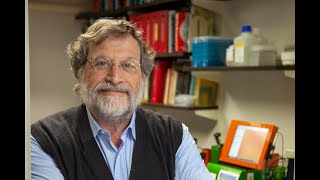
The video records Sylvia’s introductory remarks and Jeff Ravetch’s lecture titled “The Paradox of Immunity” as part of the Wall Seminar Series Dyer Lecture. Sylvia provides historical context about Dr. Roland Eugene Dyer’s contributions to the NIH, the origins of the extramural program, and anecdotes about early NIH scientists. She then introduces Jeff Ravetch, highlighting his molecular‑biology training, his pioneering work on IgG‑binding (Fc) receptors, and his mentorship style. Jeff’s talk explores why antibodies can be both pro‑inflammatory and anti‑inflammatory. He reviews the historical development of antitoxin therapy, the concept of immune tolerance, and the discovery of IgG Fc receptors. He details the molecular architecture of the Fc region, the role of the conserved N‑linked glycan, and how variations in subclass, fucosylation, and especially sialylation (α2,6‑linked) modulate binding to activating versus inhibitory Fcγ receptors and to a distinct sialic‑acid‑binding receptor (SIGNR1/DC‑SIGN). Using a series of mouse models (immune thrombocytopenia, arthritis, lupus, tumor xenografts, H1N1 infection) he demonstrates that (1) activating Fcγ receptors drive pathology, (2) inhibitory FcγRIIb raises activation thresholds, and (3) sialylated IgG (or IVIG) engages SIGNR1 to up‑regulate inhibitory receptors and suppress inflammation. He discusses human genetic polymorphisms in Fcγ receptors that predict therapeutic response to monoclonal antibodies (rituximab, trastuzumab) and to IVIG, and how pathogens (e.g., Streptococcus pyogenes) exploit Fc glycan cleavage. The lecture concludes with engineering strategies—defucosylation, Fc‑glyco‑engineering, humanized FcγR mouse models—to enhance antibody effector function, and a broader view of how IgG glyco‑modulation resolves the paradox of immunity. The session ends with a brief Q&A on receptor recycling, FcRn saturation, and clinical implications of high‑dose IVIG.
Chapters
Okay, welcome everyone to the, um, Dia lecture, which is part of the Wall Seminar Series run by the director. So as you can tell, the director is not here today, but I'm backing up for the introduction. I first want to spend 2 minutes just introducing this lecture because it has the name Dyer Lecture in honor of Dr. Roland Eugene Dyer, who was a former NIH director, as you can see, from 1942 to 1950, which were very interesting times because it was war times in the country. And as I was reading, it was kind of interesting to sort of go back to those times and how it was a really good time for NIH in terms of building infrastructure. And actually, Dr. Dyer was particularly good, not just because he was because he was a very good scientist in infectious diseases, but also because he set up the foundation for important parts of NIH. One is extramural NIH, which love or hate, particularly NIH— Jeff might have something to say— that started with Dr. Dyer, and also the Clinical Center in main campus was started in his time. And I'm also showing another slide because I find it quite interesting looking through all the information that I could find, Dr. Tayer. And it was interesting to me reading this book, which is related to the discovery of Rickettsia at the Rocky Mountain Laboratory in Montana. And the good part was to show how Dr. Tayer was giving public service a name, not just because he was a scientist, but also he was sort of an experimental subject himself. One of the rickettsias was discovered because he was visiting Montana and caught infected with one of the new rickettsias, which ended up being the Nile Nile fever disease. He's, as you can see, important for running a whole institute, but also for catching a disease that nobody knew about. It's also funny to imagine that nowadays a director visiting Montana and catching something. But hey, things can happen. So I'm going to switch. Introduce Jeff Ravitch, which I know very well because I spent quite a few years training with him. And it was definitely a great time to be with him. A lot of sort of top discoveries in a complete important field in immunology that he's actually leading by far. Out of all the scientists. Jeff actually trained particularly in molecular biology, so he's not really an immunologist by training. It's a little bit— I feel like I trained with him for the same reason, because I'm not an immunologist by training. He started just with the absolute basic molecular immunology at Rockefeller when they were studying sequencing. They were studying purifying polymerases, ligase. He always tells the story that he would trade vials. I give you my polymerase if you give me my ligase. And in those times, cloning genes was actually a very big deal. So he used that knowledge to basically start cloning genes that were probably biologically important, but people were not really sure about the function. Receptors that bind IgG immunoglobulin. The reason for finding something that binds IgG is because he happened to train here at NIH as a postdoc with Phil Leder when at the time they were studying antibody genes. And I guess the idea he took from here is that if antibodies are there and they have some constant region that might bind something that is important, and that was sort of the beginning of Many years, many important contributions, and a lot of people are interested in what ACE receptors do in inflammation and in all kinds of subjects. And Jeff has done tremendous work in basically finding out all kinds of important effects of these receptors and in finding clinical relevance, such as understanding how immunosuppression like interventional neuroimaging works, and you name it. And I want just to finish giving you a couple of just details on how Jeff's sort of view of science works. And I always find him like a really great spirit in terms of challenging the view of people. And one time I remember I went to give a presentation and then people were kind of tough with the experiments I was showing. And I went back to the lab saying, I just don't want to answer difficult questions. And Jeff said, well, I love when I have those questions, because then I always say, what, you don't believe me? Then prove it back to me. And I think that that shows that science is also about proving people that you have the right idea. And the last thing is that he's always been in love with science. And once he brought his kid to show him electrophoresis with chocolate, and he kept saying, see, science is fun, science is fun. And all of us were like, I'm not sure. So, but he's always been in love with it. So thank you, Jeff, for coming and giving this great lecture.
Thank you, Sylvia. That was a very warm personal touch for today's lecture. When I was asked to deliver the Dyer Lecture, I was quite flattered. Having spent my years at the NIH from '79 to '82, it's something of a lifetime accomplishment to be able to come back and see your friends and colleagues and talk about the work you've done since you've left and hopefully did something worthwhile during that time. Of course, I want to thank the NIH for inviting me, and I love the extramural program, so let nothing ever be said about my absolute complete conviction that the extramural program, well, truly is the unique opportunity this country offers for anyone who can come up with a good idea to get funding, and I think that is the strength of the system and the virtue of the system. You're not part of a club. You don't have to have a certain lineage or genealogy. You have to have a good idea and the ability to do the science to get the funding. And that's what I think should always be remembered about why the NIH system, with all of its, you know, occasional hiccups and bumps, is distinctive in terms of how science works and how this country's science has managed to remain at the forefront of the international efforts. So the title for today's seminar is The Paradox of Immunity. And when I thought of the title, I realized I was in trouble because the title was so good that I knew I couldn't possibly match it with a seminar up to the same quality. So I think all of us to think, think about the immune system. And as Sylvia said, I am not an immunologist by training. I woke up one morning in 1993 and realized I had become an immunologist, kind of like Kafka's cockroach. You wake up and realize you've become an immunologist because I was making knockouts in mice and studying immune responses. And what else is that if not an immunologist? And we all know the immune system, of course, has an intrinsic paradoxical nature. Of course, it is the evolutionary response to multicellularity. It's what allows us to survive in a hostile environment. Being able to deal with infectious pathogens and survive infectious challenges. Yet we know that the immune system, if not correctly regulated, will of course contribute to pathology. But I'd like to go a little further than that in today's talk and talk in terms of a specific alteration in biological function that we've been studying for a very well-known molecule, the IgG molecule, and how that, I think, in some ways encapsulates many of the issues that relate to the paradoxical activity of the immune response. So to give a little background, a little introduction, I thought we'd start really where immunology had its first significant foray into human disease. And that was the realization that for centuries, of course, mankind was subject to devastating epidemics. This is an epidemic in Wisconsin in 1874, and this is the mortality record for one family, 5 children who all died within the same 2-week period of diphtheria. That the mortality rate, as you can see from this very, I think, touching slide, often exceeded 80% in affected populations. This was truly a devastating public health response. And in 1890, thanks to the efforts of Emil von Behring and Saburo Kitasato, the responsible toxins had been identified— the diphtheria toxin by von Behring, the tetanus toxin by Kitasato. And what did they do when they found these toxins? How did they go about moving from their basic research interests into what causes diphtheria— fatality to a therapeutic intervention. They turned to animals. They immunized rabbits and mice with diphtheria toxin, with tetanus toxin, and discovered the concept of the antitoxin, that the cell-free extracts from these immunized animals were able to neutralize the harmful toxic substances produced by the bacillus. They did adoptive transfer studies, we'd say today, and were able to cure infected animals and protect animals who were being challenged with toxin. So if anything, of how modern scientific inquiry has progressed, and particularly in immunology, relying upon the mechanistic insights that animal models provide to allow for extrapolation to human disease treatment. Treatment, because within 4 years— and I find this as remarkable as their experiment— within 4 years, vials of diphtheria antitoxin were being produced by the predecessor of the NIH, the Marine Research Laboratories in Staten Island, to be used to treat children who were contracting diphtheria. And this shows the result of that treatment effort. Therapeutic vaccination with a passive antibody. And they found that the mortality rate in New York, in the United States, for example, dropped by over 70% within the decade that they introduced the use of an antitoxin. So an incredibly effective therapeutic, the result of basic research in animal models. It's irresistible not to tie this into contemporary ideas. This is a telegram that was sent in 1925 from Nome, Alaska, alerting the world that there was a diphtheria epidemic in the icebound city and that there was a request for 1 million doses of diphtheria antitoxin to be sent there. We all know how that was sent. It was sent by dogsled. This is the statue commemorating Balto, the lead dog of that, famous rescue effort in Central Park, and you can see how polished it is. That's because of generations of kids, including my own, getting up on top of the statue and rubbing Balto. And what Balto did, of course, was to deliver, together with his handler, of course, the doses of antitoxin to Nome and prevent what would have been a devastating epidemic of that community. So we begin the paradox because here by the turn of the century, an incredibly effective therapeutic had been developed for diphtheria, for tetanus, and the expectations were that the ability to make antitoxins would in fact rid the world of the scourge of infectious diseases. Ehrlich in 1900, the great theoretician and experimentalist of immunology, recognized that the process by which antitoxins were generated could in principle be able to turn on itself. And he called that horror autotoxicus because to him it was an incomprehensible perturbation of a normal physiological condition for autoantibodies, as we now call them, to be generated. So he speculated that the organism must possess certain mechanisms or contrivances by means of which the immunity reaction to generate autoreactivity or autoantibodies would not occur, that there must be a mechanism which we now would call tolerance. So Ehrlich predicted that tolerance in fact would be an essential aspect of any immune system. Of course, we know that tolerance is incomplete In the United States alone, over 8 million people have autoimmune diseases. 3% of the population develops autoimmune disease. It's truly a major public health problem, and it results from those intrinsic aspects of the immune system that allow for protection against the foreign world that occasionally gives rise to autoreactivity. But we didn't really know what the mechanisms were behind autoreactivity and the— more importantly, the pathogenesis of autoimmune diseases until 1958 when Henry Kunkel, working at the Rockefeller Institute, was the first to describe the existence of anti-DNA antibodies in patients who had SLE or lupus and thereby was able to conclude with the usual reticence of Henry Kunkel, that this was likely to be a causal factor in an autoimmune disease like lupus. And we now know this is in fact the case. For example, in lupus, we see evidence of immune complex deposition in glomeruli. We see evidence of glomerulonephritis and the sequelae, of course, of tissue destruction that give rise to disease. One of many autoimmune diseases, immune thrombocytopenia, a relatively rare autoimmune disease, but here autoantibodies are generated to the patient's platelets, giving rise to thrombocytopenia, which leads to hemorrhagic diseases, petechiae, hypersplenism, as you get engorgement of the spleen with macrophages that have ingested platelets. And the second paradox now appears. How do we treat patients with ITP? We treat them with the preparation of gamma globulin that's purified from the serum of thousands of donors. It's called intravenous gamma globulin, or IVIG, and it's— and we use absolutely massive doses, a gram per kilogram, for treatment to revert these patients to a normal condition. So autoantibodies trigger inflammatory diseases. High concentrations of serum antibodies seem to treat the disease. And this, I think, sets up the fundamental nature of this paradox, that IgG antibodies are both pro-inflammatory as well as anti-inflammatory. The IgG molecule, of course, is all well known to us. Antigen binding specificity is the most well-studied aspect of the molecule, the result of a remarkable recombination pathway that was discovered in part here at NIH by Phil Leader's group as well as by Lee Hood and Susumu Tanigawa. And the Fc domain is responsible for this collection of activities that we call effector responses. And it's the effector response aspect of the IgG molecule that I want to focus on today and try to get to the point of being able to account for both the pro and anti-inflammatory properties of an IgG molecule and how that, in fact, impacts on both homeostatic mechanisms and pathological conditions. So my own interest in IgG, as Sylvia said, began here at the NIH 30 years ago, having the good fortune to work with Phil Leder in Building 6, which is still here. And in 1980, together with Lanny Kirsch, another postdoctoral fellow— both of us needed haircuts in those days and probably some of us still do. And Phil always said, your haircut is fixed when you're 19 years old. You see, look at my haircut, look at your haircut, and I guess he's right. However, we began cloning the genes for the human and mouse immunoglobulin locus, focusing on the heavy chain genes, and together cloned the IgG molecules, looking at the constant regions, observing the interspecies homology between these molecules,— and recognizing that this was an evolutionarily conserved system on which selection was obviously operating to maintain fidelity of sequences in these molecules. The reason for that conservation wasn't clear, but we certainly suspected that the effector functions of the IgG molecule would transcend various species barriers. So we now know that the Fc of an IgG confers quite a few different inflammatory properties. Pro-inflammatory, as I mentioned, giving rise to pathogenicity of immune complexes, for example. The ingestion of various pathogens by phagocytosis, as I'll show you in a few moments. The cytotoxicity of antitumor antibodies are an Fc effector function. And even classical neutralization of viruses and toxins, now seems to have a contribution from the Fc response as well. The anti-inflammatory activity is really the result of the clinical efficacy of intravenous gamma globulin as a therapeutic. It's used to treat a variety of autoimmune diseases, approved by the FDA for that purpose. And as you'll see later on in today's talk, it's unequivocal that there is anti-inflammatory activity in the Fc fraction of IVIG. The question is, what causes that? The Fc domain is also able to contribute to the modulation of the immune response. If you think of it in terms of a metabolic pathway, this is classical feedback regulation. As the IgG molecule is made, it feedbacks on the cells making it, the plasma cells, as well as on the cells involved in triggering the response, the DC cells., and I won't be touching upon this very much today. And finally, the reason that most people think about the FC is simply half-life. In fact, the FC region was thought to be essentially there just to maintain half-life of the IgG molecule. It confers about a 10 to 14 day half-life depending upon the species and the subclass on the IgG. So that extended half-life is certainly a function of the FC domain. One way to account for this diversity of biological responses for a single molecular entity is to invoke a variety of binding partners. And we know historically that the Fc domain has in fact been shown to interact with a large number of different types of molecules. The complement cascade through C1q in the classical pathway, the Fc gamma receptors which were really first described by Benacerath in 1965. The neonatal receptor for Fc, a class II MHC-like molecule, first predicted by Bramble. And of course, lectins will interact with the glycan in this structure. And I want you to remember the presence of this very highly structured, highly resolved carbohydrate entity which is found in the pocket formed by the A and B chains. We'll come back to this as one of the features of the IgG molecule, which in fact confers many of its biological properties. So FC receptors, which I'd like to focus on, were first described by, as I said, Benacerraf and, uh, Burkian back in 1965, and they were described by virtue of their ability to directly bind to macrophages through their FC domain. So people up until this point, of course, had known that you could take an antibody and it would bind to an antigenic surface through the FAB. And what Benacerath and colleagues showed was that there was an activity on the surface of macrophages that conferred Fc binding, and they were later referred to as Fc receptors. The biological function of these receptors was in fact quite questionable, because if you pick up a textbook of immunology anywhere up until the mid-'90s, it would say very unequivocally that the in vivo activity of IgG as a pro-inflammatory molecule was a consequence of complement activation. And that's where the story ended. So why have a system by which IgGs can interact with cell surface-bound receptors? Well, to resolve that, we took a— then at that point, a pretty classical approach. Let's first define what the receptors are, clone the genes, characterize the protein, study their regulation, study their structure and try to understand what biological function they would have. And this is a little snapshot of the predicted proteins for the mouse and the human Fc gamma receptors, the IgG Fc receptors. Fc receptors for IgE characterized by Henry Metzger here at the NIH have some comparable structures. And there are Fc receptors for the other subclasses as well, for IgA and IgM. And what the receptors for the IgG family look like are cell surface transmembrane receptors, in most cases heterodimeric, with a gamma chain subunit that confers activation signaling or ITAM motifs. And unique to this family at this point was the presence of a receptor called FcRIIb, and I take full responsibility for these terrible names, but I named them as I got them, right? So 1, 2, 3, 4, it just, you know, not very impressive. My children have real names, so not to be concerned, right? So 2B is the receptor that had not an ITAM motif but another class of signaling motifs which turned out to be inhibitory. And this in fact defined before the KIRs and the NK inhibitory pathways this family of inhibitory motif receptors, that the Fc receptor family existed in a balanced signaling capacity, both activation and inhibitory signaling. The human and the mouse share significant homology in terms of the types of receptors. The numbers differ and the precise specificity will differ as well. So how does an Fc receptor engage its ligand, the IgG? Complex? Well, these are by and large low to intermediate affinity receptors, so they're not occupied by circulating IgG. Instead, they bind to immune complexes. So the first level of distinction is that they are available when an immune complex is formed, which is usually in response to the recognition of an antigen by an antibody to mediate a biological response. And the pathways are very reminiscent of what we see for the B-cell antigen receptor or the T-cell receptor, the activation of sarcon cyclinases leading to the activation of inositol lipids as well as calcium mobilization to give rise to an activation response. But what's distinctive about the FC receptors is that you don't get expression only of activation receptors. You get co-expression of activating and inhibitory receptors. That in point of fact the immune complex is co-engaging these two opposing signaling molecules. When it cross-links the activating and inhibitory receptor, it now induces the phosphorylation of the inhibitory receptor signaling motif allowing for the generation of an SH2 docking site and a molecule described by Sylvia Bowen in my lab, the SH2 inositol phosphatase, SHIP, is now recruited to the membrane surface. The function of this molecule is to hydrolyze the products of the activated PI3 kinase and thereby reduce the activation signal and in some case abrogate it. So the way an immune complex or an opsonized organism particle signals to an effector cell like a macrophage is by co-engaging activating and inhibitory receptors establishing a threshold for biological response. As you can imagine, if the FCs have different affinities for different FC receptors, that will govern the absolute threshold for response, as will the absolute density of the receptors on the cell surface. And we see both, in fact, occurring. Now, in addition to this somewhat simple modular approach to having an activating and inhibitory receptor, that interact to generate a threshold for effector response. What Sylvia and Roger Pierce discovered in the laboratory was another pathway existing only on B cells where the inhibitory Fc receptor, when extensively cross-linked, could now induce an apoptotic signal. And we really didn't know what this meant. It was somewhat of an enigma to us why there would be an inhibitory signaling pathway on B cells that will lead to their death. But in an experiment that Sylvia did, and then later Ken Smith did in some greater detail, it becomes clear that this is a fundamental mechanism by which peripheral tolerance is maintained to self-antigen. And this is the mechanism by which the Fc receptor inhibitory pathway is actually not only acting to transduce activation responses to eliminate pathogen, for example, or toxic substances, but is actually functioning to regulate B-cell response and B-cell activation. So let's return to, for a moment now, to the fundamental question we asked in the beginning, is what do the Fc receptors do? Why does this biological system exist? And like Von Behring and Keddesato, we turn to mouse systems. To try to understand the basic mechanism by which these receptors would contribute to an in vivo response. From in vitro standpoints, they seem to be redundant, but when we looked in vivo, we found something quite interesting. This is a mouse model of immune thrombocytopenia. It's a model of platelet clearance induced by an antiplatelet antibody. And what you see here is a depletion of platelets in a wild-type mouse. In a mouse that is lacking C3, which is a central component of complement activation. And a C3-deficient mouse was able to deplete a complement-fixing antibody in vivo, while a mouse lacking the activating Fc receptors was protected. So the first hint that the complement pathway in vitro was not showing the same activity in vivo came from studies like this. Admittedly quite artificial. A more representative experiment perhaps done by Diana Silvestri was the Arthas reaction, described in 1903 and has been a model for immune complex-triggered immunopathology for the last 100 years. It's a model where you inject immune complexes into the skin of an animal and then look for an inflammatory response. And that inflammatory response has historically been attributed to complement activation. Well, it turns out that if you do an opus reaction in a mouse lacking C3, these were generated by Mike Carroll at Harvard, we find that we have a perfectly wild-type type of response. We get edematous reactions, red cell accumulation, and neutrophil infiltration. But if the mouse lacks the Fc gamma receptor activating pathway, now the opus reaction is in fact absent. Despite the persistence of immune complexes and activated complement. So something clearly was different in vivo than what we found in vitro. And I think the experiment that sums up this dichotomy came from a study that was done by Raphael Kleins in the lab quite a few years ago now where we decided to look at a spontaneous model of lupus, the NZB/NZW F1 animal. Animal, these animals develop spontaneous autoantibodies to DNA and chromatin. Immune complexes are deposited in their glomeruli. They develop glomerulonephritis and they have premature mortality. When we cross these mice to a mouse lacking the activating subunit of the FC receptors, the gamma chain knockout, and then segregated the animals into homozygotes and heterozygotes, what we found was that the homozygous knockout of the Fc gamma receptor was sufficient to protect the animals from the immunopathology of this disease. Patent glomeruli extended survival despite the fact the kidneys showed abundant immune complexes as well as abundant activated complement C3. So clearly the complement pathway was not sufficient and probably not even necessary in vivo to trigger an inflammatory response. Response. In fact, it was an FC receptor. And that was a result that I believe could never have been obtained if we didn't have the ability to manipulate animal systems to look at these biological responses. This translates immediately into a clinical observation. Because in 2000, the first monoclonal antibodies were being introduced into clinical treatment for a variety of neoplastic diseases. Antibodies. Two of the antibodies I'll show here, anti-CD20 or rituximab, anti-HER2-neu or Herceptin, were being used for non-Hodgkin's lymphoma and metastatic breast carcinoma. And the question that we asked was really quite simple. How do these antibodies work? The doctrine seemed to be that they were engaging through their FAB specific cell structures that modulated cell survival in these tumor models. And for the case of Herceptin, that certainly seemed like a rational interpretation and certainly supported by in vitro data. In the case of Rituximab, it was a bit more difficult. So we asked a very simple question. Let's look at a tumor model in these mice with lacking or possessing Fc gamma receptors. So in Rituximab, I think the picture says it all. This is a xenograft tumor growing in a nude mouse. And the mouse is treated with the rituximab in the clinical doses used in patients and the tumor completely regresses. If the mouse has a background lacking activating Fc receptors, these tumors grow despite the persistence of therapeutic levels of anti-CD20 antibody. So in this mouse model, you needed to have the effector arm of the response, the macrophages binding through their FC receptors to the antibody binding to the tumor cell to kill these tumor cells. So ADCC or antibody-dependent cellular toxicity was absolutely required in our mouse models. In the Herceptin case, it was a bit more interesting because the— excuse me— the HER2-neu surface receptors certainly can be shown in vitro to modulate tumor proliferation. And that's shown in this slide. You can add the antibody antibody to a culture of human tumor cells, BT474M1 cells, and the cells will arrest in culture as you titrate in the anti-HER2-neu antibody, presumably blocking an autocrine-like growth loop. So we mutated the Herceptin antibody in the Fc domain, single point mutation that abrogated Fc receptor binding, all Fc receptor binding. Didn't affect the ability to arrest proliferation in culture. But when you look in vivo, the mutant antibody does not modulate tumor growth. The wild-type antibody arrests tumor growth, but the mutant antibody, which can still block proliferation, is unable to arrest tumor growth in vivo, showing quite clearly that, like in the case of anti-CD20, anti-Her2Nu, despite its intrinsic ability to mediate an antiproliferative activity required an FC effector pathway to show its biological effect in these mouse models. How is this a clinical experiment? Well, based on those studies, several groups decided to look at patients who were being treated with anti-CD20, rituximab, or anti-HER2/neu, Herceptin. And the question they asked was, do all the patients respond? Respond, and of course, unfortunately, they all don't. They segregate into responder and non-responder population. Here's a responder, here's a non-responder, here's a responder, here's a non-responder. What is different between these patient populations? Turns out that the single most significant difference between these populations is an allele of an activating FC receptor. Not just any allele, but it's the allele that confers a threefold higher binding affinity between the IgG1 Fc of these antibodies and the FcR3 activating Fc receptor on macrophages. So as we saw in the mouse studies, the human studies confirm that in vivo, a patient's ability to clinically respond to this therapeutic will depend upon which allele of an Fc receptor he has— she has, excuse me.— he or she has. And that leads to a significant difference in survival, which is, I think, an important criteria. Now, what else do these receptors do in terms of traditional antibody systems? Well, here's a study I took from the literature done in 2001 looking at the protection from the H1N1 vaccination in mice that were either wild type or those lacking the Fc gamma receptors, hence unable to mediate effector responses. And even though the antibody is perfectly neutralizing in culture by standard methods in vivo, its ability to protect animals from premature fatality is severely compromised if there's no effector pathway. So neutralization, which is nothing new here, Bernie Fields said it 25 years ago, includes not just antigen binding to a unique epitope but the effective properties of the antibody as well. Recent study from Dennis Burton's laboratory looking at one of the most interesting antibodies that's come along in a long time for HIV, a so-called B12 antibody. And the B12 antibody will reduce viremia in monkey models quite effectively. But what Dennis did was to take the trick that we showed for Herceptin, mutate the Fc of his B12 antibody, and use it in his— in his anti-HIV protection study in monkeys. And what he found was that if you mutate the Fc so it no longer engages Fc gamma receptors, you in fact can no longer modulate viremia in these monkeys. So part of the mechanism by which this antibody was neutralizing in vivo requires an effector response, as he states in that paper. And finally, the experiment that I mentioned before, and these are my patients, and therefore, you know, we treat them with the dignity and respect they deserve. We've been talking about the ability of antibodies to trigger effector responses, inflammatory responses if you will, against an antigen, be it foreign or self. And what Sylvia found was that the inhibitor inhibitory receptor which has this unusual activity on B cells of triggering apoptosis, when backcrossed onto a black 6 background compared to a BALB would spontaneously generate an autoimmune phenotype with autoantibody production, immune complex deposition, glomerulonephritis, and these animals will in fact die prematurely. Sylvia went on to characterize the mechanism behind the strain epistasis. The pathways that are triggered. Suffice it to say the immune complex interacting with the B-cell inhibitory receptor is an important pathway by which we maintain tolerance in the periphery. So therefore it should not be surprising that when we look in human diseases and ask do the Fc receptors show any disease associations, the answer is of course yes. That we see that same allele of activating receptors showing up in inflammatory diseases, Bechet's disease, various myopathies including RA, that the polymorphisms that occur in the inhibitory receptor are found to contribute to susceptibility to autoimmune diseases like lupus in different populations. And very recently, Ken Smith has looked at the interesting observation that the frequency of an allele of an inhibitory receptor that's a hypermorphic phenotype, meaning it gives reduced signaling capacity, is in fact overrepresented in populations where malaria is endemic. So if you look, for example, you can't see this probably, if you look in the UK between British natives and British Southeast Asians, there's a tenfold difference in the allele frequency for the allele that gives rise to a weaker inhibitory signal, which turns out to reduce susceptibility to malaria. So once again, as in thalassemia, malaria has exerted a selective pressure on the human population to select for alleles that would otherwise contribute to autoimmunity but enhance inflammatory responses and therefore are maintained in the population. So with that as a somewhat elaborate background, let's talk a little bit about the actual interaction between an IgG and its Fc receptor. And we're helped by studies done by Peter Sun and by Peter Sonderman here in 2000 solving the crystal structure of an Fc receptor, a soluble fragment of an Fc receptor with the CH2-CH3 domains of an IgG1 Fc. And as you can see, there's an extensive surface of interaction between these molecules. The glycan, which is shown in yellow, is a complex bi-antennary glycan we'll talk about in some detail. It doesn't make direct contacts with the receptor but is critical for maintaining a conformation that is permissive for Fc receptor binding. We decided to examine the contribution of Fc receptors to the in vivo activity of various IgG molecules. And we first looked at the IgG subclasses. We've known for a very long time that in the mouse and in the human, different IgG subclasses have different intrinsic biological activities. Some are more pro-inflammatory than others, attributed to a variety of factors including complement fixation and the like. So together with Falk Nemmerhan, we generated a panel of monoclonals each with the same V regions, each binding with the same specificity to an anti-tumor antigen that was defined by Alan Houghton called GP75, and then alter the Fc from 1 to 2A to 2B to 3. And now we use these antibodies, which actually have finally progressed into clinical development, in an in vivo model of metastatic melanoma where we inject B16 melanoma cells into a mouse— and look 2 weeks later for the appearance of metastatic nodules. If you treat the animals with the G1 antibody, you see very little difference in the ability to modulate this tumor growth. The 2A antibody is essentially therapeutic with the 2B falls somewhere in between. And what correlates very well with this hierarchy of activity is a hierarchy of binding affinities between the activating and inhibitory receptors. 2A has a very high binding affinity for activating receptors and a very low binding affinity for inhibitory receptors, hence it's the most potent subclass. So we can account for all the in vivo biological effects simply by looking at which FC receptors it binds to with what affinity. Now, what role does the glycan play? Clearly amino acid sequence confers subclass specificity and that confers binding affinity. But the glycan, as you see here in this crystal structure, just the CH2 domains of an FC dimer, is holding the structure in an open conformation. When we deglycosylate this, the crystal structure collapses. So one more time, the open structure, the FC receptor is binding to the site up on top of the molecule. Watch this site, it collapses. So the prediction is that when you deglycosylate, an IgG molecule, it will lose Fc receptor binding activity. And as we've said before, when you lose Fc receptor binding, you lose in vivo pro-inflammatory activity. So here is the deglycosylated antibody generated by making a mutant in the glycosylation site, N297 to Q. There's no glycan associated, and the Fc receptor binding is no longer measurable. What does this do in vivo? Well, biology is really wonderful. There's an organism that's done the experiment for us. So one of the Group A streps, strep pyogenes, which is responsible for cellulitis and a variety of other human diseases, has an enzyme that it specifies, discovered by Matthias Collins, our collaborator on these studies, that specifically recognizes and cleaves the glycan on the IgG Fc and nothing else that we've observed. So this is absolutely specific for this Fc glycan. It cleaves right after the N-acetylglucosamine, leaving the Fc backbone with a single branched sugar. So the reasoning that we had was that, well, why would this organism have this enzyme? It must confer some protective advantage. The organism itself doesn't have any N-linked sugars. It must be to somehow prevent clearance by a host. Clearance by a host for an antibody will be through Fc receptors. Deglycosylating would reduce Fc receptor binding. So let's take a system like the BXSB mouse, another spontaneous autoimmune strain of mice, and look at the survival of these animals in their untreated state. And they will all be dead by 30 weeks. Treat the animals at 12 and 24 weeks with a single dose of NOS, and the survival is extended quite significantly, meaning that if we artificially prevent the antibodies, the autoantibodies in this disease, from triggering effector cell FC receptors, we have extended survival. So the glycan clearly plays an important role in how the FC sees itself. Receptor. We can see that in greater detail now focusing on the glycan itself. This is the complex bi-antennary glycan. And as you remember from the organic chemistry classes, this is non-templated, therefore it represents a population of molecules that are driven by enzymatic reactions. We can generate homogeneous versions of these in defined strains, cell lines, and we generated two populations of molecules, one lacking— one with the fucose and one lacking the fucose in the first branch position. You measure the affinity constants and find that the ratio for the fucosylated will be 7, and it's about 3-fold higher for the a-flucosylated antibody, predicting that in vivo this antibody should have 3-fold greater potency. Well, we attach these onto the antibody that recognizes our tumor antigen This is the appearance of the animal treated with the fucosylated antibody and this is the animal treated with the defucosylated antibody. A threefold reduction in tumor metastases. So this concept has been widely adopted and second-generation Herceptin and Rituximab molecules are now in clinical trials, I think in phase 3, looking at the defucosylated versions of these antibodies that would increase their in vivo potency by virtue of increasing their ability to trigger in vivo cytotoxicity. So I thought it might be amusing at this point to take a quick movie break and look at the glycan in somewhat greater detail to reinvigorate the audience. So this is a movie I found on the internet. I don't know. Everything is on the internet. But— and I guess it does have a certain— you know, see-all quality to it as well, because we can see directly into the glycan structure. And in a moment you'll see that the glycan is in fact a very highly defined structure. This is distinctive from what we see for glycans that are on most glycoproteins, which are exposed to the solvent surface. Those are highly mobile structures. They don't resolve on X-ray crystallography. And in fact, The notion that the glycan itself has a structural contribution to a molecule is somewhat controversial. But here there's no question. By being constrained within this hydrophobic pocket formed by the A and B chains of the antibody molecule, this glycan is highly structured. In fact, here's that fructose sticking out over here with the FC receptor. We're looking down now on the binding pocket. And it certainly is reasonable to conclude that small changes in this glycan structure can in fact influence the interaction of the receptor with the antibody molecule. So if you make antibodies with enhanced cytotoxicity, how do you test it? I told you earlier that the mouse and the human have many similarities, but their FC receptors are in fact both qualitatively and quantitatively different. So In order to move it to the next level, if you will, to be able to generate variants of human antibodies that have predicted in vivo activities that can then be used in clinical studies, we generated a strain of mice shown here schematically where we delete the mouse low affinity Fc receptor locus and we replace the entire locus with a large chunk of chromosome 1 of the human that has all the human Fc receptor genes. These mice, in fact, express the human receptors in the correct pattern of distribution found in the human and not in the mouse. So B cells have only the inhibitory receptors shown here in both the human and the humanized mice. Dendritic cells, which in the human— these are monocyte-derived dendritic cells or conventional dendritic cells— have a receptor called 2A and 2B. I should point out there is no 2A in the mouse, so this is the expression of a human gene in the mouse with the tissue specificity of the human, and the humanized mice recapitulate that pattern. So you can take these mice and now test a modified antibody. We generated an antibody that recognizes a platelet determinant that leads to thrombocytopenia, those experiments I showed you early on, but now we modified modify the Fc to have a human FcG1 modified to generate a higher binding affinity to an activating receptor and compare it to a wild-type antibody G1 in a depletion assay. And here you see that the enhanced FcRIIA binding affinity leads to enhanced depletion at the same dose. So we can in fact utilize what we learned about the immune response in the mouse to generate models that I think have some clinical relevance. So to summarize up to this point, and I don't even see a clock, so I'll keep on going. What we now can say is that the Fc portion of the IgG antibody is critically important in the effector responses to generate the pro-inflammatory response. And there are rules that we've written that tell one how to engineer an antibody to make it a more effective cytotoxic agent to use in passive immunotherapy. We don't know how these rules apply spontaneously in vivo, what adjuvants generate what subclasses or what glycan configurations, but that's the obvious next step to do active immunizations and know how to manipulate the immune response to generate antibodies that have the desired activity. So I'd I'd like to now turn to this notion of the anti-inflammatory properties of IgG. I think I've convinced you, hope I've convinced you by now, that the pro-inflammatory activities can be attributed in mouse and in man to the interaction of the Fc with Fc receptors, and depending upon which Fc receptor, you can get a different biological response. The anti-inflammatory activity, as I said, came from the serendipitous observation that a therapeutic preparation of IgG pooled from plasma donors used initially for replacement therapy for children who were born hypogammaglobulinemic turned out to have anti-inflammatory activities when used at these gargantuan doses. To give you a comparison, these are doses about 100 times higher than one would use if you're using a monoclonal antibody to treat a patient. So they're gram per kilogram. It's not a typo. It's approved by the FDA for ITP, Kawasaki's, Guillain-Barré, CIDP, a variety of autoimmune diseases. And of course it's used widely off-label in about 85 other different autoimmune diseases. It's generated from plasma preparations, and everything that is purified is used. There's often shortages of the material. This struck me as being a particularly vexing paradox because we knew from all these studies up to this point that IgG could be pro-inflammatory, engaging Fc receptors. It could be rendered impotent and/or non-inflammatory, but how is it actively anti-inflammatory? How do we suppress an inflammatory response with serum IgG? And more importantly, two clinical studies done in the '90s show that the Fc the FC fragment alone was able to confer this anti-inflammatory activity. So it wasn't some fancy idiot type, anti-idiot type interaction. It was the FC fragment itself from this gemisch of IgG pulled from human donors. So we turned back to the mechanistic approach of using mice. And we established certain criteria to make the mouse models as relevant as possible. Possible. That we looked at models of inflammatory disease where IVIG would suppress inflammation, so they were treatment models. Number 2, that the FC fragment would be active but the FAB would not be active. Third, that it would require doses comparable to a gram per kilogram to see the activity. And we're using human IVIG now in these studies, so it's not mouse IgG, it's human IVIG in mice. So these are relatively short-term, 8- to 10-day studies. And finally, since IVIG preparations from every different manufacturer are used interchangeably with the same biological activity, we believe there must be a common mechanism of action. So that was the— those were the criteria we established. Here's a model of nephrotoxic nephritis, the ITP model I showed you before, the Mathis and Benoit model of inflammatory arthritis, a KB/N model. And in every case, we could induce inflammation, protect with IVIG or the Fc fragment, not Fab, induce arthritis, and protect with the Fc fragment but not the Fab. And interestingly, in all of these models, mice that were genetically deficient in the inhibitory receptor, the R2B deficient mice, no longer showed the protection with the IVIG or FC fragment. Now, that didn't mean that the IVIG was interacting directly with the inhibitory receptor. It simply meant genetically the pathway included an inhibitory receptor response. So with these models in place, we could now start to dissect what were the components that were required. Why was the inhibitory receptor possibly involved? And what we saw was that if you look now at the inhibitory receptor expression on various macrophage populations, and these are inflammatory macrophages in the spleen, the synovium, and in the glomerulus in these models of inflammation, you see that after IVIG or FC treatment, there is upregulation, enhanced expression of the inhibitory receptor. So somehow IVIG or its FC fragment was leading to the upregulation of inhibitory receptors on the effector macrophages, these very same macrophages that trigger tissue pathology. That makes sense. We raise the threshold. Raising the threshold would in fact be one way to reduce the severity of an inflammatory response. Turns out it's true in humans as well. Very recent study looking at patients who were treated with IVIG for this chronic inflammatory demyelinating polyneuropathy, CIDP, chronic EM/BA showed upregulation of the inhibitory receptor on monocytes and on B cells in those patients who respond to treatment. So what was true in the mouse is true in the human. There is a pathway by which IVIG is able to upregulate inhibitory receptor expression on a variety of cellular responses. Well, what is it about IVIG that does that? Well, the first hint came from the dose requirement itself. If it takes a gram per kilogram, it must mean that the active fraction is a small percent of that, that most of it is just, you know, carrier or irrelevant. So where is the heterogeneity in the IVIG preparation coming from? And the greatest source of heterogeneity comes from this carbohydrate, this glycan, found attached at asparagine 297. This is a complex bi-antennomy glycan shown in yellow, so-called core glycan that's found invariably. And then it's decorated with various branching as well as extensions to generate in normal human serum about 40 different N-linked IgG glycoforms on the Fc. If you take our serum, fractionate by MALDI-TOF, you'll find 40 different peaks of Fc-associated glycans. Could one of these rare populations of glycans, when associated with the Fc, confer an anti-inflammatory response? So to do that experiment, Toshi Kaneko took IVIG. This is our arthritis model once again. Here we're inducing arthritis. Here we're protecting with IVIG, a gram per kilogram. He's then and deglycosylates with an enzyme, PNGase F, cleaves off the entire glycan and we lost biological activity. No longer conferred protection. That's pretty cool. So let's now look at specific sugars. And looking at neuraminidase, which will cleave the terminal sialic acid from the IVIG, he found the same result. That desialated IVIG was no longer able to suppress the inflammation triggered by by the KN arthritis model. The same is true in the NTN nephritis model as well. So this was a general finding. Now, you should know that the fraction of IgG in IVIG that has terminal sialic acid is about 3%. It varies between 2% and 4%, about 3%. So we should be able to enrich that population by using a simple lectin affinity approach. Let's take a lectin that that selects for termosialic acid and purify a population of IgG that's now been enriched for the sialic acid and compare it to fractionated material. This is the SNA lectin that enriches it. This is IVIG unfractionated at 100 mgs per kilogram, 1/10 the dose we're using here. There's no protection. So you need a lot of IVIG to get protection. But the SNA enrichment material at 1/10 the dose is now able to suppress inflammation. So we can in fact demonstrate the requirement for sialic acid and the ability to enhance activity by enriching for sialic acid. So let's put it all together. Let's do it all in a recombinant platform. Take an IgG Fc, generate sialated forms of the molecule, test them in vivo and see if we enhance the activity. In a completely IVIG-independent fashion. This experiment shown here, here we induce arthritis. This is a control with the SNA-enriched material. So at 1/10th the dose, you see protection. Now we're sialating a recombinant backbone with either a 2,3-sialotransferase or a 2,6-sialotransferase. That gives you a different linkage between the penultimate galactose and the sialic acid. The 2,3-sialotransferase— which is fully sialated— shows no protective activity, but the 2,6 material is quite protective on a recombinant backbone, and it gives you, when you calculate, about a 35-fold enhancement of activity or increase in potency over the material that is used as IVIG. So clearly we can show an anti-inflammatory property when we attach a sialate acids. But why would that be anti-inflammatory? Well, what happens when we try to look at the binding of IgG Fc's that are either sialated or not sialated? This is an experiment shown here that Rob Anthony did. Here we see a 2,6-sialated Fc compared to the acylated Fc, and you find that a wild-type macrophage, which has a variety of Fc receptors, binds both populations. When you knock out all the the ones that give rise to the pro-inflammatory activity, you see that the acylated material loses all binding affinity. So the absence of sialic acid is critical for binding to canonical FC receptors, but the 2,6-sialated material retains binding in these FCR-negative mice. That's because they have another receptor. It's a sign molecule. It's a member of the type 2 C-lectin family. And these sial molecules have been known for a while. Their function is still being resolved. But a sial molecule, sial R1, binds specifically to the 2,6-sialated material. Knock it out, you lose binding. But the A-sialated still retains binding by virtue of canonical activity. So what happens when we knock out sial R1? When you knock out sial R1, and do the IVIG protection with the 2,6-silylated material. Here is the inducing arthritis. Here is protection with IVIG or silylated FC. In the SINE R1 knockout, you lose in vivo protection. So you must have the SINE pathway to mediate the activity. Humans don't express SINE R1. The closest homolog is something called DC SINE, which many of you might know quite well. DC SINE is expressed on dendritic cells as well as on monocytes. It's a subject that Peter Sun has extensively published on and taught us about. DC-SIGN is involved in the interaction with high mannose sugars including GP120 of HIV. Human DC-SIGN turns out to be the homolog of SIRN1 for the sialated Fc binding. These are now transfected cells expressing SIRN1 and human DC-SIGN or the human canonical Fcr2b molecule are sylated. Fc binds to both. If you can compete the binding with the canonical ligands for sialyl-R1 and Dc-Sial, mannan, for example, but fibrinogen, which is a glycoprotein that has the same structure of a complex bi-antennary glycan terminated in 2,6-sialic acid, doesn't bind to any of these molecules, meaning that it's not just the glycan.— and it's not the Fc. It's the combination of the Fc and the glycan conferring what we think is a unique structural property on the Fc to give rise to a novel binding interaction. Now, is DC-SIGN able to reconstitute SINE-R1 deficiency? So we made transgenic mice where we expressed DC-SIGN as a transgene behind a CD11c promoter. And now we repeat the experiment. Experiment I showed you before. Wild-type mice inducing arthritis, protecting with IVIG, looks good. Sine R1 deficient mice, no Sine R1, no anti-inflammatory activity. But when we reconstitute DC sine now on dendritic cells with the Sine R1 deficient background, we now restore protection in our IVIG treatment. So we're coming to the end. What we think is going on is that There is a population of IgG that contains sialic acid shown here. This is a normally occurring population. It is modulated, as you'll see in a moment, depending upon different conditions upon antigen stimulation. When it engages its FC receptor, which is a sialation FC receptor called sialyl R1 or DC-SIGN, these cells which we're calling regulatory macrophages or dendritic cells lead to the transexpression of a cytokine profile that acts on the effector macrophage to upregulate the inhibitory receptor. And by upregulating the inhibitory receptor, we raise the threshold required for an immune complex to trigger an inflammatory response, and thereby we can show a protective effect. So the features that are unique to the system is that cytokine isolated Fc binds to a novel receptor, acylated Fc binds to the canonical receptors. That there is a connection between these two cell populations but they are distinct. That the effector cell is responding to signals generated by the homeostatic pathway. So we have this notion that there is a switch that occurs during the immune response. In the steady state, IgGs exist in a more highly sialated form in order to prevent incidental macrophage activation by immune complexes that might occur and trigger an inflammatory response and by virtue of generating an anti-inflammatory signal. But upon exposure to an antigen, we find that the IgG class-switch antibodies that are antigen-specific have very low levels of sialation. Acids, these IgG antibodies are now able to bind to canonical FC receptors and trigger an inflammatory response. So there have been a few experiments that have been done in other laboratories that have begun to support this model. This is one that was just published a few months ago from a Dutch group. I probably can't pronounce the name. And it's based on the observation that women with rheumatoid arthritis who become pregnant undergo a spontaneous remission. And this graph on the bottom from their paper charts the degree of remission. About 40% of the women will undergo a remission which peaks by the third trimester, and then it begins to wane after delivery until they basically go back into a relapse. And the question was, is there any correlation between this attenuation of inflammation in these patients and the status of their IgG antibodies. And what they found was that if you look at the degree of sialic acid on that antibody, it corresponds very precisely with the attenuation of inflammatory response. So during pregnancy, there is an increased sialic acid on the IgG antibodies, and that could contribute to a reduction in the pro-inflammatory properties of the autoantibodies that are found in that disease. So to end, the paradox that I was trying to elucidate is a paradox by which the IgG antibody can have both pro- and anti-inflammatory activities. And we can resolve that by realizing that the pro-inflammatory properties come from engagement of canonical Fc gamma receptors, which are of course influenced by by the Fc and its glycan backbone. When the glycan is in a sialated form, it now acquires binding to a novel receptor, DC-SIGN or SIGNR1, and that confers anti-inflammatory activity that now allows IgG to in fact suppress inflammation. So once again, we've been talking about how the immune complex can trigger an inflammatory response by engaging canonical FC receptors and how we can modulate the activation of B cell and dendritic cells by immune complexes and finally how through the interaction of sialation and sialic acid receptor binding pathways we can suppress inflammatory responses. So I wanted to take the opportunity to acknowledge the people who I've worked with over the years, the alumni of the lab. Sylvia, of course, down here. Diana Silvestri and Astrid Samuelson. Diana for the orthus reaction. Astrid, who started the studies on IVIG and was first to show the animal models that worked. The group that you saw earlier from Sendai, who— Toshi Takai, who generated the first knockouts for FC receptors, the gamma chain, the 2B knockouts. Falk Nemrahan, I talked about, and Rafael Kleins. And the current lab members, including Rob Anthony, Jin Han, Toshi Kobayashi, Fubin Lee, and Andrew Pincetic, together with our lab manager Patrick Smith. Our collaborators really have been critical in our understanding of the glycobiology. It's an exciting new area for us. We've worked with Jim Paulson and Vern Rydhout and Dane Wittrup on some of the Fc engineering. And for many years, both Michelle and Ralph have been friends and colleagues, collaborators. That have made working a lot of fun. So thank you very much, and I'll be happy to take any questions. Jeff? Peter, yes. So you clearly showed that the involvement of DC-SINE in this anti-inflammatory property of IGG. The exquisite preference to alpha-2,6, not 2,3 sialic acid, seems to also match the Siglec, you know, another class of a— So we tested Siglecs, you know, I made the postdoc do that experiment over and over again, and we could never demonstrate binding of sialated Fc to Siglec Siglecs, either 2-6 or 2-3. So it doesn't interact with the Siglecs. We made antibodies to the Siglecs, tried to block their activity in vivo, it didn't attenuate. We looked at Siglec knockouts, it didn't attenuate. So like you, I thought initially it was going to be the Siglecs, but it's not. So we haven't ruled out them not playing a role at some level, but certainly at this level of interaction, we can't see a role for the Siglecs.
6 yet. Okay. Hi, great talk. So you associate most of the disease and the function of the receptors to their activation phenomenon, regulation and activation versus inhibition with the ITIM-bearing ones. I wonder whether any of the diseases you've looked at could be correlated to problems in recycling of the receptor, endocytosis, clearance. Sometimes those things might go together, but is there—
can you ever— can you separate out those phenomena? Yes, because mutants have been made that affect ITAM signaling and didn't affect recycling. So for example, FCR2B is a very potent recycling receptor. It recycles beautifully, but it will not compensate for the loss of the activation receptor. Receptors. We've also looked at mutations, for example, in the SIC pathway. And the SIC pathway is downstream of the ITAMs. SIC inhibitors shut off the IgG-mediated immune activation. And in fact, there are SIC inhibitors now that are in Phase 3 development for arthritis and for ITP for this very reason.
So you're saying predominantly it's the signaling effects of the receptors.
Yes.
So you don't see any problem with therapy? In some situation, or even where let's say you have a lot of immunoglobulin circulating and the failure of clearance, let's say in the liver, and means it ends up in the kidney or something, and that—
so that doesn't happen, or that doesn't happen? I think that's actually where the, uh, the complement pathway plays a more dominant role. I think the, the observations that patients who are complement deficient have higher incidences of autoimmunity, of lupus, correlates, as Mike Carroll has shown, correlates very nicely with the inability to clear immune complexes and apoptotic debris, for example. Any idea how DC signs discriminate alpha-2,6 from alpha-2,3? Yeah, I mean, it's very fine. Yes, it's very fine discrimination, and that's not known, right? So, you know, part of our problem has been to generate homogeneous populations of defined ligands that we can use in biophysical studies, and we're just now getting there. So we now have the first beta-core measurements that we can actually rely upon so we can actually start doing that. Those studies were all done by scattering, which was not as revealing. So we're making the mutations now in the DC sign to try to map those binding interactions. Yes? Phosphorylated ones are better, you know, efficient in terms of interaction with the FC receptor. What about with the zyalec acid? Because the receptor which is available is in a G0 form, not in a zyalec form, and now they produce the phosphorylated, but it's not completely zyalec. So, so what's interesting is that the therapeutic antibodies that have been made have been made in either myeloma cells or in cho cells, 293T cells, and none of those cellular systems are very good at sialating IgG, which is fortuitous because had they been good at sialating Fc, there would have been no Herceptin or rituximab or anything else. So we don't understand that. We don't know what is the constraint because you can sialate other proteins beautifully in 293Ts, like EPO will sialate like crazy in 293Ts, but the IgG-Fc is this unusual conformation that seems to resist a lot of the enzymatic modifications. So among the problems and questions that we have to address is what is in fact discriminating these various glycosylation pathways in vivo. We can collect— reglycosylate this. Yes, we do that all the time. Right. Though in vitro, it's It's fairly straightforward.
We get pretty efficient galactosylation and sialylation in vitro. We have to wrap up really fast, so two quick questions and then we have a reception across at the library so anybody can talk with Jeff. IVIG is used to bring down specific antibodies against a donor before renal transplantation, and what we observe is that really the level of antibody Goes down.
Would you elaborate on that, or how does it go in this model? So I'm not quite sure I understand that. The, the—
so this is pre-transplantation, this is trying—
you're reducing the specific antibodies against the donor, so you're depleting those antibodies, or—
and how are you depleting them?
What's the mechanism? Massive doses of IVIG. Oh, I see, using the desensitization protocols. Yes, right. Yes. So that actually might be attributed to the enhanced clearance that you might be seeing by saturating the FcRN pathway, and that leads to the indiscriminate reduction of IgG. That's certainly true in mouse systems.
If you use an antibody to, to block FcRN, you'll see increased clearance because of the inability to saturate.
Models that you have shown, like lupus, both things could be happening? Yeah, I don't think so. We, we've looked at that because sylated Fc does not distinguish between the Fc-RN binding, and only sylated Fc is active in vivo. So the pathways we're looking at really are attributed to sylated Fc. It doesn't mean that in a clinical indication where you're using high doses of IVIG, there might be some component which is saturating FcRN. But I think in the system that we're looking at and based on the dose activity, I think it is probably predominantly through these pathways.
Last question. In the H1N1 model that I thought was very interesting, two questions. A, certain adjuvants are used to specifically shift the response to particular subclasses of antibodies.
Yes.
In the outcome of infections with H1N1 and whether the other part of it is whether you can actually increase pro-inflammatory responses, let's say, in the lungs, which can be the negative outcome.
So I don't think there are any systematic studies that have been done to look, for example, at what happens to glycosylation of an antibody upon various adjuvant preparations. And that's clearly important for vaccine development where you want a cytopathic antibody to to kill virally infected cells or to kill the virus itself. So I think that's an open area, I think an important one. In terms of shifting from Th1 to Th2 responses, which is what you're really doing, the dogma has always been you want to skew towards the Th1 and the Th1 does have the highest Fc receptor binding capacity, the 2A in the mouse, G1 in the human. So it's consistent but it's important to rethink some of these some of these parameters. If we believe that vaccine efficacy is attributed in part to the quality of the IgG response, not only in terms of antigen recognition of the epitope, but in terms of FC effector properties, we have to look at that. And that hasn't been done. That's my pitch to you, Mike. Thank you.
More from Joshua







Scientist Stories: Hans Waterham, The value of phenotypic assays in peroxisomal disorders




Scientist Stories: Joel Zlotogora, Genetic carrier screening programs for reproductive purposes