Scientist Stories: Jennifer Lewis, Printing Synthetic and Living Matter in Three Dimensions
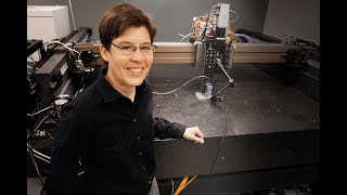
In this talk, Professor Jennifer Lewis outlines two decades of work at the intersection of materials science, 3D printing, and biology. She begins by motivating the shift from bulk fabrication to digital processing, emphasizing how direct ink writing and embedded 3D printing enable precise control of composition, structure, and function across multiple length and time scales. Lewis then details the design of viscoelastic inks, first colloidal gels that shear‑thin under pressure and rapidly re‑gel after extrusion, and later polymer gels filled with anisotropic particles such as silver platelets for stretchable electronics and cellulose fibrils for plant‑inspired shape‑morphing hydrogels. She showcases functional demonstrations including a millimeter‑scale interdigitated lithium‑ion battery, a soft stretchable electrode that lights an LED, and bilayer hydrogel actuators that mimic pine‑cone opening and wheat‑awn twisting. Transitioning to biology, Lewis describes cell‑laden inks and a fugitive ink strategy for printing perfusable vascular networks within centimeter‑thick tissues. She focuses on kidney engineering, explaining how multi‑scale architecture—from organ‑level vasculature down to nanoscale slit diaphragms—requires combining bioprinting with stem‑cell‑derived organoids to achieve filtration and reabsorption functions. The talk concludes with examples of vascularized organoid integration, functional assays (albumin uptake, glucose reabsorption), and a forward‑looking vision for organ‑scale tissue repair, while acknowledging collaborators and funding sources.
Chapters
Good afternoon. I'm deeply honored to have this opportunity to represent Class 3 and share a bit of my laboratory's work, which focuses on printing soft matter in 3 dimensions. As a young undergraduate student at the University of Illinois, my passion for material science was ignited when I first understood that how you process or make a material profoundly influences its structure and properties. And in fact, in the NAS 1989 Decadal study on materials science, this paradigm emerged where processing is intimately related to structure and properties. At the time though, the forming methods or the processing methods that we had at our disposal as materials scientists really were bulk fabrication methods like molding and casting. What is intriguing to me about 3D printing and why our laboratory has focused on that for over 20 years is that by digital processing, we're able to now control the composition, the structure, and the function function at multiple scales. So not only multiple scales in dimensions, but as you'll see, also often in time. So our lab has pioneered two main printing methods. They're both shown here, they're playing now. One involves directly extruding materials through a fine deposition nozzle. We refer to this as direct ink writing. So the materials that we're making are not like the inks that come out of your pen or out of the printer cartridge when you're putting words on paper, but they're really functional or biological or structural materials.
The other method that's being shown here we refer to as embedded 3D printing.
Here we're not printing the entire architecture, but we're using a nozzle to go deep into a matrix and write only the functional features of interest. And in this case, what you're seeing is a vascular tree being written, and I'll come back to that later when I talk about how we're making biological materials and that these vascular structures are necessary to keep— in thick tissues, keep cells alive. So what do I mean by soft materials? Just, there are many classes of materials. What we mean by soft materials are those that have some type of structure, structure that's pervasive beyond the individual building blocks themselves. So the individual building blocks in a colloidal gel are the red particles here that you see, the colloids themselves.
In a polymer gel, it would be the individual polymer chains.
In human cells, of course, it's the cells. And what we're really thinking about is how does this structure beyond that control the properties. And for soft materials, as the name implies, these are mechanically weak and deformable, and that means they can be printed. So from the perspective of designing inks for the types of printing methods that we do, as you saw in those movies, our inks maintain a filamentary shape when they come out of the nozzle, okay? So unlike the inks that you have in your inkjet printer that break up into droplets and wet and spread due to Rayleigh plateau instabilities, we're designing a viscoelastic response into this material. And all of you have come into contact with a viscoelastic material today, at least I hope you have, because all of you have squeezed toothpaste out of a toothpaste, right, onto your toothbrush this morning when you brushed your teeth. That kind of viscoelastic response is exactly the kind of response that we're putting into these inks. They flow under an applied pressure, but as they exit the nozzle, just like as they exit the toothpaste tube, they stop flowing so they don't wet and run all over your brush.
So we are engineering in this shear thinning response.
This means they flow more easily the higher we apply pressure. But as they exit the nozzle and go back into a quiescent or zero shear condition, they rapidly resolidify. And that resolidification process is inherent due to the structure that I described, this gel-type structure. So what are these inks? So the first one we actually worked on in my lab was in close collaboration with Joe Sassarano, formerly at Sandia National Labs. And these are materials that we refer to as colloidal gels in the sense that we have to introduce some type of attractive interaction between particles to drive their gelation, and that allows them to form a spanning network in 3 dimensions.
And they basically undergo this little clustering phenomena, but when we flow them through the nozzle, as you can see here, just as a small wedge near the wall of the nozzle, these clusters deform and break up under shear.
But as they exit the nozzle, that attractive interaction, which is on the order of thermal energy, kT, rapidly causes them to reform the particle network that's continuous that allows them to support themselves.
So what can we do with colloidal materials?
Well, here's one example. I'm printing, in this case, an anode and cathode that are interdigitated and out of plane. So we're making a 3-dimensional battery. This is a lithium-ion battery, much like you have in your cell phones and your laptop computers, but what's What's kind of interesting about this battery is the entire form factor, the entire size of this battery is on the order of a single grain of sand. I'll let that sink in for a minute. So salt shaker, single crystal of salt, we're talking about something that's on the order of a millimeter by a millimeter in aerial dimension and about half a millimeter out of plane.
And it works. This is the aerial capacity, it can be charged and discharged many times.
So this is what one can do with printing. One can design things that one couldn't make by conventional fabrication methods. Now the other thing that we've been focusing on in our lab is pure and filled polymer gels. So these are inks where now the structure is really to do with the fact that these polymer chains overlap and form physical entanglements that lead to this viscoelastic response, but then we can fill these materials with building blocks, in this case anisotropic fillers, and I'm going to highlight two examples. One where we're using these platelets that are conductive, so silver platelets that provide conductivity to the ink as we're printing, and the other one where we're using cellulose fibrils, which are stiff fibrils much like you find in plant cell walls, which I'll come back to in a minute, that can be used to tailor the anisotropic stiffness in the printed architecture and hence swelling behavior.
So because we have the shear field in the nozzle as the ink is flowing through, we can shear align these anisotropic particles in such a way that they orient along the printing direction.
And I'll highlight two examples. The first one involves taking these platelets of metallic material, silver, embedding them in a thermoplastic material that's an elastomer that can be stretched to very long distances. So here is an electrode that we've printed on a back a pure polymer substrate, you can see that as this is stretching, the LED light that it's connected to is gonna start to dim, and ultimately when we get to about a strain of about 200%, we'll break electrical contacts between the particles and the light will go off. And then as we relax this back to a zero strain condition, it then of course reconnects electrically. And what's connecting really are these contacts between particles that are able to slide as we stretch, and reconnect as we come back. So what can we do with something like that? So here's an example of printing a soft electronic device.
First we put down a substrate, this is sped up quite a bit. The other features that you see are silver lines, these conductive inks that I just presented. Now we're using the printer in a sort of interesting way, we're picking and placing discrete components, we're putting down connectors, And then ultimately we can pick this thing off the printer. What you see now is just interconnects between the printed conductive features and the LED components that we've picked and placed. But this is a fully soft functional electronic device. You can see this coming off the printer very nicely. We'll connect down here with power supply and then we can light that up, okay?
So this is a very nice way of printing a soft electronic device. So, okay, so what else can you do with these oriented building blocks? Well, we can use the shear, as I said, to emulate the kinds of architectures that we find in plants. So if we look at plant cell walls, they have these cellulose fibrils that are oriented along specific directions. And because they're stiffer along that axis, the long axis of the fibers, relative to the orthogonal direction, this allows for the nastic motion of plants.. So for example, we see when we immerse a pine cone in water, it undergoes a bending transition because each one of these features has this bilayer architecture where there's an orthogonal 90, 0-degree orientation between layers. Wheat awns, for example, have a 45, -45-degree orientation between the two layers, and they undergo a twisting when they, when they swell. So we were able to design an ink that essentially mimicked the cell walls of plants. So this is a hydrogel with cellulose fibrils.
We can orient those, we can measure their experimental swelling strains and stiffness anisotropy, and with Maha Devan's group at Harvard, who developed a predictive model that predicts both mean and Gaussian curvature, just based on the orientation difference between the bilayer filaments that we're printing, coupled with these experimentally measured measured values, we can create kind of complicated bilayer architectures such as the one shown here. And you can see along these different features that the bilayer design is quite different, and each of those is encoded with a specific shape morphing characteristic. So I'll let that movie play just a little bit more. You can see as we're printing just how the ink is coming out of the nozzle and building layer by layer. And then if we take that and we put it into water, it undergoes this dramatic shape change, right? So we have opposite chirality here in these regions that are twisting, these are ruffling, and this region is patterned with that 90, 0 degree to introduce bending.
And you know, this is really inspired by a natural flower, the orchid shown here. So this is some of the things that one can do with soft materials using 3-dimensional printing. Now when I joined Harvard 6 years ago, we decided to take on a new challenge, and for somebody who was trained in material science, I have been drinking from a fire hose for the last 6 years trying to learn biology at a time when this field is rapidly evolving and exciting breakthroughs are coming out almost every day. We had at our, sort of at the core at its core, this desire or drive, if you will, to use our printing methods to introduce or embed vasculature within 3-dimensional printed tissue. And when we first started out, we spent a lot of time developing material sets, both cell-laden inks, so now replacing fillers that are synthetic materials with cells, and putting them into hydrogels themselves, and directly printing tissues, and these tissues can have these features that are channels that can then be seeded with endothelium and serve as blood vessels that allow you to then perfuse them over time to provide nutrients to keep the cells alive.
And one of the things that we did was we printed a tissue that was laden with stem cells.
It was about a centimeter thick. We could perfuse it over months, and we could provide through the vasculature cues that would drive the differentiation of those stem cells down specific lineages. But if you compare, and as I will, for one tissue type of interest, the kidney, the type of architectures that you find that provide function to your kidneys over multiple length scales, it became pretty quickly obvious to me that bioprinting alone would probably not get us there where we needed to be for emulating vital organ tissue function and architecture. And so we have really picked up some of the advances that have occurred in the stem cell field, particularly using stem cells and differentiating them in multicellular aggregates to form organoids or these organ-like building blocks. But I'll kind of walk you through a progression that we made in our own lab over the last 6 years, starting with just bioprinting because it can do some things, and then how do we combine that with stem cell biology to do maybe something much more. So the inks that we developed for bioprinting include both an extracellular matrix, which is composed of a hydrogel. In this case, it's an interpenetrating network of gelatin and fibrin, and we use enzymes to cross-link this in a dual enzymatically cross-linked way. And then the sort of secret sauce, if you will, for printing the vasculature was to print a material, in this case what we call a fugitive ink, and you saw that being printed in that vascular tree in that embedded printing movie that I showed earlier. That ink comes down and we print the structure, but what's interesting is we can print at room temperature, but when we cool the material down after we've printed it to about 5°C, where cells remain viable, we can actually flush that out of the printed architecture, leaving behind a pervasive open network of channels. Now those open network of channels can be lined with endothelial cells to create vasculature, or for something like the kidney, we can also line them with proximal tubule cells, which would create a proximal tubule mimic for the kidney.
So this turned out to be kind of an interesting material, particularly because it doesn't do what most materials do, right?
Most materials, when you heat them up, they melt, but this is a material that actually when you cool it down, it liquefies, and the reason is because you're Essentially neutralizing, if you will, to some extent the hydrophobic interactions that are driving the aggregation into micelles that form this gel-like material at room temperature when you cool it down. All right. So we have become very interested in the kidney. There is many motivations for this from the perspective of using these kinds of tissues to help develop screens for— to identify drugs early in the pipeline that are being developed to understand whether or not they have any toxicity. It would be very important to have human tissue in order to do that drug testing. But also from the perspective of long-term— our long-term goal, which is tissue repair and replacement, the kidney is one of the most needed organs. So if we look at the U.S. donor organ list, There's roughly 120,000 people on that list, and 100,000 people are waiting for kidneys. Moreover, once you get into end-stage renal failure, you go on dialysis, and you roughly have about 5 years to live, and if you don't get an organ in that time, you really have a high rate of mortality. So this is a real driver for us, and we, as you'll see, want to take baby steps towards this, our aspirations are ultimately to do kidney engineering over these scales. So first of all, if we look at a kidney, it's about the size of my hand. Each of us have two, and inside our kidneys we have what are called nephrons. There are about a million of these nephrons, and they're responsible for filtering our blood continuously and also doing the resorption process.
So the way your your kidney works is your blood goes through this capsule, the glomerulus, it's called the Bowman's capsule. There kind of everything is pulled out of your blood and then it continues to go through, this filtrate goes through what's called this convoluted proximal tubule region and about 80% of what was pulled out, that's good, goes back in and there's a reabsorption process and then it continues through the rest of the nephron.
If we look at the structure of a kidney from a material science perspective, I'm always thinking about scales, we see that it goes—
there's this sort of large scale, just looking at this arterial tree that you would find in your kidney. There's the tubular compartments. There's the glomeruli.
And then even at the nanoscale, individual cells are forming this slit diaphragm that gives rise to the filtration. So very quickly, if we start with bioprinting, which is how the lab started 6 years ago, what could we do?
Well, we could take some baby steps. We could look at one of the key regions here, this proximal tubular region. We could form an analog of that through printing on a chip sitting on an ECM.
If I go to the next slide, you can see that as we're removing that ink, we can then open that channel and we can seed that channel with the proximal tubule cells of interest.
Those then form a confluent epithelium on the walls of the channel. If we take a cut through the channel and look at that, we see these cells, and in fact we see them in this cuboidal state and they have these microvilli, and essentially they look just like the types of cells that you would find in your proximal tubule in the native kidney. And we can then introduce a vascular component by co-printing these two channels alongside of one another, and you can see if we take a cut through this structure, We have these two lumens that are co-located, and that will really allow us to study resorption.
And so we've been using these kinds of constructs to study things like albumin uptake, glucose reabsorption, and of course for also disease modeling. But in my last couple minutes here, I wanted to tell you about some exciting work that brings together stem cell biology and these bioprinting capabilities, because while bioprinting might be able to do this kind of length scale, it's very difficult to imagine any type of printing process that alone would give you this full complexity of architecture that you see there. That's really required for the filtration plus the reabsorption. So we can do reabsorption, I think, by bioprinting, but filtration, we really need to rely on biology. And so here we've been very fortunate to work with Ruijie Morizani and Joe Bonventura's group at Brigham and Women's And Ryuji has a protocol for starting with iPSC-induced pluripotent stem cells, organizing them into aggregates, and then directing their differentiation. We disrupted his protocol at day 11, and we took those pre-tubular aggregates, and we put them into a chip that we bioprinted, and we're looking now at cues like how does flow influence the development of vasculature within these organ building blocks. And this has been a limitation of the organoid field for those of you who are familiar with that. Being able to generate vascularized organoids through these kinds of protocols that have been published to date simply has not been possible. So we have in fact been able to demonstrate a path forward where we can vascularize these organoids, both by introducing flow across them as they're continuing their differentiation, and also by embedding them onto an adherent extracellular matrix which we designed.
And I won't go into all the details here, but I think I can convince you that within these organoids, we've got these regions that are glomerular-like that have capillary invasion, and you can even see the semblances of foot processes much like you see again in native kidney tissue.
This is really just a starting point for the work, but it's an exciting starting point because now we have organoid building blocks that have a pervasive microvasculature This helps mature the cells that are in there, both in the glomerular compartments and the tubular compartments. And then if we take these kinds of building blocks, like I said, and then combine them with the embedded printing process that I described earlier, we now have the ability to kind of, if you will, create thick tissues that have these as their starting components, cellularly dense, architecturally rich, functional, with embedded vasculature. And this is really work that we haven't even published yet, but I think is very promising. And so I'll end with that. I want to thank a number of people, my group first and foremost, because honestly, without them, none of this work happens.
And it's a privilege to have the opportunity to work with so many talented young people, not only in the current group, but many who have come before them.
As you can also see, we're a highly collaborative group, so I would be remiss without without thanking a number of key collaborators, both at my former institution, Illinois, and now at Harvard and beyond. I also want to thank my PhD advisor, Michael Cima, who was a wonderful mentor. So I'll end there. And if there's time for questions, I'd be happy to take them. I will also thank my funding agencies and those that have provided generous support for the work. So thank you. And I guess I'll just leave that slide up. Thanks. Thank you, Jennifer.
More from Joshua







Scientist Stories: Hans Waterham, The value of phenotypic assays in peroxisomal disorders




Scientist Stories: Joel Zlotogora, Genetic carrier screening programs for reproductive purposes