Scientist Stories: Jennifer Petersen & Joshua Zimmerberg, Exosome Biogenesis & RNA Communication
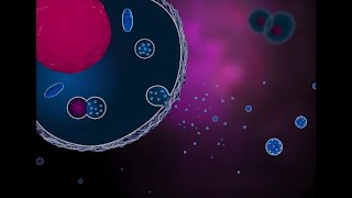
In a joint ERCC‑ISEV eB Journal Club webinar, Jennifer Petersen (now at NINDS) presented electron‑microscopy evidence that human umbilical vein endothelial cells (HUVECs) generate a specialized membrane domain that produces large, multicompartmental microvesicles (MCMVs). These MCMVs contain internal multivesicular bodies (MVBs) loaded with intraluminal vesicles (ILVs). Serial thin‑section transmission EM, using ultrafast freezing and freeze‑substitution, revealed protrusion clusters, neck‑connected vesicle domains, and the presence of mitochondria, ER fragments, and clathrin‑coated endosomes within the protrusions. Crucially, omega‑shaped exocytic profiles were observed on the surface of MCMVs, indicating that ILVs can be exocytosed from the MCMV after it buds off, creating a hybrid MV‑exosome carrier. Petersen discussed the methodological advantages of rapid freezing for preserving membrane ultrastructure, the identification of MCMVs as a distinct EV class, and proposed functional roles such as protected cargo delivery, time‑release signaling, organelle clearance, and angiogenic regulation in fetal development. Joshua Zimmerberg contextualized the work within membrane‑fusion biophysics, highlighted the need for complementary live‑cell imaging (e.g., calcium‑indicator movies in lymphoid tissue explants), and raised broader questions about polarity, isolation, prevalence across cell types, and downstream signaling. The Q&A explored topics like EV polarity, isolation methods, immunogold labeling feasibility, and the relevance of MCMVs in vivo. The discussion underscored the novelty of hybrid MV‑exosome structures, the challenges of isolating them, and the potential impact on extracellular RNA communication research.
Chapters
Hey everyone, I'm Roger Alexander, the Director of Scientific Outreach for the Extracellular RNA Communication Consortium. I want to welcome you to the last ERCC webinar. It's appropriate that today is a joint webinar with ISEV's eB Journal Club run by Ken Whitwer. Over the last 10 years, there's been some really interesting synergy and collaboration among labs within the consortium getting work done and finding new biology that we wouldn't have done otherwise. And so So it's going to be interesting to see how the field moves forward without that synergy and its forums like the EB Journal Club that will help fill that gap. Today's talk is really interesting new EB biology from Jennifer Peterson and Joshua Zimmerberg. Jennifer's now at the National Institute of Neurological Disorders and Stroke. This work was done at the National Institute of Child Health and Human Development where Joshua Elkington is still working. Ken, do you have anything you want to say before we get started?
No, I just wanted to say thanks for this great opportunity because it's a coincidence, but I think a very happy one that the EV club was going to feature this paper yesterday and ERCC was going to feature the paper today. And I think that really speaks to two things. First of all, this is a very provocative paper, very interesting science here, and we're looking forward to hear more about it. But secondly, these commonalities, these common goals and values that the NIH ERCC and that ISEP have together. And so I think even though this webinar series is coming to an end, you know, we're going to see the legacy, the influence of ERCC continuing into the future. And for those of you who want to have that regular jolt of good xRNA or EV science, please do subscribe to the EV Club and we'll make sure that you get the weekly notifications. So yeah, back to you, Roger. Thank you.
Yeah, agreed. The EV Club is a great forum. So before we start, please everybody stay muted. Put questions in the chat during the talk, and I'll try and ask those at the beginning of the Q&A session and then open it up to people asking questions live. I don't want to monopolize the Q&A. So if you want to add details or comment on your question when I ask it from the chat, please feel free to do that. And so Jennifer and Josh, please, we're looking forward to hearing about your work.
OK, well, I'll get started. Let me share my screen. And while I do, I'd love to thank you for that fantastic introduction. Joshua and I really appreciate this invitation from both of you to present our recent publication in Journal of Extracellular Biology. And we also appreciate that you arranged to make the meetings into this joint presentation. So, thank you so much for that. And thanks to everyone who's tuning in today. Can't wait to get the feedback from everybody. So at the end, I'm going to start, and then at the end, Joshua Zimmerberg is going to give some statements and open the discussion so we can hear what everyone is thinking. Okay. So, today, I'm going to present electron microscopy evidence that endothelial cells, as the title suggests, release basically EVs that are MVs on the outside and exosomes on the inside, to put it simply. And that these exosomes can get released after the MV has departed the cell of origin. So, it's going to be a lot of electron microscopy today, as you can tell from my background. I am in a vesicle. Okay. So, for this group, I don't think I need to give too big of an introduction to extracellular vesicles and particles. I think most of us are familiar with the idea that extracellular vesicles arise from two broad distinct mechanisms, exosomes that arise from the endosomal membrane system when MVBs fuse and release ILVs to the extracellular extracellular space versus microvesicles, which bud directly from the plasma membrane. And there are also extracellular particles, exomeres and suprameres, that have been relatively recently identified and are a very hot topic of research. And all these different EVs and PEs participate in intracellular signaling in both healthy conditions as well as disease states, serve as biomarkers and diagnostic tools for disease, and provide therapeutic vehicles for treatment of diseases. It's a really exciting field, as you all know. So, a few years back, I had the good fortune of entering into collaboration with Dr. Suki Kaur and David Roberts, who I think are probably on the call today. And they study endothelial cells and the EVs released from endothelial cells. They're in the Cancer Institute, the National Cancer Institute. So, the endothelial cells form the endothelium, which is a single-cell layer that lines every single artery and lymph vessel in the body. And some amazing facts about the endothelium. In an average person, the endothelium covers about 7 square meters of area and would weigh 1 to 2 kilograms if you could weigh it. So, the endothelium and the EVs that are released from the endothelium participate in the maintenance of vascular homeostasis. And some of the features of vascular homeostasis are listed there. And to look at endothelial cells and look using electron microscopy for where EVs are coming out of endothelial cells, we used a cell line which many of you will probably use yourself. Use yourselves, called human umbilical vein endothelial cells, or hUvECs. And these endothelial cells can be harvested from the— I'm going to start my laser pointer— be harvested from the umbilical vein, because they line the umbilical vein in donated umbilical cords. So, a little bit about the technique used. It was an electron microscopy technique. Where we initially preserve the cells that are grown on coverslips by ultrafast freezing, by plunging the coverslips with the cells attached into liquid ethane. So this freezes the cells from the live state in their culture medium. They were perfectly happy, having a fine time, and then all of a sudden they got plunged in liquid ethane and froze. And ideally, the medium around the cells and the water in the cells freezes so fast that you don't have ice crystal formation. So you get very nice ultrastructure, and you also freeze within milliseconds. So you can capture very rapid events like exocytosis, and it's particularly good for preserving cell membranes. So after the cells are frozen, they're maintained in the frozen state, and then they go through a process called free substitution. So while they're still very cold, the water in the sample is replaced by a solvent. In this case, it was acetone. And in that solvent are fixatives and stains that stabilize and preserve the cells before they're warmed up to room temperature. So it's— it provides really good views of ultrastructure compared to chemical fixation, which occurs over minutes and can impart some structural artifacts due to osmotic imbalances and things like that. So ultimately, the embedded— the cells are embedded in a plastic resin. Such that the cells are on the very surface of the plastic. The coverslip is removed and you have just the cells there, the bottom of the cells at the very surface of the plastic block. And that can be sectioned into 70-nanometer sections. And on the right is actual sections coming off, floating on the water of a diamond knife boat. And you could see the sections are little trapezoid shape. And they come off, if you're lucky, they come off in what's called a ribbon, and they stay in order. So for structures in the cells that would span more than one 70-nanometer section, if you pick up the sections in a row, you can examine that structure in adjacent sections and gain information about its three-dimensional organization. So let's take a look at a Huvec cell. Oh, this was just my little scheme showing you serial sectioning. Of a cell. And let's look at a Huvec cell now that was preserved using this method. This is about half of a Huvec cell. They're kind of oblong shaped often, and these are wrinkles in the plastic section, so don't get distracted by those. The nucleus is highlighted here, and just for your reference, like, the dark squiggly things are mitochondria. And the light circles are MVBs because they very often have a light electron density lumen. So, we were looking for where could EVs be emerging from these endothelial cells. And most of the plasma membrane is smooth. But we noticed in some cells an area on the cell where the membrane became more elaborated. And if we look at that at higher magnification, we see that this area here is a cluster of protrusions on the cell in a specific location. And I'll just highlight a couple of the protrusions by outlining them, showing you that they connect back to the cell by thin necks. Furthermore, we noticed that inside these protrusions, you could see membrane-bound organelles, particularly organelles that resembled MVBs. So I told you that I cut thin serial thin sections. So let's just look at a few sections through this zone of protrusions. This is the very first section right at the level of the coverslip. So now those protrusions, which I just showed you were attached to the cell, kind of look like a cloud of vesicles. But that's because where they're attached to the cells actually occurs at a higher section in Z. And I've color-coded different protrusions where I, that I can connect by looking at the serial sections showing that some of them are very branched, particularly the pink protrusion. So let me just go through a couple of these serial sections for you, if I go the right way. So here we go, we're moving up away from the coverslip and I'll just go to this one. It's about the Fourth section where you see the necks of the protrusions connected to the cell. And again, I've color-coded them to label different protrusions. And in the next section, I'm labeling at least 6 independent protrusions that make up this domain. Some of them are very branched, like the pink and the blue one. Others are simple unbranched protrusions like the number 1 and number 2. So I'll just finish up going through a couple more of those sections about— I went through about 500 nanometers of the protrusion site. So as I pointed out, some of the protrusions contain membrane-bound organelles, and we wanted to identify those organelles. So In the orange boxes here, I've surrounded some organelles in the cytoplasm that we can recognize strictly by their ultrastructure morphology. So I'm going to show those enlarged on the next slide, and then I'm going to show you what we can match them to in the protrusions. So here are the organelles from the cytoplasm. We can recognize mitochondria because they have cristae and they stain very darkly. MVDs, of course, that have a light electron, light lumen and interluminal vesicles, numerous endosomes. Some can be clathrin-coated like this one or uncoated like this one. And endosomes can also be tubular and also endoplasmic reticulum. And now in protrusions from that series of sections I showed you, We see MVBs that match, they're identical to MVBs that we saw in the cytoplasm. There's also small mitochondrial fragments like this one has a nice cristae, so it's definitely a mitochondria. More MVBs, MVBs were a very prominent component of protrusions. This protrusion had many endosomes, both the uncoated and clathrin-coated type. And tubular. And there also appears to be pieces of ER perhaps in these protrusions as well. And I just want to drive home the fact that MVBs are in the protrusions. This is another example on a different huvex cell. And in the green box is a localized site of protrusions. And it's enlarged on the right. And you can see this fabulous, big, beautiful MVB in this protrusion. So this is an MVB. Like 600 nanometers in diameter and has ILVs, and it's in one of these beautiful protrusions. Okay. And this is now just showing you across multiple cultures that we see protrusion sites, and these are the different ways they can appear. The top row shows protrusion sites that were cut in the Z height where you can see where they're connected to the cells. And the bottom row shows protrusion sites that are cut some distance from where the protrusion's cut to the cell, attached to the cell. So they actually can look like they're separated from the cell. But by looking through the serial sections, you can confirm that they're attached to the cell at a different height in Z. So the idea that on Cuvex cells, there's a specialized domain of membrane protrusions is not a new one. In this earlier study by Tara Aletti et al., where they looked at HUVEX with scanning electron microscopy and thin section electron microscopy, they identified a localized site on the HUVEX surface where they had protrusions. And we think this is the same site that we've identified in our thin section EM. And they further reported that vesicles were shed from the specialized domain that contain enzymes that participate in angiogenesis. So, if vesicles are being shed from this domain or pinching off from these protrusions, and we showed— we've seen that many of these protrusions contain MVBs that contain ILVs, could it be that they pinch off with an MVB inside? So if this is our diagram of our protrusion site, can they pinch off containing MVBs? And if— and I've also shown you that these protrusion sites go project out and away and up from the cell. And so if a vesicle is to pinch off the protrusion, it would diffuse for a while in the culture medium. But maybe if this is happening often enough and they're abundant enough, on occasion one of these diffusing MVBs, or yeah, one of these diffusing vesicles, excuse me, would happen to contact the coverslip surface where it might adhere and survive through the EM processing and still be there. So if I was to look at sections beyond— look in the section as close to the, to the coverslip surface, but look beyond the periphery of the cells, might I see membrane-bound organelles that correspond to pinched-off protrusions? And do they contain MVBs? So that's what I did, and I'll show you an example right here. So this is the periphery of a Huvec cell. And then there's blank coverslip. This fuzzy stuff is what you see when you do rapid freezing and freeze culture medium. So that's nothing. It's just telling you that it was a freeze-substituted sample. And out here we see an organelle, which I'll show you enlarged. It's a membrane-bound organelle that in turn contains many round vesicles itself, and some of these vesicles contain additional vesicles. And the— I can use the serial sections to confirm that this organelle is in— no, has no cellular extensions linking it back to another cell in the coverslip. It's an independent structure out on the coverslip. So I'll run through these serial sections, and all these sections are available in the publication in the supplement, and they're in movies that you can open in ImageJ. And really study them. So I'll just go through some of these sections and point out that every section, which is 70 nanometers thick, reveals additional vesicles. So vesicles that are smaller than 70 nanometers will only be visible in one of the sections. And we can keep going, and it's just chock full of vesicles. There's this big one here that is there for the entire series through this structure. There's also some complex internal vesicles that contain multiple vesicles inside vesicles, which I'll talk a little bit more about later. And I also want to point out that these large electron-lucent vesicles inside this organelle look like MVBs. However, we haven't seen an ILV in there yet. Okay, so let me just finish up this section, series of sections. So, since we have identified this membrane-bound compartment that is separate from cells that we believe pinched off from the protrusions, we're defining it as a microvesicle. And because it has many membrane-bound compartments inside, we've donned it the multicompartmented microvesicle or MCMV. And there was a recent EV club talk and covering the recent publication as well in JEV where people— where all these multilamellar, multilayered EVs have been described more and more that are observed by cryo-EM. And we think that that is probably some of what people are seeing are organelles such as these. And I'd also point out that these are observed without any type of isolation, centrifugation. I mean, that was just a coverslip frozen from live state. So the idea that sometimes the isolation step can create multi-layered EVs is not always the case. We see them here without any of those isolation steps. So, I pointed out that these large vesicles inside here are— have the electron density of an MVB, but we didn't see any ILVs in them in this case. Although interestingly, there are small vesicles attached to the outside of this organelle. And these observations raise the question, do MCMVs secrete exosomes? So, for that to be true, the The MCMV would need to contain MVBs that contained ILVs. And we would also need to see evidence of exocytosis somehow. So, when we look around more on the coverslip and look at other MCMVs, we saw that they do indeed contain structures that are identical to MVBs. It's a beautiful MVB. It looks like it's— undergoing an outward budding step, and it contains many ILVs. And also on this cell, on this MCMV rather, there are omega figures on the periphery. Now omega figures are a hallmark of exocytosis, and we think we're capturing them because of the fast freezing method of preservation, where chemical fixation, it may be less obvious where you have an omega figure. But they're formed when a vesicle fuses with another membrane and creates this kind of light bulb shape. So you have a pore basically opening up to the outer extracellular milieu. And there are several on this MCMV that I outline in the blue turquoise. And I also point out just this little bump here or a little indentation here, which could be a later stage exocytic profile that has now almost fully collapsed and become continuous with the MCMV outer membrane. I'll show you another example of another MCMV that also contains MVB-like organelles as well as many other vesicles and has some nice, a nice omega figure here. And these, again, you can look at in the supplement of the publication. So I'm just going to show you one more MCMV. This one was very actively exocytosing. There are omega figures in every section, and I think there are a dozen serial sections through this MCMV. But I'm just going to show you one. And in the turquoise box is an omega figure that has no ILV in it, but we see On this MCMV, some of the omega figures contain small vesicles that are identical to ILVs. So, I'll go to the next section where there's another omega figure containing a small putative ILV. And it's hopefully in the process of ejecting this ILV. But interestingly, often in some cases, the ILV looks very closely associated with the inner surface of this exocytosing vesicle. And I'll mention that again in a moment. In the final serial section that I'm going to show you here, there are 3 additional omega figures. So, I think there were something like 15 or more on this one MCMV. And 2 of them contain ILV-like vesicles, 1 of them doesn't. And in this MCMV, there's also a beautiful MVB with an ILV. So this just brings us back to the first MCMV that I showed you, and I mentioned that it looks like it has MVBs, a big MVB that doesn't have any ILVs, and it had these small vesicles attached to the outside. So now we think we understand where these small vesicles are coming from. They are the result of when one of these omega figures fully collapses on the MCMV outer membrane surface, and an ILV that was closely associated with the inner membrane of the exocytosing vesicle remains attached and is now hanging out attached on the outer surface of the MCMV. So, these vesicles on the outside could be reporting a history of exocytosis in that MCMV. So, now one last thing, If vesicles are being released from the MCMVs, is it possible that if I just look at the first 2 or 3 sections on the coverslip, will I find vesicles that are single membrane vesicles that correspond in size and appearance to vesicles that we observed in the MCMVs? And in fact, we do. So these are just— if you go to a high mag and search blank coverslip, you can find these little exosomes or small EVs on the surface. And I think they're just fascinating to look at. But you can even see their contents here, little fuzzy contents. So the fact that we're observing MVs that contain MVBs with ILVs that appear to be able to release the ILVs as exosomes. In other words, it's some kind of hybrid MV-exosome combination organelle that's dynamic. So it releases from the, the cell of origin, and then it may start remodeling, ejecting some cargo or not. But we think this may explains some of the difficulties in making and clearly separating EV populations because they could at times be all in the same carrier. And this is just a kind of diagram showing the whole story. We have an MCMV release site. MCMVs bud from this site and can release exosomes by exocytosis. They could also remain intact or partially intact. And interact with recipient cells as a whole MCMV, or their contents, MVs and exosomes, could reach recipient cells. Why might cells use MCMVs? We have a few ideas. They might shield the contents of the released MV from degradation so they can travel longer, particularly, for instance, in the bloodstream before getting degraded. They could make it so that multiple contents get delivered in a signaling kit in the right proportions and at the right time for more efficient signaling. These could be a way for the cell to eject damaged components, damaged organelles. They could also be kind of— number 4 is kind of like number 1— time-release capsules where it's spreading out its signal over a longer time instead of all at once, just going out all at once. There's a lot of questions. So, are MCMVs unique to endothelial cells? Are MCMVs preserved by the various isolation techniques that are used? Maybe they get ruptured. Don't know. Figuring out the molecular mechanisms regulating the formation of the protrusion site, where it is, does it come and go, is it upregulated or downregulated, how are cargoes sorted into the protrusions, into the MCMVs, how do they pinch off, what are the kinetics, and what are the downstream signals mediated by MCMVs What are recipient cells? So there's a lot of fantastic questions. Now, I'm going to hand it off to Josh now, but I'll just take a minute to thank everyone in the Zimmerberg Lab where all this work was done, particularly Elena, who grew the beautiful Huvec cells on the substrates. And Paul helped with some quantification that we present in the paper. Our collaborators at NCI, Suki and Dave Roberts. Thank all of you for your attention. And again, Ken and Roger for the invitation to present our work. And we really appreciate it. And now I will hand it off to Josh.
Thank you. Thank you, Jennifer. Beautiful presentation. I wanted to just give a tiny bit of background. Kind of in two directions why we got interested in this work. I think like many, many people, we've seen hints, we've seen evidence of extracellular vesicle activities in multiple different places, but in particular, we've been— have been looking at human explant tissue, tissue histocultures,. And I'll show you a movie of one of those from human lymphoid tissue. But I really have to thank the NCI and the NIH that really got some seminars off the ground to consolidate into the field. Mostly this society stepped into the breach, and really the Extracellular Vesicle Society, to get things rolling. But the huge amount of work done by the ER, the RNA, the extracellular RNA community, and the workshop that they just held was just fabulous. So with respect to some of the importance of what Jennifer's shown, first of all, I think it's very important just to connect for a minute to the actual technologies that Jennifer used to see these. We're a membrane lab. We've been involved with the membrane biophysics of membrane fusion, in particular exocytosis. The techniques that were developed to understand the mechanism of exocytosis are the techniques that go into these pictures. Freezing into vitreous materials, sectioning after going through a freeze substitution technique that preserves membranes with a real kit of compounds that Jennifer fine-tuned that allow for the vitreous frozen material to go into ultimately plastic after being fixed at -90 degrees, the temperature that acetone melts.
These—
all this work on membranes is what went into this, but there's a second thing that went into this that's very important, that this is the actual surface of a cultured cell that is being looked at directly, so that the cells are just growing the surface that's growing on is just frozen. It's put into plastic so you could look at it, but we're looking at what's right outside the cells. And boy, the best thing in the world that we could wish for is to have the recipient cell, if there's something going on, the field really needs to know what's the best model system where we could have both the giver and the receiver in, in the same, on the same coverslip and on the same slice of tissue. But that really brings up a lot of questions that Jennifer addressed in her list. We don't know that these elements will be preserved in the current isolation technologies.. But if they are preserved, then there will be exosome markers in MV. So MV and exosomes will be inseparable from the point of view of isolation and looking for markers. It will not be possible to separate something that's one and the same. It's just like we use the analogy of a Matryoshka doll, that there's elements inside of elements inside of elements. Secondly, that by looking at the surfaces that cells release MV, we've seen this preservation, but those are artificial surfaces. What about tissues? So I'll show you a confocal Let's see, I'm going to share my screen here. Hopefully you're seeing a movie now of a slice, a confocal slice through a histoculture, an explant culture of a human lymphoid tissue in which the stain that you're looking at in all of its colors is a calcium indicator. So the most common intracellular signal for giving rise to an omega figure, to getting fusion to occur, is calcium. And if calcium is going up, we should perhaps see some calcium signaling. What we see in the tissue are some cells that are just pegged with calcium. Calcium is high in red and low in green.. But there's also signaling going on between cells, but there's much more activity going on outside the cells. This is not the same as what's seen in densely packed tissue like brain. This is lymphoid tissue. It's looser and it's highly— I don't really know what gives rise to the design of those. But you can see that elements are moving. Sometimes in straight lines like this cell marching along. But at the same time, it's not just the flow of the medium because you can trace when you look at this and analyze it, things moving in the opposite direction. There is a lot going on. I'm going to stop sharing. There's a lot going on. In tissue itself. And we don't know how this relates to disease states. And we don't know what gives rise to durability. But there's— now that we understand the hypothesis, basically, the discovery work that Jennifer's done to discover this new element is a hypothesis that can be then tested with other modalities that you can all think about in your systems. And I think it's very important that this is something that probably is accessible by light microscopy. Everything we've seen so far by light microscopy is not in conflict at all with the electron microscopy. And finally, I just want to say that's very important for the Institute on Human Development, at least the developmental field. You have to look at the cell where it's growing, the umbilical vein. The vein is going back to the fetus, it's carrying fetal blood, the oxygenated fetal blood. It's going directly to capillaries, it's going to interact directly with all fetal cells. So we don't know how important that is that we've seen that in this cell. As Jennifer pointed out, we would have to— we can't see, we haven't seen this in other cells, and we don't know what isolation will bring. So there's a lot to do, and we are certainly looking for postdocs to continue the work. You have to go to the Journal of Extracellular Biology itself to look it up because it's not indexed yet.
And thank you. Jennifer and Joshua, that's really, really interesting. Thanks for the talk, and I'm sure we have a lot of questions. Just quickly about the video, Joshua, what was the scale on the, on the movie?
Well, the cells are about 10 microns.
Those are lymphocytes. Okay. So I'm just going to go through the chat questions and then we'll open it up. My first question was, what's the range of thickness, like either, you know, micrometers or number of cell layers for endothelium?
It's a single cell layer.
Okay.
And there are a lot of different types of endothelial cells. But they typically are very thin. I'm not a total expert on endothelial cells, but I would say— I'm going to say about 4 microns thick. So single cell? They could be thinner. Yeah.
But a single layer. Yeah. Ellen Millman asks, are your hUVEC cultures polarized? Do you see the clusters on the apical or the basal side?
That's a great question that we really need to answer. Because, for instance, microsomes are very similar in some ways to what we see. And they form at the trailing edge and also contain other membrane-bound compartments. So, we don't— and these endothelial cells that we looked at are subconfluent on the coverslip. So, they probably think they're trying to be together because they prefer to be together. So, they're likely and I'm sure they are quite motile. And so these protrusion sites could not only be at the trailing edge or the leading edge, they could also be in response to a semi-wound-like state. They may be trying to signal to their friends to, to form an endothelium. But the other aspect is that they do have a basolateral side and an apical side, and we don't know which side this protrusion site would form on in an intact endothelium. I suspect it's going to be facing the blood or facing the body fluid, but we don't know that yet.
Right. Our next question is from Fata Kashanshi. And I just would like to say he's the president of the American Society of Intracellular Communication, ASIC, which I believe will be one of those forums I was talking about for that'll, you know, move on with the ERCC's mandate after ERCC ends. So he asks, he says, very nice EMs. Do your protrusions settle at 2k, 2,000 G, or 10k, 10,000 G, or both? Does addition of ATP or serum starvation change the dynamics of NCMV release?
Great questions. Don't know. We haven't tried to isolate them. We haven't tried to concentrate them. So, we just don't know those things yet. And haven't added anything to the medium. These are unstimulated endothelial cells. Other than being subconfluent, they're not treated with anything to trigger microparticle release or anything like that.
So, we don't know. I want to get back to that question later. I think it's an interesting question for this group, how to look at what you're doing with EM by other methods. But let me get to the questions in chat first. So, Meenu Kumari asks, in intact tissue, the umbilical vein, which direction will the EC release MCMVs? And she also says MCMVs appear to have some amount of cytoplasm. Am I right in saying that?
Josh, do you want to say which about the blood flow through the umbilical vein?
Well, yeah. I think the first part of her question is, when you culture cells, it's not exactly the same as the apical side versus the basolateral side, which is which is what it would be in the tissue. So it's being grown in a culture dish. So what seems to be the side where Jennifer's been able to look at all these things are most prominently, as she's shown, on the edge of what seems to us when she looks at cross-sections to be a cobblestone kind of appearance on the— on her special plastic surface. That's been studied by other people as well. There are a number of papers looking at the geometry. If it's on that side, is it on top? We haven't been able to look, but there's already evidence in the literature from scanning EM that it's on that apical surface. Yes, the protrusions, which are, we think, allowing these multi-compartmented microvesicles to pinch off and be released, are going directly into the fetal vein containing the oxygenated blood. But it's also, you know, if we're just fantasizing, important to understand that that blood has just been as intimate as it gets with the mother. Right in the placenta itself where the oxygen— they have very, very thin membranes separating to allow for oxygen flow between the maternal circulation and the fetal circulation. So it's also possible that there's signals that could be looked for from the placenta in terms of vesicles that are also moving. And this idea that it's protected, and it's moving a great distance is consistent with that kind of view. Also, it seems to be, like you point out, there's cytoplasm inside. Yes, these are like— I think we joked that these are mini-me's, people know that movie— that this is— there's no nucleus, but all the activities of cytoplasm we could— we suspect might allow a jackknife kind of vesicle, that the vesicle, what the vesicle does and what signal it gives, may depend on the receptors at the receiving end as opposed to what the cell is trying to send out. So it may be able to do more than one thing even. But yes, it has cytoplasm inside. We, we, we tried really hard to look for specific packaging and specific isolation and specific organelles, but nothing we could ever do allowed us to convince ourselves that there was any special packaging or any special difference between cytoplasm and a pinched-off multi-compartmental microvesicle.
Next question is from Rebecca Hecking. Thank you for this great talk. I'm very new to EM microscopy. Are these omega structures specific to exocytosis, or could they also indicate endocytosis?
It's a really good question that we We propose that they're exocytic profiles because if— well, they don't have a coat, so if it was an endocytic process, it would be not clathrin-mediated. And since they contain the small vesicles, the idea might be that they're capturing exosomes or small EVs from the culture medium, but that seems unlikely that they would catch one and endocytose it. So we don't think that they're endocytic, but it's not impossible. And we have to do some tests to try to figure that out. That's a great question.
Thank you. Alex Schmeyer asks, says, great talk. Given the advanced technique, is there any reason to think MCMVs are actually the norm rather than exception for regarding MV release in general?
I like that question.
I feel like Joshua may have been saying that near the end there.
No, no, we don't have any data. Yeah, we don't know. We just know what we can— we only can do this kind of work with any great certainty in the, in the, in the vicinity of the cell itself. And we would welcome somebody, and any, we'd have to work with anybody who wants to help start to look at isolated extracellular vesicles, and from different cell types or to try to look on cells, and everybody should try this, I think.
I will jump in just quickly to say, you know, you mentioned at the end it would be great to be able to see cells releasing and target cells accepting these vesicles in the same setup, and it seems like endothelium, where it's going to get— where the vesicles are going to get whisked away by the bloodstream, isn't going to be the system to do that in. And if you can find these MCMVs in a system that's more quiescent, I suppose you might find it. I wonder if there's any reason to believe that you wouldn't find MCMVs in such a quiescent system, why they might be in areas where they would get whisked away to somewhere else.
Yeah, absolutely. I mean, given that The cells have the ability to do this. That is shown by at least one example. So given this, the ability and how useful it could be, there's no reason not to suspect it, but it requires, it would, at this point, it requires the kind of dedication that Jennifer's had to this one cell type.
I saw you laughing, Jennifer, when you said, and you can look in blank slides for individual exosomes. I'm sure that was painstaking. Jenny Kang says, thank you for the great talk. For the flash frozen technique, can it be paired with gold antibody immunostaining to look into specific proteins?
Yeah, and what's nice about that is, is you can label this if you can, and we've done a little of this, if you can label the cells live, in such a way that it doesn't perturb anything and watch things get internalized and sorted, then you have kind of achieved the holy grail where you have very, very good ultrastructure and very, very good labeling. So that's an exciting direction that we've— we're already embarked upon. It seems to work really well. So we can start to get at the molecular composition of these domains and the MCMVs.
Yeah. Arefa Kardani asks, says, thanks for the talk. Would integrins connecting to ECM inhibit EV release in HUVECs?
I'd be really interested to know things that either cause protrusion sites to pop up or maybe go away. So something like regulating the extracellular matrix could very well be involved in that kind of regulation. So that's just a really cool thing to look into.
Yeah.
Uh, Svetlana asks, it says bloodstream will help to move the vesicles around. However, if these protrusions are multiple, would they interfere with blood flow?
I think we have to distinguish between the protrusions, which are on cells, and we don't think are getting ripped off and traveling around wholesale. But we think that's the organelle of MCMV production, versus these tinier things that are much smaller than red cells that shouldn't include anything. We don't seem stuck to each other at all.
So I'm going to say, so that was the last chat question except Mina just put one in chat, but let's get into discussion. Mina, do you want to just ask your question live?
If not, I can ask it. Yeah, thanks for answering my questions. I, you know, it makes sense that these exosomes present in MVVs within MCMVs have a predictive role of angiogenesis. So it makes sense that the developing embryo requires angiogenic process So, this may be helpful if they are moving in the direction of fetus. And my second comment, which I didn't get a chance to write, memory cells also probably are releasing this kind of vesicles, but I don't know if anybody has looked at it. And they will be engulfed by gut epithelium, so there is a system that you can use then. And in addition, Sertoli cells also release this kind of, this kind of structures. I have done it myself many years ago, and they are engulfed by developing germ cells. So, these are the two systems that you can look at.
That's very interesting. We should talk more offline. Okay. Great. Do you mind sending me an email? Sorry.
We've not met before.
No, we didn't. I don't want to lose you when the thing goes away.
That's very interesting. Any other questions? I have one. We touched on this a bit earlier. In my opinion, and I'm sure in yours as well, EM is the gold standard for understanding structures smaller than cellular, where, you know, above molecular and smaller than cellular. But have you thought of ways, and Fata was asking about this, to isolate these MCMVs for like more bulk cargo analysis, something specific about them? I guess as he was asking, maybe you'd find them on a different piece of a gradient.
We just don't know anything about where they would centrifuge out at or what height. I don't do the isolation stuff myself. So just in my intuitive sense, one of these more gentle methods that I've heard about, the different flow filtration flow and fractionation where you don't do harsh physical spins. Maybe you could find them that way and enrich for them that way.
So I would, I would urge people on this call who have ideas or experience with these methods to contact Jennifer Elkington and Joshua Elkington, and then Ken can bring everybody back to talk about their collaborative paper in a year or two.
Roger, you know, we contacted some of the people making the devices that were highlighted at your workshop, and yeah, I think it's a good thing that they're focused on exosomes because that really seems to be a more, um, uh, something you're going to be able to actually get your hands around and use. These are, as we see, even directly outside the cell changing as time is progressing. So they're changing with their release, because we don't see omega figures in the protrusions, we see them outside on the coverslip. So things are changing. They're dynamic structures, and I showed a little bit of what I saw with signaling, you know, why would you need— if the only reason for a piece of a cell like an immune system where there's so much cell-cell communication by just ligand epitope binding, there's so much recognition going on, why would you need some, something to signal inside of an exosome? We can't see that, but there'd be lots of reasons for signaling inside of a larger piece of a cell that was sent out on a mission to do things that we, you know, who knows what's happening out there after it's released.
I'll say two things. One is this field likes the challenge, and two, you listed a number of you know, hypotheses about why these things might exist and what they might be doing. And the one that I latched onto was time release. I think something about exosomes or something about vesicles that I think maybe it's not underappreciated, but I think it's important, is some of these vesicles have enzymes in them which release signaling molecules. And what that enables you— this— the body to do is to generate signaling gradients that are more complex than just a simple diffusion gradient of a signaling molecule being released from one location. And I think an order of magnitude higher, doing the same thing, you know, releasing exosomes at different places is an even higher level of complexity and sort of ability to, you know, it could be important in development, a number of things.
Other questions? I'll just say it's really sinking. I really like Mina's first comment that, you know, the blood gets to the end of the line and then it can't go any further. Maybe it's releasing angiogenic factors to get the line to go keep on going.
Yeah. I was wondering if it's a, if it could be a weak point for the formation of cancer, you know, that's one of the hallmarks. But I mean, I suppose it's in HUVEC, it's supposed to be doing that. So one question, Jennifer, on the first solo vesicle that you showed, it looked to me as if it was like smooth on one side and fuzzy on the other. And you had shown earlier like, oh, here's the clathrin-coated vesicles, and you can sort of see the fuzziness. Around it and then non-clathrin coated. Do you have any thought about like, you know, polarity, what that fuzziness on that one vesicle might be? On the vesicle? Yeah, the first image you showed of a solo vesicle.
Oh, some of that fuzz is that culture medium fuzz. But if you really look at those vesicles, you can see what look like actual proteins, little protein embedded in the membrane. And I have to, I'd have to go back and look at that exact vesicle that you're talking about. I think I know the biggest one in that gallery and it has some fuzz.
I think that's fuzz. I mean, it could be an artifact of the depth, but I'm just curious.
Right, it could have been. Yeah, but I think it's, Some of it is stuff in the culture medium, but maybe that's sticking for a reason. But you can— there are proteins on the surface if you look really closely at those little vesicles, a little bit like the corona, although not as dramatic as the corona that has been shown. Maybe that doesn't stain well or get preserved.
Somehow in these. I think we're at the hour. It's been a really good conversation, and I hope you get some feedback offline from people. There's a whole new area to explore, and people can maybe come at it from complementary perspectives and help you understand the biology better.
Thanks.
Thanks so much for the opportunity, Roger. I really appreciate it. And Ken too.
Thanks.
It's been great.
Thanks, everybody.
Thanks, everybody.
More from Joshua







Scientist Stories: Hans Waterham, The value of phenotypic assays in peroxisomal disorders




Scientist Stories: Joel Zlotogora, Genetic carrier screening programs for reproductive purposes