Scientist Stories: Kevin Dean, Imaging Metastatic Cancer with Light Sheet Microscopy
Kevin Dean (UT Southwestern) presented his laboratory’s integrated approach to study the metastatic cascade of patient‑derived melanoma using cleared‑tissue light‑sheet microscopy. He described the biological problem – the rarity of successful metastatic colonization and organ‑specific tropism – and the need for a model that faithfully recapitulates patient tumor behavior in mice. Dean introduced the technical platform that combines (1) multi‑immersion objectives and aberration‑free remote focusing to enable axially swept light‑sheet microscopy (ASLM), and (2) a four‑axis, multi‑scale cleared‑tissue light‑sheet microscope (MCT‑SLM) that can switch between mesoscopic (2–21 mm FOV, ~5 µm resolution) and sub‑micron (≈300 nm) imaging. He detailed the software stack (Python‑based MVC, server‑client architecture, REST API, recipe‑style acquisition) that drives adaptive, intelligent imaging pipelines, including automated detection of candidate metastatic events, targeted high‑resolution re‑imaging, and integration with machine‑learning segmentation tools (Cellpose, Mesmer, etc.). Dean discussed practical challenges such as tissue clearing across diverse organs (lung, liver, kidney), autofluorescence, dye selection, and throughput (hundreds of cleared organs, multi‑time‑point studies). He highlighted biological insights already obtained (organ‑specific metastasis patterns, stem‑cell niche imaging, vascular proximity of metastatic cells) and outlined future goals: scaling to hundreds of samples, multiplexed labeling, robotic sample handling, and deeper phenotypic profiling (proliferation, DNA damage, metabolic activity). The talk concluded with a call for community adoption of the open, modular platform.
Chapters
Kevin, who's our first in-person speaker, possibly ever, but certainly in 3 years, and I'm so excited he's here. Kevin got his BA in chemistry, just like myself, at Willamette University in Oregon, and then he did his PhD in biochemistry at the University of Colorado in Boulder, where he stayed on to establish the first campus-wide light sheet microscopy facility. At the BioFrontiers Institute. And today, since 2022, he's a tenure-track professor at the Linda Hill Department of Bioinformatics and the Cecil H. and Ida Green Center of Systems Biology at UT Southwestern in Dallas, Texas. So Kevin, I'm super excited. Thanks for coming.
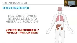
Well, if I am the first since the dark times, it's really nice to be back.. And I'm just saying how much I really appreciate kind of getting out of my bubble and talking to people and actually seeing human emotion on the other side of this display. So I really appreciate it. I love this place. A long history with several people here. And so it's my pleasure to be here. All right. So today I'm going to talk about something that's a little bit green and essentially is Brian said is I started my lab essentially last fall. I'd been at UT Southwestern for a long time. I got there in 2014. I did my postdoc. They kept me on as a research track professor. And then like, like a very stubborn obstacle, I eventually forced them to give me a tenure-track position. And so this is now the research that's going on in my own lab. So we're really focused on developing technologies to study the metastatic cascade. Now, this is a prototypical process that is superficially or stereotypically shown as this linear step or sequence of events. Of course, the first thing in the metastatic cascade is the cell actually has to have some formation of an oncogenic behavior. It has to continue to proliferate irregardless of its environment, at which point it can then locally invade. Away from that site, that primary tumor formation. It can enter the circulation, at which point it can then travel very long distances, essentially for some types of cancer throughout your entire body, where only a handful of cells, very select cells, can then leave circulation and enter a new site where they can then survive and ultimately try to proliferate. Each one of these steps is kind of— you could think of it as like a seed. What may provide you with the ability to do very well at step A or step B may be very different than what you have to do at step D, E, or F. And so if we really want to study this cascade, I think it's valuable to think of this from a perspective of microscopy. We're all very much microscopy people, I assume, in this audience. We want to look at the subcellular localization of signals and activities and, and things like metabolic activity. We don't want to reduce these cells to just bags of proteins like we might do with next-gen sequencing. So from a microscopy perspective, it's very challenging to look at step A. Needle in a haystack. Let's say you're looking at melanoma. You actually don't know necessarily where to look to see where's this first cell that's becoming locally invasive. Likewise, if you're actually looking at this part right here, this is also incredibly challenging. And we believe that this is actually one of the major gateways that acts as like a significant barrier towards cancer progression. So this is actually a very rare process where only 1 in 100 cells or less actually survive this, uh, transport in the vasculature and successfully colonize that distant site. So the question is, why is that cell popular? Why is that cell successful? What allows that to occur? And to begin actually asking questions like this, you have to have a very good model. And so we reached out to Sean Morrison at UT Southwestern, and we started working using his melanoma model. This is a library of 22 patient-derived stage 3 melanomas that were isolated in clinic. Now, very interestingly, when you put these cells into a mouse, in a very specific type of mouse that's essentially had its immune system abolished, then what happens is that these cells phenocopy what they did in the clinic. So if they metastasized in the clinic, they then metastasize in the mouse. And one of the— and you can see one-to-one correspondence with the exception of just one single cell line here. The other thing that's very interesting about this model is that they're highly tumorigenic. So a lot of people studying metastasis will inject maybe a million cells into the tail vein. These cells are delivered directly into the blood. They circulate, they get trapped right in the lung. In contrast, we're doing a subcutaneous injection and we can do it with just a handful of cells and you can still create tumors. So it's not necessarily a cancer stem cell or a very special cell type that can create this primary tumor. By doing limiting dilution analysis, you can actually see that 1 in 2 cells or 1 in 4 cells for some of these cancer types is all you need to inject into a mouse to create a primary tumor. What I find perhaps the most interesting is that they all display kind of distinct organotropisms. What is organotropism? This is what I find to be a major goal of our research right now. If you have a, say, from patient 487, a melanoma that metastasized preferentially to the kidney, you put it in the mouse, it metastasizes to the kidney. In contrast, patient 481 has preferential metastasis to the liver. Why is it that some go to the kidney and some go to the liver. And so, as I mentioned earlier, this is— and for melanoma, the assumption at least is that all of these cells are entering general circulation. So they're actually going throughout the entire body, yet they differentially colonize and metastasize in different tissues. So why is that? So our goal is to kind of do the simple microscopy thing and just look. And so, you know, from a thought experiment, we can start to say, you know, if we're looking at, say, metastatic colonization in the lung, is that colonization linear with time? So is it kind of like the primary tumor is constantly shedding cells and they're, they're, you know, colonizing that distant tissue at a steady frequency? You know, is it that only a handful of cells colonize at which they exponentially grow? Or do we see like an initial bolus of cells followed by essentially clearing out of cells that could not survive, then perhaps a latency period followed by an exponential decay? I think what's very important is that you might come up with different chemical strategies targeting these populations. If it's this one, maybe you need to target the actual leaving of the primary tumor. Whereas if it's this one, maybe we need to start to look at these latent cell populations and identify what molecular features they have. And so we're going to just do the most obvious thing, which is just comprehensively catalog colonization site cellular abundance and cell state. We're going to do that for 3 different tissues. Now, of course, we're all non-transparent, which is mildly problematic from an imaging front. So if we're looking at something like the lung, a functional lung has huge complexity. And from an optical perspective, we have regions of mucus, air, you know, extracellular matrix, highly epithelialized tissues. And so from an optics perspective, Every one of these interfaces will scatter and aberrate your light. Now, of course, we could use an optical window, but that's going to very much limit us to superficial imaging in this otherwise very large and complex tissue. So the approach that we're using, which is also quite popular, is to use tissue clearing. In this case, what we do is we chemically fix the specimen, so we give up on our live cell imaging capabilities. We then strip the tissue of all of its lipids and leave behind essentially a proteinaceous mass that's just a phantom of that tissue. And we immerse that protein mass into a solution that has a match refractive index. So all of a sudden you can take this large brain slice here, which is actually a human brain slice, and make it transparent. So you can now go in and look at that at very high resolution. Now the challenge is that there's a lot of different clearing methods, and each one of these methods has been typically optimized for brains, although increasingly they're being expanded to other tissues. So you actually have to have an imaging system that's capable of imaging in all these different solvents, whether that be a hydrogel-based method like expansion microscopy, which is water-based, or Clarity, which is about 1.45 refractive index, or BAV, which is about 1.56. So us and others in this room have adopted these multi-immersion objectives. These are essentially specialized objectives that have a, a very concave final element here that minimizes refraction at that final optical interface. So you can put it into any solvent of varying refractive index, and the rays leaving that optical objective all go straight. What's great is that this has a very long working distance. You can image deep into tissues. And it maintains this diffraction-limited performance between 1.33 and 1.56. So you can image, say, water, 54% glycerol, and in this Pegasus, which is about a 1.56 solution, and maintain this nice diffraction-limited performance. So we're also all very familiar. So if you have these objectives and you can do nice diffraction-limited imaging, you have only a couple choices that remain. The first is, should you use, you know, the workhorse of biology, which is the laser scanning confocal microscope? But we all know the advantages and disadvantages here. Essentially, we're going to illuminate above and below our plane of interest. We're going to typically acquire our voxel information in a serial format, so you might have a 1-microsecond dwell time. And acquire voxels at a megahertz. But ultimately, that's still quite slow. On the plus side, you get confocal detection, which makes you robust to scattering and— or on the detection sense, robust to a lot of aberrations. Now, we and everyone else have opted to go with a light sheet microscope, which typically confines the illumination to the field, the plane of interest, and most importantly, provides massive parallelization. So a modern CMOS camera easily gives you more than 4 million pixels. Allows you to reduce your laser power substantially and still have an exposure time that's only a few milliseconds and fly through the imaging. Now, of course, you do that, there's no free lunch. And so us and many, many others have tried to come up with ways to get past this one critical limitation, and that is your, your Z resolution depends critically on how you— on both the detection optics and the illumination optics. Specifically, the simplified way to think of this is for very skinny beams, your resolute— if your beam is skinnier than the depth of focus of your detection objective, the resolution is limited or is dictated by that beam waste. However, to get a reasonable field of view, you typically need to use a much larger field of— or much larger beam. And unfortunately, that beam waste which grows nonlinear with the field of view, begins to dominate your resolution. So a good example of this is just this cleared tissue image of cortical neurons. We see very nice detail in the lateral perspective. However, if we look at it in the axial direction, it's significantly worse. And so that is essentially just demonstrating that the lateral resolution is dictated by your detection optics, but if you have to have a very large beam that covers that entire field of view, you're going to have pretty poor resolution in that along the optical axis. So Reto Fiocca and I back in 2015 came up with this strategy. And this is what we call axially swept light sheet microscopy. And what we do is we use aberration-free remote focusing. So this is a method developed by Tony Wilson and Bachrabi that allows you to scan a beam in its propagation direction and maintain a very tight diffraction-limited beam waste over a very large scan range. So in this case, a lot of the work I'll show you today, we can maintain an NA 0.7 beam waste over our plus or minus 170 microns from that nominal focal plane. So we can now deterministically position the beam at the edge of our sensor. Kind of shown schematically here, and then we sweep it across synchronously with an array of active pixels in the light sheet mode. We do that, like I said, using this aberration-free remote focusing. So here we bring the light in, we focus it onto a mirror. This mirror is conjugate to the specimen. Scanning the mirror here scans the beam here. Any aberrations introduced by imaging this mirror outside of the nominal focal plane are equal and opposite to the aberrations necessary to create a diffraction-limited beam outside the nominal focal plane here. The advantage here is that we can now decouple this trade-off of field of view and axial resolution. We do give up something. Typically, we've been a little bit slower. We can only scan this mirror so fast. And, you know, even though we're only collecting photons from the beam waist, we are still illuminating the specimen here. So we also give up a bit of confinement. But for something like, you know, clear tissue imaging, that's something that we're willing to tolerate. Now, of course, there's other ways to do this. Most notably, the best way— you know, this is one mechanism for decoupling this field of view and axial resolution tradeoff. The other is joint deconvolution and imaging from multiple perspectives. So the first system we built was using this NA 0.7 objective. The resolution was— it varied with refractive index. So at high refractive index, we could achieve a lateral resolution about 310 nanometers at a Z resolution about 260 nanometers. This provided very nice imaging throughout our entire field of view. The original microscope at our highest refractive index was about 267 microns, and then water is about 320 microns. This allowed us to do already some pretty beautiful imaging. Here's an entire hematopoietic stem cell niche that was also imaged with Sean Morrison's lab. If we zoom in on just that square, And then zooming on that square again, we get this level of detail. And so what we can see in gray is the fenestrated sinusoids. In green, we see the peripheral nervous system. And in magenta are these stem cell or progenitor cells that are undergoing maturation in the stem cell niche. So really beautiful. You can begin to see things like these cells going into and out of circulation. At these fenestrations in the sinusoids. And you can see kind of provocative things such as the formation of what appears to be synapses between the peripheral nervous system and these stem cells. So there's been a lot of talk about whether or not the nervous system directly or indirectly modulates stem cell maturation, and that this is just begging us to come back and do actually something more with it. We also see just what I consider absolutely stunning cell shapes, morphologies. So on the left, we have these highly specialized leptin receptor positive cells that exist within the stem cell niche. They have these delicate finger-like protrusions. And so this is kind of a daisy chain of cells. You see a nucleus, nucleus, nucleus, nucleus. And these protrusions essentially wrap around the stem cells and locally secrete growth factors that modulate their differentiation. Now on the right, we actually see a specialized epithelial cell type from the kidney that to this day is, by my understanding, described incorrectly in physiology textbooks. But these finger-like protrusions, you can see the cell body here, and finger-like protrusions essentially interdigitate and create a very tight single-cell tubule that is important for kidney function. I'll maintain a little bit of intentional obscureness for the sake of my collaborator. But we were able to then isolate or image single cells of this in this tubule network and then segment it and evaluate it for cell shape. So actually what you see in red is areas of flat curvature and areas in blue are high curvature. All right, so everything's good. We have everything we need, right? Yeah, we typically will. So can you repeat that? Yeah, so the question is what dyes we're using for this. I think that's, that's a good question. Depends on the tissue. Certain clearing methods will use endogenous fluorescent proteins like GFP, or I mean, a lot of the biologists created these mice that have tdTomato or dsRed reporters. Most of the stuff I'm showing you, we're actually doing secondary immunofluorescence against those endogenous fluorescent proteins with dyes like CF555 or CF647 or Alexa Fluores, these sort of dyes. Okay, so if we can image these cells with this sort of detail, then apparently, I guess, you know, We should be good to study cancer. And the challenge that immediately comes up is that, you know, typically when we're doing imaging with the CTAS LIMB, we'll acquire data that's about 320 by 320 by 200 microns, and we'll do it with a sampling of 167 by 167 by 200 nanometers. And by comparison, these tissues are just astronomical. And so the problem that we essentially face is now if we try to image this kidney, lung, or liver with this sort of tile size, it's going to take absolutely forever. So a back-of-the-envelope calculation saying that if we wanted to do diffraction-limited Nyquist sampling of a lung, it would take you decades to image this with a confocal microscope. It'll take us a year with our CT-SLM. So we need to do something a little bit more intelligent than just brute force the acquisition. And so the approach that we are doing— oh, that brings up one other problem. So not only do we have to— we have massive volumes that we need to acquire, but then there's the fact that this process is actually biologically very noisy. So if we look at one cell type here, that's been— all these mice were subcutaneously injected with the same number, same conditions. You can see this one has very nice preferential— or essentially every mouse identified at week 15 and 13 is positive for METs in the liver as measured with bioluminescence, whereas many of these other tissues are much more heterogeneous. So these cells have very weak metastatic burdens and very high metastatic burdens. So you get kind of a non-unimodal distribution. So, but the key is, if we really want to start to try to find the first cells that actually colonize this tissue, we're going to need to be sacrificing these mice at week 7 and 9. And there's going to be a good chance that we have to image the entire tissue and not actually see anything. So it's kind of, we get into this photon counting regime where you actually don't want to see things most of the time. You want to see the rare events that come in. And so not only do we have to image these large samples, but we have to image a large number of them. And so the crazy approach that we've come up with is we're going to do 5 time points and we're going to do 7 to 10 mice per time point, each with negative controls, which at this point is coming out to around 500 lungs, livers, and kidneys. So when I read that, then I think, you know, what did I get myself into? But we're going to take that cancer biology model We're going to take the tissue clearing processes which we've optimized in-house. We're going to develop computer vision with Gaudens' team so that we can really accurately identify these cells in large biological contexts. And we're going to come up with more of an adaptive intelligent imaging approach. And so that leads me to this multi-scale clear tissue, actually, so a light sheet microscope or MCT-SLM for short. So this is essentially what happens when you, you have a biological problem and you just take the most pragmatic approach forward. What we did is we created a 4-axis imaging system. It has 2 imaging modules that operate independently. We have a macroscale module here, which is essentially a mesoSPIM. On this side, we have our high-resolution cleared tissue ASLM. And they're essentially welded together. The sample is positioned at the center of these 4 optical axes, and that allows us to then quickly switch imaging modes between these 2 different systems. So you guys are probably familiar with Fabian Voigt's work. So he was able to use a single— he created mesospin. We're taking the single-sided illumination variant of it. We do— it is an ASLM-like microscope. They're doing low NA illumination beam scanning, so you can do that with an electro-tunable lens. And then by using a stop-shifted digital SLR lens, you can then create very nice telecentric illumination over a field of view that goes from 2 to 21 millimeters. On the detection axis, we use this 0.63 to 6.3x mesoscale imaging system. This provides that field of view of 2.1 to 21 millimeters. And in general, this will provide us with about a resolution of 5 microns regardless of that field of view, although sampling gets to be a little bit sparse and we're not at Nyquist at the larger field of view. And this whole thing is positioned on a focusing stage here. So we combine that with the already, you know, proven CT-SLM that I discussed earlier. More recently, we were able to improve the aberration-free remote focusing optics and improve that field of view by up to 60%. And we were able to increase the bandwidth by using a more recently developed actuated or pneumatically actuated voice coil. So historically, we've done most of our imaging with about a 200-millisecond exposure time, which is actually quite slow by our standards. And by looking at this Bode plot, we're thinking that we should be able to actually increase this up to about a 50-millisecond or 40-millisecond exposure time, which should drastically accelerate imaging. So then equipped with this, we can now do simple things. We can essentially look at this large tissue here, in this case, the liver, and within a single acquisition, we can image this entire field of view. So 21 by 21-millimeter field of view. We can go through and identify all the tissue boundaries perhaps not at a resolution that's sufficient to really detect single cells. So we could then automatically identify the boundaries and then do an exhaustive search at our moderate magnification, say 6x magnification, do automated identification of candidate events. Those events can be whatever you want. So the software we've developed is pretty versatile. It could be something like, I want to find every cell that's within X microns from the vasculature. Or I want to see every cell A that's in complex with cell B. In this case, we're starting simple. We just want to find METs, and then we switch our imaging modes and we do local targeted imaging with that low 300-nanometer resolution. So this is the sort of scope that you can actually achieve. And so this is a mammary gland shown here. You can see the entire ductal tree on the left in a field of view that's upwards of 13 millimeters. And then we can go and select a single terminal end bud, switch imaging modes, and go in and see nice single-cell events. We can see adipocytes here, these kind of large, empty-looking cells, and we see this kind of terminal end bud shown here with the nuclei shown in magenta. And just because I find it beautiful and I love everyone else's setups as well, I wanted to show what the system looks like. Essentially, here's that large travel range stage, so XYZ theta. It's best— puts the specimen in the center, central chamber. We have the mesospin illumination here and detection here. We then have the entire CTA-SLM elevated in a very compact assembly, so we're able to get this down to roughly 18 inches by 24 inches, um, by folding it a couple times and using some cage optics, which was, um, you know, we learned what we do not like about cage systems, but they're take it or leave it. We can actually see also the chamber from a different perspective here. And so yeah, so now we can build up— we've spent a lot of time building the optics. We now also need These microscopes are no good if you can't control them, which is kind of a catch, right? Like, we align all these optics, now we need to learn software engineering. So we've put a lot of time into this as well. We started from scratch, borrowing code from a couple of different people and building up our own code base. It's all Python-based, and we're trying to adopt standard leading software principles. So we want it to be nice, nicely organized into a model-view-controller. We want to be able to do the processing in real time. So if possible, we're going to use high-performance CPU libraries. But if necessary, we can easily grab some of these nice GPU libraries as well that are readily available via Python, like TensorFlow or whatever. One of the more exciting things is that We created this server-client architecture to really be able to grab other libraries out there without creating conflicts within our own repository. So we can actually communicate to separate environments that are running elsewhere and send it data and have it send us back results, which we then use to drive the microscope. And perhaps most ideally is that we've tried to put all the actual steps for acquisition in this highly reconfigurable architecture that allows you to, to do any sequence of events that you want. So that could be, like I said, you could do find cell A in complex with cell B, do autofocusing, measure, you know, do segmentation. And, and or if you want to do a completely different order, you know, maybe find boundaries. But you can essentially, We jokingly call it like create recipes from essentially acquisition ingredients, and that allows you to do pretty much whatever you want. So you can be creativity limited, not software limited. Of course, the architecture is something that has taken a lot of time to build out. Everything is displayed in GUI. The controller acts as a communication node between the GUI and the model, which can run independently. And as I mentioned, We have this REST API interface that allows us to interact with external things. So whether that be something like Cellpose or Mesmer or whatever this new Meta segment, anything, we could basically just put that into their own environment and try to build up communication channels with that. So this is what it looks like when it runs now. So on the top left, we have our mesospem illumination, our mesospem detection. We got our sample hanging into the chamber. We've intentionally spread things out a little bit. We initially had everything aligned so the sample was co— so that the optical axes were perfectly coincident. But now we've given ourselves a little room to breathe. We basically acquire, find our feature of interest, and switch over to our high-resolution side here fully automatically. And then image that same event here with a known offset between these two paths. So then we can do something like this, and this is really where we're getting down to, you know, getting closer and closer towards the goal. So we can image an entire cleared mouse lung, and we can see that already— and this is an inverted image— you can see that there's kind of some interesting features here. So you can see autofluorescent structures here like bronchioles, Now you begin to say like, all right, this is a lot of space to cover, but there's something interesting here. I'm not sure what this is here. We have a large nodule here, small nodule here, and then something else happening down here. So by doing this whole tissue imaging at 21mm field of view, you can get a global over— like overview. We just zoom in a little bit to our 6x magnification. We can see that that initial small metastatic nodule is actually quite likely to be a real event. And we zoom in completely with our CT-SLM side, and you can now see features as small as like what appear to be little vesicles that have been shed from that tissue. All autofluorescent? This is actually fluorescently labeled. A lot of these tissues are highly autofluorescent, but this is substantially brighter than this background autofluorescence here. And the tissue is— that's something we've had to work around a lot, is that different tissues have different autofluorescence spectra, essentially. And so to get this to work in the liver took a lot of work, which is highly, highly autofluorescent. And so now, yeah, we were able to kind of essentially get this global picture, zoom in, and do local imaging. And now we ramp up and start to do this for a lot of different tissues. So a huge amount of optimization that goes into this one slide. But in green, we have nuclei. In magenta, you can see the metastatic cells here, even single cells popping in and out. In the kidney, we begin to see already what I consider anecdotal but slightly interesting features, and that is that we don't actually see a lot of mets inside the kidney. You'd expect, you know, the kidney is a filtration unit. You would think that these glomeruli, which are tight filtration regions in that tissue, is maybe where you just lodge metastatic cells. We actually see these cells kind of more in the supportive tissue outside of the kidney so far. In the lung, it's a complete free-for-all. These cells are everywhere. So you'll see them in bronchioles, in alveoli, in the vasculature. And so now we can begin to try to ramp up and do things like actually segment and quantify these features. So on the left is an image of all the nuclei in a lung tissue, and then we have our staining, which is with this anti-RFP-CF647. And what we see is kind of a cytosolic fraction of this immunofluorescence in the cells that are actually metastasis. Now, by going in, we can then segment out all of our green channels using nice machine learning methods that have been generated here and that we're trying to expand upon at UT Southwestern, as well as our standard VS Red channel. Then we can assign each population based upon a distance metric. We can say you are host if you are negative for this DS-RED and you are met if you are positive for this DS-RED. Ultimately, you can nicely see almost like a column of metastatic cells that go around the periphery with a core of lung tissue here. You can begin to already see quite a bit of detail. To do this at scale will be the interesting part. So we're developing better methods with Gans' lab to try to combine these nice 2D machine learning packages into robust 3D estimates of cell abundance. And so by doing some of that work now, we can take something like CytoxGreen. We then have our method to say, are you host or are you cancer?. And we can begin to actually count the total number of cells. Now, this is not perfect, but it's pretty good. And I would bet it's within a few percent of— if we were crazy enough to go in and try to annotate a ground truth. We have a total of 18,394 cells, and of which about 11,166 are cancer. This brings up a point that we face. The 2D annotation is painful. The 3D annotation is a nightmare in our hands. Now that we have the microscope, we're starting to get the clearing to be really good. We can really begin to do things like look at the environment. We're now looking and asking simple questions like, once we find a MET, are you inside or outside of the vasculature? And so, for example, we can see in this liver this very beautiful capillary architecture, as well as in the kidney. You see the glomerulus, and then the lung, it's everywhere. So we're currently under, you know, working to improve this labeling. It's a little bit dim by my standards, but the goal would then be to be able to segment every nuclei, the entire vasculature tree, and then quantitatively measure distance to and from specific tissue-like architectures at the whole tissue scale and eventually for hundreds of tissues. So we'll be filling up hard drives for a little bit. The next key is also that we don't want to just count cells. So we probably could have done something like this if we wanted to just count cells. We maybe could have just digested the entire tissue and done flow cytometry or something. Something. It would have been nasty. But we want to start to look at context. Where in the tissue are these cells? What kind of molecular features? What, what defines their cell state? Are they pre-apoptotic? Are they undergoing proliferation? Have they recently undergone proliferation? What's the cytoskeletal architecture and how does that organize some of the signaling scaffolds that we're interested in, whether that be GTPase signaling or kinase signaling? Have they experienced DNA damage? These cells have had to metastasize and squeeze through a lot of very tight constriction points. So you can begin to ask, do some of these microfluidic models that people use that cause DNA damage— are we seeing similar things in a liver or context tissue format? And of course, everyone seems to be incredibly excited about metabolic activity. Now, that's a much more challenging thing to go after, in my opinion. If there were good ways to look at, say, true metabolic activity at a subcellular level in a tissue, I think we'd be all for it. We can essentially go in and count mitochondria. We have resolution that's sufficient to do that. And if we could have some nice biosensors that are amenable to clear tissue imaging, I think there's some good opportunity there. And so I think I went a little fast, but not terribly. And I'd rather have more questions than not enough, but I'm out of practice. So, you know, in summary, I think we've really put in the last, you know, really just, you know, 7 months. That's essentially what we've done. I'd love to see what the next 7 months do. Currently where we're at, we have essentially the platform that we think we can grow with. We can essentially ask questions that you can't otherwise really ask. We can try to find needles in a haystack, or we could do things like comprehensively evaluate otherwise very convoluted, challenging architectures. If we look at that mammary gland, we could essentially go through and do comprehensive mapping throughout only the ductal tree rather than imaging the whole volume. So we essentially kind of augment our data overload by selectively imaging the whole region rather than just brute forcing the whole thing. Um, and as I mentioned, I didn't talk about it too much, but we have this completely reconfigurable software architecture that I think, uh, just has totally opened up new opportunities for us. So we can essentially have multiple imaging modalities We can interface with robotics, which we're going to start doing, because if you have to load hundreds of samples, you want that to be done robotically. And I really hope that we put in the effort that someday, you know, I'm not trying to release it like a general software package. I've, I've learned what effort that is from some of my colleagues. But I would love to see some of the other labs take some of this up and, you know, see that kind of impact grow. So with that, got a great team. So Zach is the first postdoc brave enough to join the lab. He came from Jorg Würstersdorf's lab, and he has been instrumental. Hazel has done all our tissue handling. Jin Long is our, like, optical microscope building guru. Annie, I found her in the most creative ways. She works in California. She previously worked for IBM. She just wants to be able to stay at home with her parents or with her children. And I said, yes, please program when you can. Dax was an intern. He set up essentially all of our GUI. Torquil is helping us out with the mouse work. Sean Morrison. And of course, we've got the, the, the key group of people here that are incredibly influential. So Andy's been has always been awesome to just chat with and bounce ideas off of. And of course, Reto, Gowns, and Beau Ray. And Felix has been really spearheading some of the analysis. And apparently Gabriel's here for the Hannah Grace, and I'm going to go find him later today. So with that, I think we got a little time for questions. Appreciate it. This needle in the haystack idea is really interesting to me. And can you— do you think that you're set up both mathematically and microscopically to think about whether or not you're examining tropisms or just whether or not cells go everywhere and happen to continue to survive in particular sites? Yeah, so I think the hope is that we can eventually gauge, you know, things like if you see a colony with 1 cell, 2 cells, 4 cells, can you start to build up kind of a historical perspective of how many cells came in the first place, right? Like you could almost get to kind of classical methods there. And so And I would like to combine that with molecular ways. So maybe we could do, say, a tamoxifen pulse chase at a certain moment before, and then we get a window where we said, yes, in the last 24 hours, the cell actually divided. And so, uh, I think my perspective is, and it's probably simplified, is that like if the cells can actually proliferate in that tissue, then I would consider that like an effective tropism to some extent. What I'm curious about are the ones that make it and then don't.
Yeah.
And what happens to them? Do they actually just go into like a, um, a stationary, um, you know, train? Like, do they go on hold for a while? If that's the case, that's also interesting. Like, because, you know, we're not going to try to study that, but, you know, some people have cancer for 8 years and you got this latent population of dormant, um, nightmare-causing populations of cells. And that's a hard thing to study. So, you know, I think that's kind of the cool thing is we might be able to go and see in these certain cases where there was no cells detected by bioluminescence, are there cells just there? And if so, I think that's, that's information that you can't get really right now. Yeah, this seems tailor-made for dormancy.
Yeah.
Chris, this is amazing. It reminds me a bit of some of the pipelines we have in this building, though, which are huge amount of work for each sample, right? You get awesome data out, but each— it's going to be low throughput, right, in terms of large-scale, you know, how many times you can do it. Since you aren't trying to go live anyway, you're already working in prepared, cleared tissues. Do you have sort of some vision for hyperspectral expansion to give yourself more and more and more labels in each sample? Because you might be able to gain— rather than having to do the same experiment 10 times, if you can do the experiment once and pull all of these other colors out, Yeah, that, that might be a way to sort of increase your power. I mean, it's the way we've tried to deal with 3D EM and things in this building because we just can't do them frequently enough. So the more information you get out of each sample, the more powerful it is.
Yeah. Well, I think there are certain approaches, but then— and that's where I think we're, we're fortunate that if you want to try to do some sort of multiplexed imaging locally in a tissue, then the classical mechanisms are probably going to fail. So you need a more creative approach that maybe uses things like hydrogels or active methods to drive antibodies into tissues to speed things up, because you got all these things that are against you. You can't work in bab. You would have to rehydrate the specimen, relabel. So we are—
so sample prep is the problem, not actually optics?
Is that what you're saying?
I would say that we've scaled up sample prep to the point now where we can handle lots of mice at the same time. It's just a reagent problem. It's a lot of waiting. We'll stain for a week, we'll rinse for several days, we'll stain for another week. We're hoping to get the robotic arm in so that we can truly do the full pipeline. We put a sample on, it finds the boundaries, it does exhaustive imaging, and it only images what it thinks is right. There's a little bit of ways to go for that. For you, where you're— I think you're trying to say is how can you get more molecular information for your buck per specimen? And I think that requires a very creative approach from the methods we're using won't work. Like, in fact, you like, you know, I submitted an R01 proposing ways to that work, and I hope that we can talk you into joining us so we can do that together. But ultimately, I think it requires a comprehensive approach that takes imaging, probes, clearing, and you will only be able to multiplex chemically if you have all those things in-house.
Kevin, can I add to this? I mean, first of all, awesome job. Can you describe to me how different— you said it was different liver, kidney, lungs. Can you describe to me how different that really is and how do you optimize even from a labeling and from an imaging perspective?
Yeah, so it was very interesting. It was like we started out with the lung and things worked pretty easily and we thought, oh, we're making great progress. Well, the lung is predominantly empty space. So antibodies penetrate well, your reagents get in and out. And, and then we went to the liver and it's this highly epithelial tissue, these tight, tight cell-cell junctions, and it's loaded with P450 and all these hemes that just elevate your, like, the red region of your spectra. And so it really was like we got great signal in all our lung, and then we go to the kidney and liver, and it's a disaster. So we took the most obvious approach, which is just go further red, and that worked. Now, in our hands, some of our far red dyes don't perform as nicely. But, you know, if, if I were to say, like, the photophysics of these dyes in these weird solvents is also not very well appreciated, you know, you get these absorption emission shifts Perhaps we're getting some quenching, I don't know. But like otherwise these dyes are like great in aqueous environments are just not as good anymore. So I think there is opportunity there for probes if they want to kind of— it's a very niche probe application, but I would love to learn more about that too. But going further to the red helped us out a lot. So speaking of probes, you use this eFluor 660. Did you need that like flow cytometry super bright polymer-based dye to get signal, or can you use other things? So that eFluor 660 was just what we happen to have. So that vasculature labeling is a little bit sadistic. What we do is retroorbital injection into a living mouse to label the vasculature with directly conjugated primary antibodies. So we don't get the amplification that we would like from a secondary antibody. And so far it has proven to be quite valuable to have that. Now we're exploring different fluorophores for that. Um, that E-4660 is not ideal because while we got good vasculature labeling, it now took up the channel we need to see METs in livers. So, uh, we have to kind of shuffle things around And so, so far, that is the one good result we've had. We're hoping that by moving to the green, we'll have some good results there too.
All right. Fantastic talk, Kevin. I was wondering a little bit about your sampling strategy. Because going from the mesoscale to microscale, I would be tempted to go look at all the things that look interesting. But I think actually what you might want to do is sample different distinct regions. I'm wondering what your strategy there is going between the two scales.
Um, I mean, I could see perhaps if I'm reading the question that perhaps we should sample some other spaces too, just as a nice internal control to make sure we're not missing anything with the mesoscale imaging. I think that's a good idea. The plus side is that because we do the mesoscale imaging, I think it is possible to do a lot of local targeted imaging and it not be detrimentally long. You know, if we go in and we image, say, a 40-micron volume in 50 different spots, it's not a big deal. It's when we try to do, you know, 30,000 tiles that becomes a problem, and then we have to rely on all the other tools to actually deal with that data. So I think that's an interesting idea that maybe we should actually introduce in our sampling, um, where we have directed sampling as well as indirect kind of random sampling as well. It's just a built-in control.
Kind of following along the lines of what Mark was saying, I'm wondering, like, have you thought about like brute forcing just a few? It might be kind of interesting, right? I mean, because you have the capability, right, to do it and you probably would learn things because you could do the whole organ.
Yeah, I think, um, we are definitely doing that. And I think from an optics perspective, there's some interesting stuff there too, is like how well can we perform over that scale. Now, of note, we have had to resort to slicing some of these tissues as well. So the lung, we can do very large tissues, but we now slice the lungs or the livers and kidneys. Um, yeah, I think our most initial approach was to actually do some brute force imaging as well. And the hope is that with the faster bandwidth, maybe that factor of 4 or 5 really does kind of save us a little bit. You know, some of the data I showed, like the stem cell niche, I think took 5 days to image. So, but from a software perspective, that would absolutely test the limits of like how reproducible we can run this, how like do we have enough have we done a good job engineering the system? Yeah, I think from both a biology hardware and a software perspective, we would have things to gain. From Mark's perspective, we would be able to say, did we actually detect all these cells? So we do this macro scale imaging, we detect 25 pop-like nodes, and then we do exhaustive imaging. And we probably would have to do like a human in the loop to make sure that it passes the muster from a biologic motivated perspective? And then likewise, can our optics and our software actually drive that acquisition process?
And sort of the other thing I was thinking about is like there's the sort of approach that was taken here with like Drosophila brain, we just brute force by parallelization, right? So if you had 10 of these things that were operating continuously, yeah, you know, that's sort of another strategy, which, you know, I mean, it seems like you're—
just tell the NIH this strategy. Yeah.
Okay.
And then the last thing I was thinking about as I was listening to you is like, it seems like, you know, another way to do this would be to like sort of like resin embed and then slice. I mean, for the kind of brute forcing thing. I mean, like, what are the pros and cons of that approach of like taking these organs, you know, basically making them hard, slicing them and then imaging it that way?
So from the resin embedding, I think that the advantage there is that perhaps you could do Let's say you took a different strategy and you really wanted to push even higher resolution. I think you could use higher numerical aperture objectives. You could do some imaging and say like an oblique plane microscope format, and then you take it over, slice it off, and image it again. So there's been some beautiful work on that. I think this— yeah, combining serial— to have resin embedded with serial sectioning allows you to push the optics a little bit harder. We basically compromise and say, you know, we're— for now we're happy with our low 300-nanometer resolution. That's good enough to see what we need to see. But there are a lot of cases where we wish we had more. What else? So resin embedding. Are there any other advantages? I would say, you know, if you do resin embedding, then perhaps some of these cyclic immunofluorescence methods could work too if you did serial sectioning and then had just a huge array of tissues that you could then cyclically go through. That's another advantage I think that you would have. You wouldn't have to worry about having to penetrate through 1.5 millimeters of tissue with an antibody. You could just do it on a 5-micron section, right? So there's definitely advantages to that. And Yeah, the hope I think is that with the robotic loading and unloading that we would be able to have the capacity to image this with throughput we need, but we'll find out. Still not entirely sure how long it will take, but if we have to build another, we'll try to dig up some money. Thanks so much, Kevin. Thank you. We have one more question.
Yeah, so this is just kind of following on with what everyone else is saying. This kind of methodology for automation is kind of near and dear to my heart, and one of the big benefits that I see is being able to, you know, build up these statistics for where things are, and you have this really nice mesoscale imaging that maybe you don't get a hit on. But, you know, one of the things that I don't know how much signal you're getting for your autofluorescence, but I think it's kind of important to see in these kind of cases, not just where things are, but where things aren't and what you can learn from that with the additional. It's really hard with kind of like what Chris was saying with the spectral space that you have. But can you, what can you learn from that? Are you thinking about ways to kind of leverage that white space?
Yeah. So, more comprehensively cataloging the entire system. Like, yeah, presence or absence. Yeah. I'm not entirely sure what the— I think our first approach is to really just acquire the data and then stare at it and then come up with the next approach. As a very interdisciplinary project, we're working with cancer biologists. They're not used to dealing with imaging data like this. So I think by just being able to point at cells and say, you know, I trust that their, their knowledge of why some cells would be adjacent to a bile duct versus not would be— I trust their opinion more than mine at this point. But, um, um, it's a good point, uh, building up the automation to more exhaustively do that. And for these low copy number— or not copy number, but events is, I think, the whole goal though, because that sort of thing can pop out, hopefully.
Yeah, thanks.
Great talk. Yeah, thank you. I appreciate it.
More from Joshua







Scientist Stories: Hans Waterham, The value of phenotypic assays in peroxisomal disorders




Scientist Stories: Joel Zlotogora, Genetic carrier screening programs for reproductive purposes