Scientist Stories: Kevin Eggan, Treating and Potentially Curing ALS
In this talk, Dr. Kevin Eggan, now Vice President and Head of Research at BioMarin, reviews his laboratory’s journey from basic stem‑cell research at Harvard to translational ALS therapeutics. He explains how the ubiquitous TDP‑43 protein, whose pathology is present in most ALS patients, directly regulates the neuronal gene STATHMIN2 through a splicing mechanism that, when TDP‑43 is lost, triggers inclusion of a cryptic exon and reduces functional STATHMIN2 mRNA. Using human embryonic stem‑cell‑derived motor neurons, exon‑level RNA‑seq, and flow‑sorted TDP‑43‑negative nuclei, Eggan shows that this mis‑splicing mirrors what is observed in ALS patient spinal cords. He then describes the development of a quantitative qPCR assay for the cryptic exon in blood‑derived exosomes, positioning it as a potential biomarker for TDP‑43 dysfunction and target‑engagement in clinical trials. To assess functional relevance, Eggan’s group generated STATHMIN2 knockout mice, which develop age‑dependent neuromuscular junction denervation, muscle pathology, and motor deficits without early lethality. Humanized BAC transgenes expressing the full STATHMIN2 locus rescue these phenotypes, confirming that loss of STATHMIN2 drives the observed neuropathy. Mechanistically, loss of STATHMIN2 disrupts microtubule dynamics, shifting the free‑to‑polymerized tubulin ratio in motor neurons and mimicking the effect of nocodazole, thereby linking STATHMIN2 to axonal stability. Therapeutically, antisense oligonucleotides targeting the cryptic exon restore STATHMIN2 expression in TDP‑43‑deficient human neurons, and an early dose‑escalation study is underway in ALS patients through BioMarin’s Curalis program. Eggan concludes with open questions about why phenotypes emerge only in aged mice, potential compensation by other stathmin family members, and a tentative biochemical interaction between STATHMIN2 and the axon‑degeneration regulator SARM1 that warrants further study.
Chapters
Kevin is currently the Vice President and Head of Research at BioMarin. We knew him for many years as a professor of stem cell and regenerative biology at Harvard and as a director of the Stem Cell Institute at the Broad Institute. His work over the years has had an enormous impact both in terms of our understanding of the pathobiology of ALS and also, of course, in the development of stem cell analysis, stem cells themselves, and the use of stem cells in studying ALS. And I can't resist adding that he's also the 2006 recipient of a MacArthur Award. So, Kevin, thank you so much for being with us. We're very appreciative. And I'm not hearing Kevin.
He's on mute.
There we go. I think you're muted, but I certainly see you. Thank you again, Kevin.
Go.
Fantastic.
Coming through okay? How does everything look?
Looks great. Thank you.
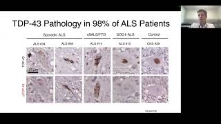
Great. I have the same throat bug that Bob does, so I'm going to do my best to muddle through this with occasional sips of iced coffee here from California. As Bob said, you know, we have been in the process of winding down my research operation at Harvard over the last couple of years while I've been making the transition to BioMarin, where we're looking carefully at the ALS landscape and asking how we can get involved in bringing new therapeutic ideas forward. So I've really enjoyed what I've heard so far today, and I'm looking forward to providing you all an update on work we've been doing collaboratively with Bob's group and Matthias Verhagen's group in Amsterdam. And that has led to a couple of different therapeutics efforts to rescue STATHMIN2 expression in the context of ALS models and now increasingly in the clinic. So first, you know, the story of how we got into working in STATHMIN2 really comes directly from the original observations that TDP-43 pathology is really present in the vast majority of ALS patients. And this discovery has really led to a cottage industry of groups who have been trying to understand the many different roles that TDP-43 plays in neurobiology, specifically in cell biology more broadly. And I think those studies, which really show now that TDP-43 is an active regulator of many stages of RNA metabolism,. And whether or not it ranges from regulating transcription to processivity to RNA metabolism at the level of splicing, or even how RNAs may be regulated at the translational level upon initiation of stress, it's become apparent that this is quite an important protein with respect to how cells integrate environmental information and respond. And in particular, we became interested in the idea of whether or not a reason that we may have failed to find changes in RNA metabolism in animal models that were predictive of changes in RNA metabolism in the ALS patients themselves might have something to do with the evolution of RNA metabolism between those species. And that while TDP-43 might be doing the same types of things to RNAs in animals and in humans, the highly divergent nature of DNA sequence space between animals and mice might mean that the places that we were looking specifically might mostly not be conserved. And, you know, an easy thing to think about this is the role in pre-mRNA splicing, where if you look at the regulation of splicing between humans and rodents, this is largely a divergent process, with there being dramatic reassessment over evolutionary time of where splicing should happen at a nucleotide level. And this would mean that, for instance, if you looked at TDP-43-dependent splicing changes in mice, even if those were occurring in humans, TDP-43 might be acting at very different places. And so that could be part of why we had been striking out. With that in mind, see if I can— we really began to wonder whether or not even initial, I would say, less mature motor neurons in culture that were from human sources and of course, made human transcripts and had a human regulatory environment might be better predictors of the changes in RNA metabolism that we might see in patients. We set out to carry out a very simple experiment at the time, which was to take human embryonic stem cell lines that had been targeted to carry a motor neuron reporter gene. Within those, we differentiated them into spinal motor neurons and then exposed them to either control siRNAs or siRNAs that knocked down TDP-43. We're then able to use flow cytometry to produce pure motor neuron to purify those motor neurons to homogeneity, and then examine changes in transcript abundance. And not surprisingly, given the pervasive role that TDP-43 plays in regulating RNA metabolism, we saw many changes. And as you can see in the upper left-hand corner in this RNA sequencing experiment, predictably, the thing that was most reliably changed was TDP-43 since we had knocked it down in the experiment. And while all of these are, all these changes are potentially fascinating with respect to understanding the changes in gene expression or RNA metabolism that may be happening in ALS, you can see in that upper left-hand corner, tracking very closely with the change in TDP-43 is a transcript encoding the protein stathmin-2. And because of this very close association between the amount of TDP-43 alteration and the amount of STATHMIN2, and as I'll tell you in a moment, because of its function, which seemed reminiscent of the sort of thing that is altered in ALS, we began focusing first on trying to understand the underlying mechanisms that connected TDP-43 alterations to STATHMIN2. Indeed, to test and play out this idea that these changes identified in human neurons when TDP-43 is altered be more predictive of those in people. Stathmin-2 is a protein that's selectively expressed in neurons. In fact, histopathological and gene expression studies in the spinal cord suggest that it's very highly expressed in alpha motor neurons, the ones that are most sensitive to degeneration in ALS. It's encoded by a very abundant transcript, which, as I'll show you in a moment, is subject to what we're increasingly realizing is a mechanism that is regulated by TDP-43 in a variety of contexts. It, I think, interestingly, with respect to the last talk, is a member of a family of proteins that are involved in dynamic regulation regulation of the microtubule cytoskeleton. And it localizes to both the Golgi apparatus and the growth cone where it has an important role in regulating the microtubule skeleton in those contexts. And early experiments looking at the Stathmin family of proteins, because there are several of these, seem to indicate that Stathmin2 played an important role in axonal outgrowth. [INAUDIBLE] Now, to summarize a great deal of molecular biology work that was done in both my group and in Don Cleveland's lab, both groups identified that Stathmin-2 is subject to regulation by TDP-43 through a mechanism in which TDP-43 directly interacts with 5' regulatory units within the gene of stathmin-2. And in the presence of stathmin-2, the transcription is processive and splicing omits an early cryptic exon that allows processivity of the transcript to occur and for a full-length transcript and a stathmin-2 protein to be made. But when TDP-43 levels wane, there is premature transcriptional termination and splicing into a cryptic exon in the 5' region of the gene in the early intron. And this may or may not result in the production of a 17-amino acid peptide, although that's not yet been empirically detected to my knowledge. So you can see this phenomenon really clearly when you look in RNA sequencing data, not just in all of the models that we've examined thus far, certainly, but also in patients. Patients. And so here you can see on the left RNA sequencing reads from the spinal cord of a number of control individuals that passed away either in ways unrelated to ALS and were pathologically shown to be free of TDP-43 pathology. In contrast, on the right, the individuals here all have TDP-43 pathology. And you can see that many of the transcripts splice into this premature cryptic exon reducing the total amount of transcript which is being produced and splicing into downstream coding regions of the gene. A paper in which— a beautiful paper in which TDP43 negative nuclei were flow sorted and then RNA sequencing demonstrates that in that fraction, this process is almost complete. And the heterogeneity that you see here at a tissue level is probably the result of some cells having TDP-43 pathology and others not having TDP-43 pathology. Now, just as we were winding down the lab, Leslie Nash, a former postdoc who's now at the Canadian Health Authority, sought out to design sensitive and quantitative qPCR methods for measuring these changes in RNA metabolism in different biofluids, and in particular from exosomes. And here you can see on the left-hand side that relative to healthy controls or a couple of individuals with hereditary spastic paraplegia, that levels found in either familial ALS or sporadic ALS, are much higher with respect to their content of cryptic exon in the RNA. And I really want to thank the ALS community, and in particular, Marek Kuciewicz and Niels, for access to these samples. On the right-hand side, you can also see that in these same biofluids derived from blood, that there's a reduction in the amount of coding transcript that we can detect in the circulation. And again, this is, I think, highly encouraging because this would be not only a potential measure in patients of TDP-43 dysfunction within them that could be used for evaluating the success of any TDP-43-modifying therapy, but also an important potential biomarker for target engagement for any Staphman2-specific intervention that one designed. And Curalis, which is a company that I was involved in founding, has been taking this forward and has been attempting to turn it into a qualified assay that could be used in clinical studies. Now I want to tack to a related question, which is, Now that we have begun to find targets of TDP-43 that seem to be changing in patients, is there a credible role for their misregulation in motor neuron degeneration or the motor neuropathy that we begin to see unfolding in those patients that might suggest that correcting expression of those genes could be beneficial to them? And we began this journey by knocking out the stathmin-2 protein in the mouse. And so here you can see Irina Carreras, while she was in the lab, successfully produced cohorts of animals that completely lacked stathmin-2 protein expression. And as she aged those animals, she found that they were subject to an age-dependent decline in their motor system. I'm going to show you a couple of aspects of this quickly. Here you can see whole-mount immunostaining of the neuromuscular junctions in these and control animals. What I hope you can appreciate is that while though you can see the postsynaptic apparatus stained with bungarotoxin in these experiments, that after knocking out STATHMIN2, the number of those that also have a presynaptic neuronal partner is decreased. And so you can see that because in the controls, more of these pretzel-shaped neuromuscular junctions are yellow. But in the bottom panels, you see that many of them lack their innervating motor neuron. This is severe enough and prolonged enough in this model that you also begin to see another clear hallmark of motor neuron degeneration and the effects of muscular degeneration. Namely that postsynaptic neuromuscular junctions, if they lack a synaptic input for long enough, begin to degrade. And you can see this by a loss of complexity and the simplification of those denervated neuromuscular junctions. And this is very, very quantitatively apparent in this mouse model over time, as you can see both in the pictures on the left, but also in the quantification in the bottom right-hand corner. Now, this is sustained enough that we also begin to see evidence of muscle injury and a muscle regenerative response. And I apologize for some of the histopath and the— and the processing in this, what you're looking for in these images is less the difference between the red and the white space in this picture, which is an artifact of the processing. But instead, what I want you to be looking for is where are the black speckles in these muscle fibers? And what I hope you can appreciate is that in the controls, both early and late, those muscle nuclei are present near the periphery of those muscle cells, which is what one would expect from a well-differentiated and stable muscle. In contrast, on the right-hand side, while early in the lives of these animals, we see this peripheral localization of the nuclei, later in these animals, after neuromuscular denervation has occurred and we see evidence for degradation of the neuromuscular junction, You can see that those nuclei are now more likely to be found in the center of those muscle fibers. This is a classic observation when muscle injury and regeneration is occurring and might be expected as a result of muscle denervation, which can result in a response like this. So we see multiple lines of evidence to support changes in the neuromuscular axis. When we look at the behavior of these animals by either rotorod or hanging wire, we see a very significant decline in their performance in both of those assays. We've looked at this in several different cohorts of animals, both an initial cohort of animals where we had made many different alleles by knocking out the gene with CRISPR, and then looked directly at those animals we produced. And then in the bottom panels here, in independent cohorts where we had bred out a specific allele that we knew was a loss of function allele, and then recapitulated these studies. So it's very clear that loss of STATHMIN2 in this case can have very severe effects on the motor system. Now, we do not see in our hands those animals being subject to premature lethality. That is to say, in our hands, the animals are born at a normal Mendelian frequency, and they seem to live a normal lifespan, albeit with a greatly reduced motor neuron functionality. Now, we don't know why, for instance, this doesn't progress to fulminant ALS. And the story may be that other targets of TDP-43 are important mediators of the complete picture of motor neuron disease. But I think that this does underscore that the loss of even a single TDP-43 target could be contributing quite substantially to motor neuropathy in patients. So with this in mind and with the thought that rescuing STAFFMEN2 would be important not only for validating the phenotypes that we had seen in these models were dependent on STAFFMEN2, but also to create a model for studying human transcript regulation in an in vivo setting, we set out to produce Bac transgenic models, which carried a humanized Bac transgene, and to cross those into the staphylococcus mutant background to ask whether or not they could rescue the phenotypes that I just described. Leslie Nash was able to identify such a Bac, and produce founder animals initially that demonstrated expression of all of the portions of the STATHMIN2 transcript, which you can see here. Now, one of the early questions we wondered was, if we induced TDP-43 pathology in a mouse, would we see alteration of the regulation of the human transcript from this back? Which is something that we had never seen for the mouse STATHMIN2 transcript, explaining why it hadn't been identified before as a potential regulator of axonal function. To do that, she crossed the early back transgenics to the TAR44 animals that overexpress TDP43 resulting into TDP43 pathology. In the left, what I hope you can appreciate is that in this model of TDP43 expression, where TDP-43 inclusions form, you also see nuclear clearance of TDP-43. Consistent with that, and similar to our observation of knockdown of TDP-43 in human neurons, you can see a dramatic induction in the cryptic splicing event from this human back, suggesting that TDP-43 could regulate these sequences in Stathmin-2, had the human sequences been there. And now introducing them subjects those sequences to regulation by TDP-43. We found that when we crossed these human-back transgenic animals to the loss-of-function animals, we could rescue STAFMIN2 expression within those knockout animals. And restore expression of STATHMIN2, now human STATHMIN2 from that human back to levels at or above those seen in wild-type animals. When we did that, we were able to rescue neuromuscular phenotypes within those animals, both at the NMJ level as well as at the level of motor behavior. So this gives us a great deal of confidence that the deficits that we're seeing in the knockout animals in the first instance are a result of loss of STATHMIN2, but also indicates that human STATHMIN2 can complement the mouse deficiency and can be properly regulated in that context. And now offers an opportunity for exploring pharmacological impacts on STATHMIN2 activities. So, I want to just close with one last study and a couple of parting thoughts. And those are around what the molecular mechanisms might be that contribute to motor neuropathy when STATHMIN2 levels wane. And I think there's not an accidental, you know, sort of alignment between these two talks. Because what we have found is that when STATHMIN2 levels wane, there's a very substantial effect on microtubule stability, the important cytoskeletal elements that we heard about in the last talk as important mediators of spatial regulation of autophagy within neuronal cell types. And here you can see in human motor neurons that if we eliminate STATHMIN2, this dramatically affects the ratio of free to polymerized tubulin within these neurons. We've replicated this experiment also with further controls. And what's really quite striking in this experiment, you can see here now, and I'll just point out that the ratio in the representation of this has been flipped to be polymerized to free. You can see that lacking STAFMIN2 in human motor neurons, is rather similar to hitting them with a substantial dose of nocotisol, which is a fairly striking finding given the effects of that small molecule on microtubule stability. We've followed this from human neurons into the loss-of-function animals. Interestingly, we don't seem to find that eliminating STAFMIN2 has a big effect on the total amount of tubulin. Within those neurons. And it did not have a significant effect on the total free to polymerize tubulin when we looked at tubulin that was extracted from the total spinal cord. Now, this wasn't necessarily surprising to us. And in fact, I would say that we were surprised to see even such a strong trend towards effect. Because I'd like to remind you that when we looked at the localization of STATHMIN2 in the spinal cord, we find it most specifically expressed in alpha motor neurons. And it's largely unexpressed in other cell types of the spinal cord where presumably a lot of the free tubulin is coming from. Now in contrast, if we look at this in beta-3 tubulin, which is the neuronal-specific form of tubulin, which is only expressed in neurons in the spinal cord, we see an absolutely huge effect on the ratio of free to polymerized microtubules in that setting, suggesting that STATHMIN2 plays a very, very important role in regulating microtubule dynamics within the neuronal compartment, and probably even most specifically in alpha motor neurons., which is, I think, a very credible mechanistic explanation for the motor neuropathy that we observe in these animals. In closing, and I think something that I hope that the ALS1 community is going to continue to hear more about, we've worked closely with Bob and John Watts at UMass Medical School to identify oligonucleotides which bind to regions surrounding this cryptic exon. And after TDP-43 knockdown, we've been able to show that these restore normal expression of STATHMIN2. And Bob has been overseeing an in-a-few clinical study exploring dose escalation in an initial patient. And that lead oligo, which has dramatic effects on STATHMIN2 in culture, has been shown to be very tolerable up to what we would anticipate from these culture experiments to be an efficacious dose. And Bob, as I understand it, is going to be working with Curalis to understand whether or not it also leads to restoration of STATHMIN2 expression and quelling of the presence of the cryptic exon. Just in closing, you know, we've been working hard to make sure that we get these animal resources into Jackson Labs. We've also been able to distribute them to Bob and John's lab at UMass for use. We've now published most of this work in Neuron this year. And, you know, I'd like to thank Bob for this opportunity to speak today on this work. And we've managed to shutter the Harvard lab now, and both myself and my family are ensconced here in Marin County at BioMarin. And I'm really looking forward to future interactions with the ALS community as what I hope will be a future of deep engagement with the community and BioMarin will continue. Bob, you probably didn't know this, but the CEO of BioMarin was the regulatory lead at Sanofi-Aventis for Rilazol and was central for getting Rilazol across the line all those years ago when nobody thought it was possible. And has been a big believer ever since and was one of the big reasons I decided to come and join BioMarin.
Terrific, terrific. Thank you, Kevin. That's a great summary of brilliant and really, really exciting work. There are a number of questions. I think we have time for a few. The first comes from James Gregory. Great talk, Kevin. Can you speculate on why STASMIN2 phenotypes only manifest in aged mice? Then the other question is, do the human iPSC motor neurons have a phenotype if you knock down STAT1 and STAT2?
Yes. Interesting. There had been a question early on in the community because some early experiments at JAX had suggested that there might be a that the inheritance might not be Mendelian in young animals and that animals might be being lost. We actually found that there's probably something else going on in Staphman-2 heterozygous and homozygous moms that makes them terrible mothers. And as a result of that, we were losing a lot of pups. And as we blew up our cohorts more and more and more, We found that we were able to overcome that with further breeding and just a larger colony. And I think that some of those findings might have just been incidental findings with small ends. But that question about whether or not there was an early phenotype actually led us to carry out an early time course to when denervation begins. The youngest animals, as they wire the neuromuscular system, seem to have an early normally wired system. Then as the animals grow and become full-sized, this problem becomes apparent. We don't know exactly why it becomes apparent in these young adult animals. We have several hypotheses that we haven't had time to follow up that I hope the field will. One is that it could be that the demands on the motor neuron for a stable pool of, or a properly regulated pool of microtubules is not as great until the neurons become long. That's one hypothesis. Another hypothesis is that early in development, Stathman proteins show a broader distribution of expression in the brain. Another intriguing idea, at least in this context, is that other family members may cover for the expression of Stathmin-2 until aspects of motor neuron specialization become more apparent. That is to say, it could be that early in motor neurons, Stathmin-1 is also expressed, which covers for Stathmin-2. It's not until later that this is resolved. We don't know for sure. I think both of those are worth testing as ideas, and there are probably other ideas that could be true as well. Then with respect to the human phenotypes, yes, there are quite strong phenotypes. If you eliminate STATHMIN2 and you culture those human neurons for a long period of time, in addition to these changes in microtubule dynamics, one also sees a dramatic reduction in the complexity of those neurons. They have fewer branches and are much simpler in structure, which is consistent in our view with some changes in functionality in the growth cone in those and as those cells grow and mature. Terrific.
Thank you. And I had another question for you. Which concerns a relationship between STADMIN2 and the gene whose protein can trigger programmed axonal death, SARM1. And my question is, when you look at the mice that lack STADMIN2, do you see activation of SARM1 or not? I mean, the death, the process that you described seems so indolent that it's hard to believe that SARM1 is activated.
Yeah, you know, we had just begun to look at this, Bob, as the lab began to wind down. You know, it's such an interesting intersection, and we had not looked at it at all in the mice. We had begun to look at it in a different context, actually, in the human motor neuron systems, because We did actually carry out experiments where we confirmed that knocking out SARM1 in human motor neurons does lead to axonal preservation after axotomy. And so, you know, those experiments did confirm that that biology is indeed conserved in humans. Yeah. And interestingly, we identified a biochemical interaction between SARM1 and Stathmin2, which was never followed up on. And I think would be quite an interesting thing to do. You know, it makes sense given the SARM1 phenotype that there would be ultimately some tie-in to microtubule regulation. But, you know, validation of that interaction through independent means beyond the initial mass spec experiments or the pulldown found the putative interaction and functional follow-up are all wanting, but I think fascinating.
Thank you so much. Well, again, deep thanks to you and to Erica for these really stimulating and incredibly topical cutting-edge talks. We deeply appreciate that.
More from Joshua







Scientist Stories: Hans Waterham, The value of phenotypic assays in peroxisomal disorders




Scientist Stories: Joel Zlotogora, Genetic carrier screening programs for reproductive purposes