Scientist Stories: Mathew Garnett, Functional Genomic Approaches to Guide Cancer Drug Discovery
Matthew Garnett describes a platform for finding cancer drug targets using 1,900+ cell models and CRISPR screens. His team identified Werner helicase as a dependency in microsatellite-unstable tumors and validated it across 60+ colorectal organoids. They're now scaling to 145 patient-derived organoids and a public database linking genetic and functional data.
Chapters
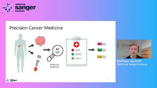
Hello, everybody. My name is Matthew Garnett, and thank you for joining this webinar today. So approximately 50% of people will develop cancer in their lifetime, and precision medicines are increasingly being used to personalize their treatment. But unlike chemotherapy, these precision medicines use our understanding of the genetics of cancer to selectively target cancer cells while ideally sparing normal healthy cells in our body. And so in the example shown in this figure, sample is taken from a patient with cancer. For example, as shown here, could be a piece of tumor biopsy or a blood sample. And this is used to perform some kind of molecular diagnostic test. Which a doctor can then use to select the best medicine for that patient. And precision medicines can lead to more effective and less toxic treatments, and in the future are a route to treating some of today's most deadly forms of cancer. And we need precision medicines because not all cancers are the same. So cancer is caused by genetic changes in the DNA of cells, that cause those cells to divide and grow excessively. Now, these genetic changes actually vary quite a lot by a cancer type, and even within a single cancer, they're not all the same. So there are many kind of almost like different forms of things like breast and lung cancer. And so today, as shown here, many breast and lung cancers, in fact, can be divided up into different subgroups. Based on their genetic changes. And this information is then used to select a treatment that's most effective for a particular patient. And despite some really great clinical successes using this type of precision medicine approach, unfortunately today most patients don't actually receive this type of precision medicine approach. And In fact, for many cancers, we still lack effective medicines at all. We know this is all done in the context of a very difficult drug discovery process. So success rates for oncology drug development are less than 10% when you consider drugs going from Phase 1 all the way to clinical launch. And that's just showing here on the left side of your image where you can see those success rates over time. And you can see in many respects they're not really changing. Let's just look at the bottom line, which talks about that percentage from phase 1 to clinical launch. We know that the leading cause of failure is lack of efficacy in phase 2 clinical trials. But the good news is, is there's increasing evidence that genetically informed targets are more likely to proceed through each phase of drug development. And that's just illustrated in this table where they've done a meta-analysis of the success rates of different drugs and also shown that overall the targets that have a genetically informed target are twice as likely to actually progress all the way to clinical approval compared to targets without supporting genetic evidence. And so increasingly, the emphasis in drug development is on the development of medicines really guided by genetics and a mechanistic understanding of disease. So to facilitate the development of precision medicines, my lab studies how the molecular features of cancer cells impact on disease mechanisms as well as therapy response. And over many years now, we've been very fortunate to develop what I sometimes describe as a preclinical discovery ecosystem. This includes the ability to make new cell models at scale, to perform very deep genetic analyses of cell models, as well as the establishment of functional perturbation platforms in particular large-scale drug screening, as well as more recently large-scale CRISPR screening to perturb gene function. Now to analyze these datasets, we develop new analytical tools, and we also make web tools that allow us to present these data to the community as a resource and to mine those datasets as well. And ultimately what we then do is take forward these analyses into more detailed mechanistic studies with the aim of coming up with new therapeutic strategies that will hopefully benefit patients in the future. So as part of our work in recent years, we've launched what we call the Cancer Dependency Map initiative. The Cancer Dependency Map aims to use large-scale functional genomics to identify vulnerabilities what we sometimes call dependencies in every cancer cell. We believe this type of map of cancer dependencies and the biomarkers that predict those dependencies could serve to help accelerate the development of precision cancer medicines. This is a very ambitious effort and involves multiple collaborators in the US and Europe, including the Broad Institute, the Human Technopole, and the European Bioinformatics Institute. So to create a cancer dependency map, we're using large panels of cancer cell models for both genetic and functional studies. And this just shows some of our recent data showing we now have aggregated and curated well over 1,900 cancer models representing over 43 different cancer types. And I really want to emphasize that we use this large collection of cancer models to have as much possible representation of the different cancer molecular subtypes that occur in patient populations. And we know that these molecular differences can underpin differential therapy response. And so to date, the Sanger Institute and as well, particularly the Broad Institute, have been working together and have generated over— data for over 1,900 cancer cell lines. And this includes genomic data, epigenetic data, proteomic data, as well as functional datasets. And these are just illustrated in this map here. Now you can see, for example, the coverage of cell lines for each data type is variable as shown in this image. And we And of course, one goal is to kind of fill in these gaps. And as I'll show in my presentation today, we continue to expand these datasets through the development of new cancer models and their enhanced annotation with new molecular or phenotypic data, as well as the addition of more functional data for each cancer model. So one such effort is what we call Project SCORE, which is using genetic screens to identify cancer dependencies. Now, there are many unexplored potential cancer drug targets that we just don't know about today. But for most proteins, there's not a drug that could be actually used for screening to evaluate their suitability as a therapeutic target. And in what context that protein might be a good target. And so the recent advent of CRISPR-Cas9 screens provides a really exciting opportunity to identify new drug targets in a way that was not previously possible. And so to identify new drug targets, we've exploited this programmable nature of the CRISPR-Cas9 system to investigate gene function in multiple cancer types. So just to give a little bit of background, CRISPR is sometimes called molecular scissors because it allows scientists to specifically cut DNA and inactivate virtually any gene in cancer cells grown in the laboratory. And so we use CRISPR in this case, to turn off genes in tumor cells one by one to determine which genes are essential for cancer cell survival, with the idea that a gene that is required for cancer survival might be a good drug target of the future. And the power of CRISPR is that we could screen thousands of genes in hundreds of cancers grown in the lab And so, for example, in the figure shown, an activation of gene B in this particular cancer is selectively required for tumor cell growth, indicating a drug that targets the protein product of gene B may be efficacious in this cancer subtype. And so a kind of useful analogy to think about here is imagine that you didn't know how a bicycle works. You could take that bike apart, removing each part one by one, see what the effect is on how that bicycle works. And this is effectively what we're doing. We're using CRISPR to deconstruct cells to determine which proteins cells need to work, and if blocked, could make a good drug target. So to do this systematically, we've actually now performed large-scale CRISPR screens in hundreds of cell lines from different tissues and identified hundreds of genes required for cancer cell survival. The challenge we then face is we identify these hundreds of genes, we need ways to filter through these lists of genes to find the best drug targets. And so to try and do this, we've developed a computational pipeline which combines this CRISPR gene essentiality information with other orthogonal datasets, including patient clinical data. So for example, is that gene mutated in cancers or differentially expressed? As well as information about things like biomarkers. So for example, can we identify a genetic event in the cancers that confers the dependency on a particular gene? And do we have pathway-level information that supports a dependency on a gene? And we combine all these pieces of information to come up with what we call a priority score for each gene in each cancer type individually and when looking across all cancer types together. And I really want to emphasize the kind of systematic, biased, and genome-wide nature of this approach. And the idea here is really allowing us to identify a much wider range of candidate targets which could be the targets of drugs in the future. And so using this approach, we assessed and ranked thousands of genes as potential therapeutic targets. So in the image shown, each circle in this plot is a candidate target. Now, not all targets are equally druggable, and so we've also assigned each target into groups based on the likelihood of being able to make a drug to target them. So for example, some of these targets that we have found are actually the targets of existing drugs today. These are the ones particularly in the group 1 targets. And of course, it's great that we find these because in many respects these are the positive controls in our screen, showing that our screening approach and our prioritization approach is working well. But I think more excitingly, we've identified many new targets. And particularly, we think the targets in group 2 are of particular interest because these are targets where we have high priority scores suggesting that they would be good targets, but there's also evidence supporting their tractability for drug development. And so these are great starting points to identify the most promising new drug targets, and particularly for cancers that lack effective treatments today. And one exciting target is a protein called Werner, or WRN, which is specifically required for the survival of a subset of cancers that are defective in a DNA damage repair process. So Werner helicase is a strong and selective dependency in microsatellite unstable cancers, or MSI tumors, from ovarian colorectal cancers and also went looking pan-cancer. So what you're seeing here in this plot is each circle represents an individual cell line and the fitness effect when you knock out Werner helicase. And what you can see hopefully is those tumors that are MSI, when you knock out Werner, it has a more profound effect on the fitness of those cells. So you see a very strong loss of fitness effect. Equally, in cells that are not microsatellite unstable, depletion of Werner seems to have virtually no effect on the cell fitness. And this is a very strong signal in the sense that the fitness effect that you see when you knock out Werner is similar to what would be observed if you knocked out a core human essential gene. So for example, a component of the ribosome. So microsatellite instability is caused by defects in mismatch DNA repair process. And in cancer cells, this leads to the accumulation of microsatellite repeats and a very high mutational burden. So mismatch repair is actually mediated by a cascade of proteins which identify mismatches that occur during DNA replication. These are then excised, um, by this mismatch repair machinery, and this allows resynthesis and repair of the lesion. And I think critically for this presentation is just to note that microsatellite is instability occurs across many different cancer types. And that's just summarized in this table here. And that actually at a very high frequency in a subset of cancers. So for example, in the setting of colon cancer, stomach cancer, and endometrial cancer. It also occurs in other cancer types, but at a much lower frequency than these other cancer types. And so based on our initial discover— discovery of a Werner dependency, we've gone on to further interrogate Werner dependency in the setting of colorectal cancer. And so to do this, we've created the largest set of microsatellite unstable colorectal cancer models ever assembled. And this includes over 62 different cancer models of MSI colorectal cancer. And I think very strikingly, we've found that 92% of MSI tumors are dependent on Werner helicase. So there is a very rare and small subset of resistant MSI tumors, but for the most part, they're highly dependent on Werner. And what's striking about this is that this Werner dependency seems to be largely independent of many other factors. So for example, we see it in cell models that are derived both from primary and metastatic disease. It's independent of the total mutational burden. We see it whether we look for this effect in a 2D cell line, a 3D organoid culture, or a cell line derived from a patient-derived xenograft culture. And in addition to that, it appears to be independent of the driver mutations that are present within that tumor. And so collectively, these data really strongly support Werner helicase as a target in MSI cancers. Now, Werner itself is an extremely interesting protein. It has what's actually quite a poorly understood role in genome stability. Including processes like DNA repair, DNA replication, and telomere maintenance. And it's also associated with a human syndrome called Werner syndrome, or also known as progeria. So this is a rare autosomal recessive disorder that's characterized by premature aging and predisposition to cancer. So Humans or people with this syndrome tend to age normally through childhood, but as they enter early adulthood, their aging accelerates. And ultimately, this can lead to early death with a median age of death for these patients of around 54 years old. Now, what's, I think, significant for um, when thinking about Werner as a target in the setting of cancer, the finding that germline loss of Werner is actually compatible with viability of a human until adulthood suggests that a targeted therapy against Werner delivered for relatively short periods of times might not be overtly toxic to tissues. Of course, the development of a drug will be required to test that hypothesis. So we've gone on to look in more detail about the mechanism of what's happening when you knock out Werner in cancer cells. And I won't go into a lot of detail, but just to say that knocking out Werner causes massive DNA damage to chromosomes in these MSI tumors. And that's, I think, really nicely illustrated on the image in the left, where you see the chromosomes in a cancer cell, just 4 chromosomes are shown for clarity. And then what happens if you take those cancer cells and you knock out Werner is you get this massive fracturing of the DNA with many chromosomal breaks. And this ultimately leads to apoptosis and cell death. And when actually microsatellite unstable tumors are growing in the lab on the back of a mouse in what we call a tumor xenograft, if you take an established tumor and then use genetic tools to knock out Werner, this can actually stop those tumors from growing. And so collectively, these data have really nominated Werner helicase as a potential candidate drug target. Now today, there are no drugs that target Werner. But based on these studies, as well as now supporting studies in the literature, multiple pharmaceutical companies are developing drugs to target Werner in this subset of cancer patients. So since the publication of the Project SCORE data, we've continued to really expand and refine our analysis. And then just briefly touch on some work that actually led by my colleague Francesca Oriorio at the Sanger Institute, who is now based at the Human Technopole in Milan. And this work was actually done in collaboration with the DepMap teams at the Broad Institute. And so we've actually together developed a method to integrate CRISPR-Cas9 screening results generated at the Broad and the Sanger Institutes. And of course, there were batch effects in these data, but we've now been able to correct for that and bring these two datasets together. And this now includes coverage of over 16,000 genes knocked out in almost 800 genetically unique cancer cell lines. And having this larger dataset really is empowering and does a number of things. First, we have a larger data set, increases the number of cell lines for each cancer type. I think that's nicely illustrated on the left-hand side. It also gives us more statistical power, allowing for the discovery of novel cancer and subtype-specific analyses and unveiling additional biomarkers of gene dependencies. And so an example is shown on the right-hand side of your image where we've looked at the dependency on ERBB2 knockout in ERBB2-amplified breast cancer cell lines. And then the left-hand side is what you observe when you only look in the Broad's data. You can see that the relationship between HER2 or ERBB2 amplification and knockdown is not significant. But if you then combine that with the Sanger data in the integrated dataset, this association becomes highly statistically significant. So in the second half of my talk, I'd like to discuss something we call Project GROW, which aims to generate a new generation of cancer models as a community resource. So We continue to use the cell lines that we have in the laboratory. Nonetheless, though, we recognize that there are a number of limitations with this current set of cancer models. We know that the cell lines have adopted 2D culture in poorly understood ways. In many cases, we have no patient genetic data, clinical data, or pathological data. For those samples, which makes some of our analyses difficult. For example, we don't know whether those patients may have responded to certain therapies or not, which would be helpful and informative in our studies. And I think critically, we lack representation of some tumor types and cancer subtypes. And I think if we really hope to achieve precision medicine for the majority of patients, we need a set of models that really reflects the diversity of those different patient populations so we can study them in the laboratory. Now, historically, the development of new cancer cell lines has been quite difficult, often taking many months and with very low success rates. And when I mean very low success rates, this can be often less than a 1% success rate. Now I think very excitingly, recent advances in cell culture methods, and particularly the use of something that's called organoid cultures, have really improved the efficiency of deriving cell culture models. So there's an image shown of an organoid growing on the right-hand side of this slide, and organoids are grown as three-dimensional in vitro cell cultures using a basement membrane extract together with very specially designed media that contains niche factors required to support cell growth. And because of this new methodology, it's now possible to derive tumor organoid cultures representing different tissue and molecular subtypes of cancer, as well as from different disease stages, with a much greater efficiency than was previously possible. And this is really transformative for the community. And so about 5 years ago, we were very excited about this technology, and we actually reported a pilot study that set out to evaluate whether we could establish a biobank of colorectal cancer organoid models. We were able to establish a small bank of 22 organoids from colorectal cancer, and we demonstrated that they were genetically similar to the tumor from which they were derived and could be utilized for downstream functional assays such as in vitro drug testing. Now, since then, we've now performed very similar studies in the setting of esophageal cancer, pancreatic cancer, and liver cancer, and there's also been multiple other studies from other groups reporting the development of these types of biobanks with very similar results. And so based on this literature and our own experiences, we've now set out to generate a much larger biobank of tumor organoid cultures with initial focus in the setting of esophageal cancer, colorectal cancer, and pancreatic cancer. Now this effort is actually part of a much larger initiative that's called the Human Cancer Model Initiative. And key to this is that all the models that we're generating and the datasets associated with them are being made available as a community resource. So within the UK, to develop organoids, we've established what we call the Cell Model Network UK. This includes clinicians and academic scientists from across the UK who are really providing their expertise and serving as champions within their local organizations to help guide this project and develop the most relevant disease models. And wherever possible, we've actually coupled the derivation with things like clinical trials or other research programs to generate the richest possible datasets for each of the models that we're making. So working with these collaborators, tissue is actually taken from surgical resections and couriered to the Sanger Institute. Now, this is fresh tissue, and where organoids are derived in a dedicated facility by a team of expert technicians following very rigorous quality control. The organoids are then banked and sent to the repository ATCC for distribution as a resource for the, for the community. And in addition to that, increasingly these organoids are now entering into our downstream analysis pipelines for genetic analyses, for things like drug testing, and increasingly also for CRISPR screening. So to date, we've processed over 500 samples of patient tissue, the majority from surgical specimens. And overall, this has led to the successful banking of 145 organoid cultures from colon, pancreas, and esophageal cancers. And already this is really having a big impact. So For example, for esophageal cancers, in the past, there were very few good models where you could really study this disease. And we now made this unique set that has massively expanded the number of esophageal cancer models that are available for the community. Similarly, in the setting of colorectal cancer, in the past, we had about 50 cancer cell lines that we would use to study colorectal cancer as a community. But now we have over 80 new models. And so this is really increasing the representation of the different subtypes of cancer, of colorectal cancer that we know are important clinically. And again, having this larger sample size gives us more statistical power for some of our functional studies. Now, I should say, this is hard work to make these models. It typically takes over 100 days to go from a piece of tissue to a set of banked cryovials, and we have an overall success rate of about 30%. So about 30% of the pieces of tissue that we accept are then— ultimately lead to the successful banking of a living model system that we can use for future studies. And the number one cause of— the major causes of failure are really just failure to propagate in the culture system and low tumor cell number in the sample that we actually receive from the clinical sites. So beyond building the resource of this next generation of cancer models, of course, we really want to begin to use these for some of the systematic perturbation studies similar to what we perform in our cell line collection. And so in addition to things like drug screening, we're also now able to have really robust protocols to perform genome-wide CRISPR knockout screens in these 3D organoid cultures, and using effectively a pipeline that was established for Project SCORE. And just to give you a sense of these datasets, We're able to detect, for example, known human essential genes, which is a really critical quality control metric for these screens, with a kind of similar efficiency as we can when we screen a cancer cell line. So that's just shown on the left-hand side of the image. This is our ability to recover known human essential genes in 4 different organoids that have been screened with a genome-wide library, and in the comparison of what— how well we can do that within cancer cell lines. And so at the moment, these screens seem to be very similar in quality to what we can achieve with the cell line. And of course, we can then look for specific dependencies in these models. So that's shown on the right-hand side of the image. These are all the fitness effects of knocking out thousands of genes in this particular colorectal cancer organoid. And just notably, for example, this is a KRAS mutant colorectal cancer organoid, and indeed KRAS is one of the top gene dependencies within this model. And so this, again, this is just a really kind of proof of concept that these screens work efficiently in the setting of the organoid model system. But because organoids are grown in 3D culture, this actually does make them more challenging to work with them. And so we've been trying to develop new methodologies and reagents to improve our ability to really work efficiently with these organoid cultures. And so one of the ways we've done that is to really optimize our CRISPR screening libraries, and in particular making smaller genome-wide libraries that use two optimized guide RNAs as compared to the 5 guide RNAs that we used in the original Project SCORE CRISPR library. And so what we actually did is we mined the Project SCORE data from screening hundreds of cell lines to identify the 2 best guides per gene. And we can then make a new library that's 60% smaller than the original library and 42% smaller than other genome-wide CRISPR libraries. And what we've been able to go on to show through screening in both cell lines as well as in 3D organoid cultures is that the minimal library retains the specificity and sensitivity to identify gene dependencies compared to the original library in both the cell line and the organoids. And so we think this new minimal library, or MinLib as we call it, is a really valuable tool for screening cell cultures that are more difficult to grow or when measuring more complex cellular phenotypes. And we're just in the process of depositing the vectors and the library in Addgene, so they'll be publicly available. And so trying to bring all this together, our long-term vision is to build a passport for each patient's organoid. And I think this is just exhilarated— exhibited here in this one example, where, for example, we have a sample from a woman who has colorectal cancer, and we're able to actually get a bit of her surgical specimen, make a tumor organoid that we've been growing in the lab, We then perform a genetic analysis on that tumor and also on the organoid that was derived from that particular sample and make that data available. In addition, the model itself is actually able to be purchased from ATCC. And so you can access all these datasets. In addition, we're increasingly adding on functional data through things like drug testing through things like CRISPR screening and making that data available to the community. And in fact, this organoid from this patient is the one where I showed that genome-wide CRISPR screening data from previously. And so to make all these datasets available, we've actually established a new database that we call the Cell Model Passports, which is a hub for information on preclinical cancer models, including genetic data and functional data for over 1,900 cancer models, including both cell lines and organoids. And you can actually come into the Passports and get a wealth of information on a particular cancer model. So this is just one example shown here for a colon cancer cell line called HT-29. And the actual passport itself for the cell line contains patient and clinical data, links to data downloads, word clouds showing the cancer driver mutations present in it, top CRISPR dependencies, as well as links to the Project SCORE database, drugs that are active in this cell line, and links out to our drug database, as well as information about mutations and gene fusions., as well as links to commercial suppliers where you can purchase this line. And so we think the cell model passports are a really useful tool to empower scientists to identify the most suitable model for their research and access genomic and functional datasets that are available. And so finally, I'd just like to close by thanking everyone that's been involved in this work. It's a collaborative effort, both from a team at the Sanger Institute as well as the core facilities at the Sanger, which would really make these large-scale efforts possible, and a wonderful group of collaborators who I've worked with for many years. And of course, also to thank the funders who have contributed to this work. Thank you very much.
More from Joshua







Scientist Stories: Hans Waterham, The value of phenotypic assays in peroxisomal disorders




Scientist Stories: Joel Zlotogora, Genetic carrier screening programs for reproductive purposes