Scientist Stories: Mikala Egeblad, Neutrophil Extracellular Traps in Disease
In this seminar, Mikala Egeblad presents a comprehensive overview of neutrophil extracellular traps (NETs), detailing their discovery, biology, and emerging roles in disease. She begins with intravital imaging of mouse lungs, showing that highly metastatic breast cancer cells (41) are surrounded by DNA‑rich NET structures and active neutrophil elastase, whereas non‑metastatic cells (407) are not. Using DNase‑coated nanoparticles, she demonstrates that degrading NETs markedly reduces lung metastases, establishing NETs as promoters of metastatic colonization. She then describes how NET‑associated proteases (neutrophil elastase and MMP9) cleave laminin, exposing an epitope that activates integrins on dormant cancer cells, thereby awakening them to proliferate. Stress‑induced NET formation is explored through chronic restraint and transport stress models, which increase plasma NET levels and dramatically boost metastatic burden; DNase treatment mitigates these effects. Shifting to infectious disease, Egeblad reports rapid collaboration during the COVID‑19 pandemic that identified abundant NETs in autopsied lungs and patient plasma, linking NETs to acute respiratory distress, mucus accumulation, thrombosis, and poor outcomes. She outlines ongoing clinical trials targeting NETs via DNase, PAD4 inhibition, IL‑1β blockade (Anakinra), colchicine, and dipyridamole, emphasizing the need for mechanistic insight to improve specificity. The talk concludes with a discussion of open questions, challenges in studying short‑lived neutrophils, and the broader relevance of NET research across cancer, autoimmune, cardiovascular, and respiratory diseases.
Chapters
Hello everyone. My name is Ron Vale, the Executive Director of Janelia, the research campus of the Howard Hughes Medical Institute. I would like to welcome you to Life Science Across the Globe, a sister institute seminar series. This series is open to viewers anywhere in the world. More information on the series is found on this slide, as well as the website lifescienceacrosstheglobe.org. And videos of prior talks can be found on this website as well. Today, I would like to welcome the second sister institute, Cold Spring Harbor Laboratory, and would like to welcome its director, Dr. Bruce Stillman.
Thank you, Ron. I want to welcome everybody to this series and also to Cold Spring Harbor Laboratory's turn as one of the sister institutes. I'm president of the laboratory, but I'm going to introduce Dr. Limor Joshua Elkington, who is going to be coordinating this series for Cold Spring Harbor Laboratory. She got her bachelor's degree in chemistry at Tel Aviv University, and then a PhD at the Weizmann Institute, also in chemistry, and then did postdoctoral studies at Caltech before joining Cold Spring Harbor Laboratory, where she is now a professor and a Howard Hughes Medical Institute investigator. And she's also former dean of the graduate school. So I want to turn it over to Limor, who will introduce the Cold Spring Harbor speakers.
Thank you, Bruce, and welcome everybody. Hi from beautiful Cold Spring Harbor lab right behind me. I'm thrilled to introduce my colleague Mikala Egeblad as our first speaker today. Mikala got her PhD in cancer biology from the University of Copenhagen in Denmark. She then moved to UCSF to work with the great Zena Werb, who unfortunately passed away just a few weeks ago. Then she began developing— there, I should say, she began to develop intravital spinning disk confocal microscopy to understand how the microenvironment influences tumor progression. She came to Cold Spring Harbor as an assistant professor and was then promoted to associate professor. At Cold Spring Harbor, she continues to study the tumor microenvironment and how that affects metastasis. She discovered the role of inflammation and neutrophil extracellular traps or NETs, what their role that they play in metastasis. Most recently, she discovered the role NETs are playing in COVID-19, and she's heading an international team to tackle this issue, which may lead to treatments. She received an Era of Hope Scholar Award from the DoD and the Pershing Square Sohn Prize for young investigators in cancer research.
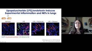
So thank you for that introduction. Good morning, good afternoon, good evening. I'm gonna talk about NETs inflammation. What are NETs? NETs is short for neutrophil extracellular traps, and you can see them here on this first slide. This smear of green stain is NETs in the lung of a patient that succumbed to COVID-19 here in New York, and I'm gonna return to this image in the end of the talk. Because we didn't start looking at NETs in COVID-19, we came from it by trying to understand the process of metastasis, spreading of cancer from a primary tumor, for instance, a breast, to different tissues in the body, such as, for instance, the lung. So that these tumor cells will leave the primary tumor, get into the vasculature, travel with the blood, and then enter a second site,. And there these cells can then start proliferating and forming metastasis, what we call the process of colonization. In terms of cancer patients, it is this metastatic step that is the metastatic stage that's really hard to treat. The primary tumors can be surgically removed or treated other ways with radiation, but once you have metastasis, treatment is, is difficult. And my lab wanted to try to understand more about the colonization step to to try to understand how to treat it better. We took a little bit unorthodox approach to get into that by using intravital imaging, imaging in living mice, and we were helped by Max Krummel at UCSF who had developed this lung imaging technique. You can see here the mice are intubated and there literally is a glass window you can see through to get into the lung and get images of what's going on there. So we did images with 41 breast cancer cells. They are highly metastatic. We labeled them with red fluorescent protein and injected them intravenously into the mice and then watched what was going to happen as they reached the lung tissue. Here using a mouse that's expressing GFP in the neutrophil population. What you're seeing here is over time that these— there's a blue stain, DNA stain. We were using DAPI, and we have really used it only to get a counterstain for the lung tissue so we could see where we were. But you're seeing this development of clouds of DNA around some of these metastatic cancer cells. So, it also overlapped with regions where we were seeing some of these green cells stop moving. So, it was kind of peculiar, and we were wondering, of course, if it was the cancer cells that were dying and releasing their DNA. However, when we compared the metastatic 41 cancer cells that are able to form metastasis with a sister cell line, 407, that are not able to form metastasis but die off upon injection, we actually found that the— these clouds of DNA were almost always only found around the 41 cells, as scored here blinded in a variety of these movies, versus in the 40/07 where we only found one example. Moreover, when we used a fluorescent probe against an enzyme called elastase, neutrophil elastase, this is a probe that only becomes fluorescent when the enzyme is activated. And you can see here, we see activity with the metastatic cells around this area of the clots, but not with the non-metastatic cells. So what is this extracellular DNA with associated elastase activity? Well, I knew something about that only because I had been to a Keystone meeting years earlier. So this was back in the time where we could go to real meetings and journals were being handed out. Here it's a journal from JCB that was handed out as an example, and I really remember this beautiful cover image of a neutrophil spilling out its DNA into the extracellular space. And you can see here the bacteria that is captured in this sticky DNA. And this is work by Artur Słuczynski's group. They really discovered the NETs originally in 2004. So what is now known is that neutrophils, which are most prevalent immune cell in blood, have three major means of eliminating pathogens. They can do it through phagocytosis, Probably most of you have seen the classical movie where the neutrophil is chasing through a petri dish like Pac-Man trying to capture bacteria. They can also kill pathogens through degranulation, releasing these toxic enzymes to the extracellular space. And then they can form the neutrophil extracellular traps, the NETs. They form these structures from activation of an enzyme called PAD4, at least in most cases. PAD4 citrullinates histones, leading to chromosome decondensation. And then this DNA is associated with enzymes otherwise found in the granuli, but enzymes like elastase or myeloperoxidase. So you have an extracellular space DNA, granular enzymes, and the citrullinated histones. And that allows us to detect these NETs in tissue sections. So you can see here the faint extracellular DNA around one of these neutrophils, also citrullinated histone, as well as the myeloperoxidase, one of these enzymes. Again, this is a high magnification view from a lung of a COVID-19 patient. So neutrophils, as I mentioned, are capable of eating, phagocytosing pathogens. This is a movie of a neutrophil coming in here, and you can see it phagocytosing bacteria in a mouse we have infected with red fluorescent bacteria. So why would they need neutrophil extracellular traps? That seemed to be a very efficient process. Well, this is an example of a movie generated from a group in Germany that have here visualized what happens when the neutrophils encounter a larger pathogen an Aspergillus spore that's labeled in blue. You can see this spore is bigger than the neutrophils. 4 or 5 of them are ganging up on it, but they can literally not eat it. One of these neutrophils is now first condensing, then swelling up, and then propidium iodide was added to the medium, so in a moment you'll see this neutrophil exploding essentially and releasing its DNA to cover the spore. And so that is another way of of taking care of the pathogens. My students like to call this that the neutrophils are spilling their guts. I prefer thinking of it like Spider-Man that's casting a sticky web to capture the pathogens, but I will add that Spider-Man in this case commits suicide. The neutrophils do not survive this process. So we return back to our cancer cells. This is what neutrophils look like in scanning electron microscopy when they are left alone in culture.. But here they've been exposed to the cancer cells, and you can see that the cancer cells have hijacked this system. They are expelling the— the neutrophils are now expelling the DNA strings. And we now know something about the cytokines that are doing that is, for instance, G-CSF and other cytokines can, can mediate this process. So the cancer cells can get neutrophils to form NETs, but does it matter? So we were able to test this using nanoparticles coated with DNase 1 to chop up literally these DNA nets. And you can see here with control nanoparticles, there are metastases all over in the lung of these mice. But with the DNase-coated nanoparticles, the lungs are mostly clear, also quantified over here. It's really a quite striking difference with this treatment. So this tells us that the NETs are important for metastasis, but how does it work? And the clue from that came from a project we started around this time. We were trying to focus specifically on this step, on how one cell become too many cells in the case of the situation known as dormancy. You may know that some cancer patient survivors can live with their cancer after what looks like a successful treatment for years or even decades, and then suddenly the cancer comes back. And it's quite unclear why the cancer can be dormant for so long and what the signals are that then awaken it, as we call it. So we wanted to understand that, thinking that there was an opportunity for intervention. A new postdoc joined my lab around this time. He was watching the lab meetings from the other project, and he started reasoning that if NETs can be induced by the metastatic breast cancer cells to promote metastasis, maybe maybe if inflammation caused the NETs to be formed, that could affect such dormant cancer cells that otherwise were sitting in the tissue and doing no harm. And so he set up a model system to test that using LPS or lipopolysaccharide, also known as endotoxin. It's a component of the bacterial wall of gram-negative bacteria. And he got that into the airways of the mice to cause inflammation. You can see lots of neutrophils, they're forming NETs. And if he inhibits the PAT4 enzyme that I mentioned, The neutrophils are coming in, but they're not forming nets. Similarly, he can digest these nets with DNase treatment. So he used this system in a context of a dormancy model, building on model systems from many other groups, injecting the cancer cells with luciferase so he could track them, imaging every 3 days by bioluminescence, and then inducing inflammation at day 7. If he does that, you can see here in the control animals, there's no, uh, metastasis visible from bioluminescence. You can find single cells at the end, but they are few and far between them. They're not proliferating. And with LPS, you have large metastatic tumors and they are proliferating. This turned out to be dependent on NETs. Here he's tracking by bioluminescence. Again, the controls, there's below the detection level. With LPS, the tumors are coming back. But treating with DNase or PAD4, you can see a significant reduction and delay in the occurrence of these metastases. How do the NETs then cause awakening of dormant cancer? So to summarize a lot of data just in a cartoon, what Sean found was mechanistically what happens is these dormant cancer cells are sitting in the lung, they have their integrins but they're not engaged, neutrophils are coming in, they form the NETs, in response to inflammation. On the nets are two enzymes, the neutrophil elastase enzyme that we had detected with this imaging probe, as well as matrix metalloproteinase 9. Together they are cleaving an extracellular matrix protein called laminin, changing the structure of this molecule so there now is an epitope revealed that can activate the integrin, leading to proliferation of the cancer cells. So this led us to think, well, maybe there was some other external stimuli that can drive metastasis from disseminated cancer cells. Maybe this was more of a theme. And the sort of clue to what that could be came from what really was a rather stressful event in my lab. We had a period where we had unreproducible, irreproducible results. A graduate student in my lab had worked on the role of a gene called CCR2 in cancer cells. She had found over and over again for over years that the key phenotype was that these cancer cells grew very slowly. Being controlled by the immune system compared here to the wild-type cells. A few experiments needed to be done before the story was completed. New postdoc came into the lab, repeats this experiment, and as you can see, sometimes you would even see the opposite. She could not repeat this key phenotype of the project. As you can imagine, this was very frustrating. We spent a lot of time trying to figure out why, countless of meetings going over the protocols, comparing line by line what could be wrong. And finally, after 10 months, I realized that I had forgotten to teach Suyang something important. We're getting our mice shipped to us from Jackson Lab. It's about a 10-hour drive on bumpy roads from Jackson Lab to Cold Spring Harbor. And Suyang, of course, wanted to finish these experiments as quickly as possible, so she took the mice more or less immediately out of these cages and started doing experiments. Of course, the mice looked something like this when they came out of these boxes, and incidentally, so did I at this point. Into the project. So, Sooyoung tried to see if it really was this stress of transportation that was responsible. And as you can see here, when cancer cells were being injected in parallel either to mice that were straight out of the truck versus mice that had acclimatized for 2 weeks as they were supposed to, there was more than a doubling in tumor phenotype. Sooyoung and I realized that this was actually really interesting because chronic stress has been related to poor cancer prognosis, but the mechanisms are not clear. There is a nice review here reviewing that the higher incidence and poor survival in a variety of different cancers are associated with stress. And transversely, there is longer survival if you can socially support patients that are cancer survivors. Now, we realized that maybe trucking of mice was not the best experimental model. So we teamed up with Linda Fennell, a neuroscientist here at Cold Spring Harbor, and used a classical model of stress, the chronic restraint model, where the mice are sitting in conical tubes for 2 hours every day and can't move, which stress them out as it would do me too. So Yang built on this model. So she first inoculated tumor cells into the mice in the breast. That gave the time to the cancer cells to naturally disseminate to the lung. She surgically removed the tumors, and then she started stressing the mice similar to the stress that a cancer patient would have upon being diagnosed with cancer. Finally see evaluated metastasis. As you can see here, there was a huge effect on metastasis from these disseminated cells upon stress. It's about a fourfold increase in metastatic lesions and number of lesions. And as you can see in this image, not only are there more lesions— these are the small lesions in the control, you can barely see them— but in the stressed mice, they are much, much larger. Building on our previous work, Suyang had the insight to measure NETs in the plasma of these mice. This can be done through an ELISA that detect DNA with bound enzymes from these NETs. And you can see that the NET levels also increased about fourfold in the plasma of these mice. So she used again DNase to try to digest these NETs, and as you can see, it reduces metastasis, it reduces the level of plasma NETs, and the lungs look much better. These mice can survive longer. Now, of course, we want to know a lot more about the mechanism, and back in March, I was sitting and working on a grant to at least get to start to apply for money so we could understand more about this mechanism. But I couldn't concentrate. I think like many of you, I was very stressed myself. And this is indeed why we are here today and watching this by Zoom instead of meeting in person, because the COVID-19 pandemic was happening. And I was starting to think, well, maybe NETs had something to do with COVID-19. And reaching out to people I knew had worked on this area, turned out that I was not the only one that had this idea. We were a large group of people that connected via Twitter, via emails, via personal connections. All of us had had the same idea. These NETs could have a role in COVID-19. And we put out a perspective in Journal of Experimental Medicine early April to explain our rationale for this. The reason is that NETs have been shown in a variety of different studies to be associated with acute respiratory distress syndrome, which you see in severe COVID-19. They see accumulation of mucus in cystic fibrosis mediated by NETs, and that's also seen in COVID-19. There's a variety of cardiovascular diseases that's linked with elevated NETs, and of course, then there's thrombosis, something we now understand is very much a part of the pathogenesis of COVID-19. And NETs there are known to play a major role in types of thrombosis. So we stuck our necks out to say that there was a possible role for COVID-19, but we really didn't know at this point. Are there nets in COVID-19? Well, I was very delighted and excited when earlier Thursday morning, I got a text from my colleague and now friend, Andrew Weber, that is treating COVID-19 patients here on Long Island in the Northwell Health system. They— Northwell Health and Cornell and the other hospitals here in New York were sharing pathology reports, and one of the first cases were being evaluated by Dr. Alan Borsuk, shared with Northwell's pathology and what he had seen was it was surprisingly neutrophilic. There was neutrophil debris in this case. And when I hear neutrophil debris, of course I'm thinking NETs. So I contacted Alain Borsuk, and within hours he had sent me examples of these histology, and he also sent us 3 cases where we could stain for NETs. And as you can see here, in all 3 cases, there were simply NETs everywhere in the lung from these patients that had succumbed to COVID-19. You can see here example of the double staining for myeloperoxidase and the citrullinated histone that is marking neutrophils in the process of forming NETs. I also really want to highlight the fellows that did the actual autopsies. Many places not able to do the autopsies due to the risk of infection, so this was done by some risk to them personally, and I very much appreciate that. In addition to finding NETs in the lungs, members of this network, Jason Knight, Jobin Kanfi, and Ray Xu were able to quickly get remnant serum, serum samples that had been taken as part of the clinical course and otherwise were going to be thrown out, and measuring again using this ELISA assay for DNA conjugated with myeloperoxidase, measure in the remnant serum that there were indeed elevated level of NETs in the COVID-19 patients. Moreover, they could see that the patients that were on ventilation versus those that could stay on room air had the highest levels. Of NITs. This was done very rapidly and communicated rapidly, even though the samples were perhaps not ideal, because they really wanted the information to be spread to the community. In parallel, Christian Cornu's group at Utah, scientists I had just had dinner with in February when I was visiting Huntsman Cancer Institute, were able to quickly repurpose one of his IAB protocols. And Beth Middleton, her fellow, was able to go and consent the patients and collect fresh plasma. These are really the samples you want to look at to prove to yourself that NETs are important. And you can see here, she found a very consistent result with that early report. Intubated patients had the highest levels, and when they got better, the level of NETs dropped down. Importantly, the patients that succumbed to COVID-19 had the highest level of NETs in blood. So what are the NETs doing? We think that one of the things they're doing is forming microclots, microthrombi, in, in the blood. You can see here that when we stain for platelet factor 4, there are microthrombi in the lung. This is from case 3, and they are positive for neutrophils in the process of forming NETs, citrullinated histone, and myeloperoxidase is present there. So based on this, we are proposing to target NETs in COVID-19. This is examples of the ways of targeting NETs that are ongoing in clinical trials. You can target NETs with DNase, and several trials ongoing with that. We have tried it in a couple of patients locally in Long Island and have a case report out on that. You can try to target the mechanism responsible for induction of NETs. It's not clear what it is with COVID-19. It's clear there's something in blood that can induce it, and one candidate is IL-1 beta, based on work from, from many other studies on NETs pre-COVID-19. And Akinra is a drug that can be used to target IL-1 beta, and case reports have suggested that that actually has some efficacy in severe cases. Now you can also target neutrophils as well as other inflammatory factors with colchicine, an old drug used in rheumatology. Again, case reports are suggesting that under the right circumstances this actually works. And then very intriguingly, Jürgen Kampff and Jason Knight's group have proposed that dipyrimidole, an old antiplatelet drug, can be used because it inhibits both the platelets and the NETs, the formation of NETs, and this way really target these microthrombi So all of these approaches are in clinical trials, randomized, and hopefully we will know within a few months if any of it works. So with that, I'd like to conclude by really saying that for, for the role of NETs in metastasis, we now know from a variety of different mouse models, starting with work by John Kolschatyk, John Spicer, and others at McGill, really were the first to show NETs playing a role in metastasis. And in animal models, I think we can feel kind of confident that they can drive metastasis. In humans, we're starting to see strong studies linking the presence of NETs with tumor stage or poor prognosis. Of course, in the animal experiments, we can target the NETs as I've shown you, but have we really proven that they play a role in vivo? And I must admit that the approaches we have taken— I showed you DNase treatment or inhibition of PET4, but we have also used inhibition of other molecules important for NET formation None of these approaches are honestly specific for NETs. The reason we still believe that the evidence is there is because there's so many different approaches targeting NETs give the same result. Nevertheless, I think the field really recognizes that we need basic discovery science to determine exactly how the NETs are formed and how they work so we can better target them. And this is something, of course, there's a huge effort to do. I should add that one of the complications is that neutrophils only survive 6 to 24 hours in culture, so a lot of standard techniques cannot be used. I want to just end by saying that what we are learning in one disease, everybody coming together to try to understand COVID-19, I think will end up helping us as we go back to our primary research areas like cancer, lupus, antiphospholipid syndrome, cardiovascular disease, and acute respiratory distress syndrome. It's been extremely valuable, I think, to have so many different views on how NETs work in this group, and it's something I'm enjoying a lot. And with that, I want to thank the people that did the work. I mentioned the people in my lab that did a lot of the COVID-19 work and stress work, Soo Yang and David. Shang-Yu Pringles did work on dormancy, and I want to give a shout out to Juan, Bobby, and Laura that did the first work on metastasis in my lab. As you can imagine, studying these DNA structures was highly controversial at the time. I want to thank my collaborators at Cold Spring Harbor, particularly Linda van Elst and Tobias Janowicz, who's working with me on some of the COVID-19 stuff, and my different collaborators on the COVID-19 projects. And here, of course, my group. We are socially distancing, but we have a shift around 2 PM when this photo was taken. One of our lab members, unfortunately, is stuck in Germany, and we look forward to having her back eventually. Finally, I want to thank the patients' family, the families that were allowing us to get access to, to the lung of these COVID-19 cases. It was really helpful to try to understand the pathophysiology of this disease, and I thank them for that. And with that, I Thank you for your attention, and I'll be delighted to take any questions.
Thank you very much. Thank you so much, Micaela. That was a wonderful talk. I think that it was some really exciting and elegant studies that you showed, and I think we're all looking forward to hearing how the clinical trials work out, particularly in COVID-19. The first question I have for you is you know, this mechanism of extruding DNA into the extracellular environment and net formation, is, do you know, is something like this evolutionarily conserved, and is it seen in other animals, organisms, plants?
It is seen in other organisms and plants even. One of the slides I took out to stay on time was showing an example of extrusion of DNA in plants. To protect plants from bacteria in the soil. So it seems to be a very conserved mechanism, and one of the things that's been proposed is that the histones actually can be toxic to pathogens when present in extracellular space. Excellent.
So we have a ton of great questions also from our audience. I'm going to call in a few people and see if they are willing to ask their questions to you directly. I'm going to start with Gurudat Patra. Gurudat, are you there? Would you like to ask your question?
Hi, can you hear me?
Yes.
Hello everyone, thank you, Mikhala, for the wonderful presentation. And my question is, uh, how much time does neutrophils take to reach the site of inflammation? And is there any deciding factor for NETs to be formed or not?
So your question was how much time does it take them to reach the site of inflammation. I think it really depends. Sometimes you can see it in the intravital imaging within 20 minutes, and sometimes it is— it takes longer. They have different chemokines to recruit them and different signaling peptides to recruit them, so it depends a little bit on the mechanism, but it can be as early as 20 minutes.. And once they are there, they have different pathways that can lead to formation of NETs. One pathway, it takes maybe just minutes, and another pathway takes hours. And we also see both processes in vivo. Thank you.
We also have a question from Yvonne Sun.
Yvonne, are you there? Yes.
Yes, can you guys hear me?
Yes, please go ahead. I'm just wondering, so with— for people with cystic fibrosis that suffer long-term and chronic inflammation, are they at higher risk for lung cancer then? Yeah, I don't know that that is clear. There is a link with lung fibrosis, so not cystic fibrosis but lung fibrosis, where we also know that there are a lot of NETs. They do have a higher risk of of lung cancer. Cystic fibrosis is a little trickier. They also at least tended to die earlier, younger, before they might be able to develop the cancer.
Great.
Thank you.
We do also have a question from Eric Zao, which I'm going to read. What substance derived from the 4T1 tumor cells is responsible for triggering formation within the tumor niches.
So we identified granulocytic colony stimulating factor as one of the substances. It's probably not working alone. We think that there might be at least two factors working together to get optimal response, but targeting G-CSF, granulocytic colony stimulating factor, can dampen the response. Other studies have, have verified that. There's also chemokines like CXCL8 in humans or CCL2 that's been linked to NET formation.
Great, thank you. We also have a good question from Abhishek Bhardan. Let's see, Abhishek, are you there?
Would you like to ask your question?
Can you hear me?
Yeah, we can hear you.
Yeah, I was just wondering how do the neutrophils actually decide which pathway is to be turned on during the immune response? Like it has to turn on the phagocytosis pathway or the NET formation?
How does it decide? I think— so you were a little hard to hear, but I think the question was how did the neutrophil decide whether to do phagocytosis, degranulation, or NET formation? Correct. So it's not entirely clear, but some of it seems to be that they can sense whether or not the pathogen is too big to be eaten. So size is a factor. They have sensors for that. Another potential mechanism is that some bacteria has developed ways to avoid being phagocytosed, and so those bacteria can then be taken up by NETs. And then conversely, some bacteria actually express DNAs themselves, and so those would be better taken up by phagocytosis than by NET formation.
Okay, thank you.
Thank you.
We also have a question here from Pat Ting. Pat, are you there? Would you like to ask your question? If not, that's fine. I will go ahead and ask it on behalf of Pat. Pat was wondering, The DNAs that are extruded out from the neutrophils, are they somehow modified to become even more sticky?
Yeah, probably. So there's this citrullination of the histones that is modified, but another question is, of course, is there any specific sequences that is increased on these nets? Is it just random genomic DNA that's out, or are there specific DNA sequences. This is something we are looking into right now with a collaborator at Cold Spring Harbor, Tom Gingeras, where we're trying to sequence what is actually the DNA and what is the nature of the DNA.
Thanks. And also a follow-up question from Pat was, is there any way to differentiate extracellular DNA found in coronavirus-infected samples, although I lost the question. So is there any way to differentiate extracellular DNA found in coronavirus-infected samples as the DNAs, for example, the NETs spilled out by neutrophils, and the DNA spilled out from cells dead accidentally that are other than neutrophils, for example, like in necrosis?
Yes, so a major way of looking at that is whether or not myeloperoxidase or neutrophil elastase is associated with the DNA. If it's just spilled out from, from a cell that doesn't have those enzymes. They're not— the enzymes are not found on the DNA. Citrullinated histone is also fairly specific to a NET-forming cell compared to a cell that's just dying from necrosis. So those are being used to measure NETs in plasma. Wonderful, thank you.
Miquela, I think that is all the time we have right now for questions, but I want to direct everybody to to our Slack channel, and I will put a link to it in the webinar chat so that you can all head over there and ask additional questions and have more conversation.
More from Joshua







Scientist Stories: Hans Waterham, The value of phenotypic assays in peroxisomal disorders




Scientist Stories: Joel Zlotogora, Genetic carrier screening programs for reproductive purposes