Scientist Stories: Muzlifah Haniffa, Decoding the developing human immune system
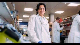
Muzlifah Haniffa presents the Human Cell Atlas, a global effort to map the developing human immune system at single-cell resolution across organs like the fetal liver and bone marrow. The team identified how immune cells emerge and mature over time, and found that developmental programs reappear in adult skin diseases like psoriasis. The atlas accelerated COVID-19 research by revealing which cells the virus infects.
Chapters
Thank you very much. Hello everyone, and welcome to the seminar series. Today I'm going to be talking about the work that we've done to try and understand the developing human immune system within the Human Cell Atlas initiative. So what I'm going to talk about is primarily focused on human development, but also some data on adult, which will be focused on the Skin Cell Atlas all of which is funded by Wellcome. So a bit of background about the Human Cell Atlas. It has an ambitious mission to create a comprehensive reference map of all the cells in the human body, and there are 37 trillion cells that make up the human body. And the idea is that this would be the reference to understand, diagnose, monitor, and find new ways of treating diseases. So anyone can join the Human Cell Atlas. It's a global initiative and it's free to join. And you can see here, we now have representation from 79 countries, more than 1,000 institutions, and more than 2,000 researchers globally who are contributing to this massive effort. And one of the fundamentals of the Human Cell Atlas initiative is essentially to use single-cell genomics technologies that will allow us to really map the cells that form that form the human body. And if you were to imagine the human body or tissue as a fruit salad here, the idea would be that you can then use either the gene expression or protein expression to help identify what are the cells that make up this fruit salad and give you a census. And this really has, you know, been a revolution, primarily from the technologies that have allowed us to measure gene expression or RNA at single-cell level. But more recently, you can also combine the measurement of RNA with surface protein, for example, or the regulatory elements of the DNA itself. And all of this done on single cells, and the technology allows you to scale and analyse many thousands, hundreds of thousands, and also millions of cells at any one point. But what is also coming into the fore is the idea that we now no longer need to sort of break apart the fruit salad or break apart the tissue to get single cells to profile, we can actually probe these single cells within that tissue or in this context within the fruit salad itself with a lot of spatial genomics technologies. We recently outlined how we would use these technologies, how it would come together across many disciplines, including the biological sciences, computational, clinical specialties, and also technology method development to really use these cutting-edge single-cell technologies to understand what are the composition of the human tissue, not just from the cells, but also really how they form the tissue architecture, and at a bigger scale, the organ itself in terms of the anatomical structures. And all of these are going to be critical datasets, which will be integrated alongside the metadata relevant to the tissue that's being analysed. And we outline how we would use these technologies and working together to achieve the Developmental Cell Atlas, which was published a few weeks ago. And this was a combined effort from many researchers across the globe who were part of the Developmental Biological Network. And what can we do with this atlas? So these are the sort of types of information that we'll have in terms of the cellular composition, the interactions, the molecular mechanisms. And you can then use this types of information, such as cellular programmes, to then, you know, better innovate. How can we build organs from stem cells in a more faithful way that recapitulates the in vivo organs? How can we engineer stem cells that are used in therapy so that they can actually, you know, perform better as a form of treatment? And also using this reference information to identify what could be potential therapeutic targets, particularly in childhood diseases. So the Human Cell Atlas initiative really approaches the problem in a completely different way, and I've done this analogy of like how one would build a shard, where you'll have many people and many groups working on one project. And this is slightly different to how science has conventionally been done, where you have one person or one group kind of working on individual projects, and they all come collectively together to form the bigger picture. But to do something Something like the Human Cell Atlas really requires a different type of infrastructure, a research framework, people coming together to kind of like work as a team and using the cutting-edge technologies and the ability to then disseminate the data that has been generated so that it has broad utility and applications for human health and benefit. I'm now going to focus on the specific research relating to the developing human immune system. And as I described, you'll be able to see how, you know, coming together as a group of researchers working collaboratively and as a team science has allowed us to construct the framework of the developing human immune system. This is quite a complex process that occurs across many organs and across developmental time. The first progenitors that are capable of making blood and immune cells arise from what's called the yolk sac, which is an extraembryonic structure. Thereafter, you have the hematopoietic stem cells themselves, which are then made in the aortogonad mesonephros, or AGM, found within the embryo proper. These stem cells then seed and can occupy the liver and the bone marrow, where they begin to undertake haematopoiesis, i.e., the production of the blood and immune cells. The liver is the dominant site of haematopoiesis from about 4 or 5 post-conception weeks all the way to 20 post- conception weeks. And the bone marrow begins at about 11, 12 post-conception weeks, but really becomes the dominant site of haematopoiesis after 20 post-conception weeks, and is the main site of haematopoiesis following birth. You have the progenitors that seed the thymus and undergo T-cell differentiation, and you've also got the B-cells that are made in the bone marrow seeding the spleen, where they then undergo further maturation and differentiation in the form of germinal centre formation and somatic hypermutation, and both of those really occurs after birth. You've also got all of the immune cells seeding the skin, the kidney, and various peripheral organs, such as the gut, the lung, and where they then form the immune network surveillance of those peripheral organs. So, all of this is exquisitely coordinated. Across tissues and time. And these are some of the publications that we have actually put out for the community, you know, relating to the respective individual organs. And all of this research has been made possible also because of the Human Developmental Biology resource that's available or existing in the UK, funded by both Wellcome and MRC.. And the ability to kind of like study many cells at scale across many organs allows us to then reconstruct the immune system, not just as individual cell types or individual lineages, but as a collective, so that we can begin to understand the emergent properties of the immune system, whereby the whole is greater than the sum of its parts, as illustrated by this migratory bird pattern. What I'm going to do is focus specifically on our recent work, which is looking at the development of the blood and immune cells in the bone marrow, and I'm going to contextualise that in relation to how the blood and immune cells develop before the bone marrow, i.e., in the liver, which we described back in 2019, and also how this, this relates to the development of T cells in the human thymus.. And as I said, this is very much the coming together of many disciplines. So here we've got immunology, haematology, developmental biology, and bioinformatics, and work across many, many individuals collaboratively. All of the datasets is available through a web portal that you can browse, and here's the portal site, developmentsalathlast.ncl.ac.uk. And these are some of the things that you can do. You can probe for the gene expression of the cell types that you're interested. So for example, this is the gene that encodes for the protein CD20, which is expressed on B lineage cells. And there, you know, you can also look at the metadata associated with it. Another way of visualising this data or browsing this data is by looking at the gene expression for the specific cell types, which again, you can, you know, type in the gene that you want, and/or a list of genes that you want, and it will show you the expression of those genes in the individual cell types. For the fetal bone marrow, we also have a website, and fundamentally what we've now included is the ability to interrogate genes that are known to cause diseases for better clinical utility of these datasets. So any gene that has been implicated in inborn errors of immunity or childhood leukemia can be browsed in this dataset whereby you click and you can actually see a dropdown menu which allows you to search for diseases, and you can see the different diseases and look at the genes that are implicated and the expression of those genes in the dataset. So, coming back to the science, what we've done is profile more than 100,000 cells of all of these different organs that I've mentioned, and generally looking at donors of 10 or more for these developmental datasets. And the really take-home message from what we've learned so far is that blood and immune cells develop by 20 post-conception weeks. And there is a big distinction between the fetal liver and the fetal bone marrow. So in the fetal liver, you have all of the lineages that you know, the lymphoid, myeloid, and erythroid. But within the myeloid lineage, you don't really have any granulocytes, so there are no neutrophils.. And neutrophils really emerge in the fetal bone marrow, as you can see here, alongside other granulocytes such as eosinophils and basophils. And I mentioned before that the technology that was initially scalable was measuring RNA in individual cells, but we can now combine this. And for this work and also ongoing projects, we use a combination of RNA and protein, which allows you to measure up to 200 surface proteins. And here's the RNA on the left-hand side, indicated by this red panel, and then the blue panel shows you the protein expression that corresponds to the cell states that we annotated based on the RNA expression, showing how you can now begin to resolve the expression profile of individual cell states. And what we've done in the publication is performed a decision tree analysis, which allows an algorithm to basically say out of the 188 or so surface proteins, what will be the best combination to actually identify these cell types? And because these antibodies are commercially available, they're extremely helpful for future downstream isolation of those cells for functional validation. Now that we've analysed cells in terms of blood and immune cells across many of the developmental organs, we can then compare it with what happens in the cord blood and adult bone marrow. And these are datasets that are already available in the Human Cell ATLAS data portal. We don't have paediatric datasets, but it would be incredible to begin to see the evolution of blood and immune cell development across the human lifespan. So, just an example of the development of the myeloid lineage by, you know, comparing developmental datasets with the adult bone marrow, what you can see is, as I mentioned, the emergence of granulocytes in the foetal bone marrow, also the heterogeneity of the dendritic cell subsets in the foetal bone marrow,. And also, the contrast to the adult bone marrow, and only in the adult bone marrow do you see the CD16 monocytes and monocyte DCs that have been described. So, what about the granulocytes in the fetal bone marrow? We can find them by gene expression, but can we also validate their existence using orthogonal approaches? So, you can do that by essentially using some of the predicted markers from the gene expression profile in the discovery dataset, and then using that to isolate those cells to perform morphological analysis, and additional single-cell transcriptomics analysis, which confirmed by gene expression profile the identity, their identity, if you now isolate based on the surface marker expression, but also by the morphology from cytospin preparation and Giemsa staining, the classical appearance of the granulocytes here shown here with the polymorphic nuclei, and also the eosinophils and basophils. So, these are really useful validation tools that allow you to kind of like confirm what you found. But why do we see granulocytes in the fetal bone marrow? Why do we not see it in the fetal liver? And if you look at the progenitors in the fetal bone marrow, particularly the granulocyte/monocyte precursor, and interrogate for the transcription factors that are known to drive neutrophil differentiation in contrast to monocyte differentiation, you will find higher expression of the transcription factors driving neutrophil differentiation in the fetal bone marrow compared to the fetal liver. So, there is this intrinsic imprinting of the progenitors in the fetal bone marrow that allows it to differentiate into granulocytes. And if you then look at the transcriptome profile of those GMPs, you will see that some of those cells are already expressing monocyte signatures, and the cells not expressing monocyte signatures are already expressing some of the neutrophil signatures, supporting this kind of early imprinting of progenitors to differentiate into neutrophils. Another vignette that we learned from the study of the developing fetal bone marrow is B lymphopoiesis, which becomes prominent in this site. And what you can see is, although the percentage of B lineage cells as a proportion of all haematopoietic cells is the same in the fetal bone marrow and adult bone marrow, the composition is very different in that the fetal bone marrow primarily has pre-pro-B, pro-B, pre-B cells, progenitors of the B lineage cells, with a very small representation of these progenitors in the adult bone marrow, which has many more naive B-cells, transitional, and memory B-cells. B-lineage differentiation is accompanied by heavy chain and light chain acquisition, and by sequencing the B-cell receptor VDJ chain, we can actually see how this occurs in the acquisition of heavy chain first, followed by light chain, and that accompanies a phase of the cells undergoing cycling, which you can also see during heavy chain acquisition and further downstream differentiation following positive selection after successful rearrangement of the heavy and light chain. With regards to the T-cell differentiation in the lymphoid, for the lymphoid cells, you can see the thymus and liver here. These are the progenitors in the liver and the progenitors in the thymus. And cells that are transcriptionally very similar and independently annotated as lymphoid progenitors, suggesting that the fetal liver progenitors contribute to the thymus, you know, seed thymus, and then undergo differentiation into T cells. And subsequently, a similar analysis shows the same for the fetal bone marrow. Then you have these differentiations of the various T cell subsets in the thymus, including unconventional T cells and the sort of conventional CD8, CD4, CD4 and regulatory T cells. Then you can ask the question, what about the cells that you find in the fetal bone marrow with respect to the T cells? Are they actually differentiated T cells, or are they more similar to the progenitors in the thymus? And what is very clear here is that in the fetal bone marrow, you have differentiated T cells. So, the lymphoid progenitors from the liver and bone marrow seed the thymus, T cell differentiation takes place, and the T cells can then subsequently seed the fetal bone marrow. And reside there. So there are a few lessons that we learned from these studies as well, particularly related to hematopoietic stem cells and progenitors, and they can be divided into two compartments relating to the stem cells. One is the intrinsic potential of the stem cell itself, and the second is its extrinsic environment, which is provided by the different tissues where hematopoiesis occurs. And a big difference is between the liver and the bone marrow across gestation.. So, to study this, we leveraged the single-cell transcriptomics data and the single, you know, and the side-seq data to look at the molecular profile, but also supplemented that with functional assays whereby you can isolate individual cells, which, you know, you can confirm their identity and whether they are primitive stem cells or more committed progenitors, and look at the ability to differentiate into the blood and immune cells. So, from one single cell, what types of colonies can you get? And then you can measure that by flow cytometry analysis. So, just looking at the progenitor compartment, what you find here is the HSC-MPP, or the most primitive stem cells. Here are progenitors that are committed to the myeloid lineage, the lymphoid lineage, and the erythroid lineage. You can now compare fetal liver with the fetal bone marrow, and what you find is that there are many more cells proportionally in the fetal liver that are committed towards erythroid lineage relative to the fetal bone marrow, where there is a bigger proportion of cells that are committed down the lymphoid and also the myeloid lineage. So, the progenitor compartment itself allows certain, you know, cell types to sort of like differentiate, so there's an intrinsic potential has been, you know, has occurred that allows the cells to differentiate down the different lineages. So, the functional assay, if you took hematopoietic stem cells from the fetal liver at different gestational stage, you'll find that their ability to differentiate down the erythroid lineage declines, their ability to differentiate to B lineage increases, and their ability to differentiate to myeloid lineage increases. So, this is, you know, what happens across gestation within one organ, but you can now compare paired fetal liver and bone marrow. And if you did that, what you find is that the proliferative capacity of the hematopoietic stem cells in the fetal liver is much higher than the fetal bone marrow. And if you then looked at their differentiation capacity, you find that the fetal bone marrow hematopoietic stem cells has a higher propensity to differentiate into myeloid colonies only relative to the fetal liver hematopoietic stem cells. So, there are intrinsic properties that are very different for the hematopoietic stem cells in one hematopoietic organ across gestation and across two hematopoietic organs comparing for the same gestation stage. Now, coming to the extrinsic environment, and this is particularly looking at the fetal bone marrow, this is the first data set looking at the single-cell profiling of of bone marrow stroma in human, and this is a fetal bone marrow, and we were able to identify all of these cell states, which aligned very well with the counterparts of the mouse bone marrow. And in fact, the interspecies comparison was extremely helpful in allowing us to identify the cell states in the human data. What we were very struck by was the two different types of endothelial cells that we identified based on initially gene expression, which was then supported by the differential protein expression. One was the tip endothelial cells and the second sinusoidal endothelial cells. And these are the genes that characterise them. The distinguishing features, the higher expression of CD34 in tip ECs relative to VEGFR2, and for sinusoidal endothelial cells, the higher expression of VEGFR2 relative to CD34. So, this is protein staining of the fetal bone marrow, and you can see here these cells that have high expression of CD34 really found in the metaphysis of the bone marrow compared to the blood vessels which are lined by the sinusoidal endothelial cells, which are mainly formed in the diaphysis, forming the sinusoidal vessels. And the relative distinct geographical distribution of these endothelial cells lining blood vessels resembles what has been described in the mouse bone marrow, of Type L and Type H vessels in the diaphysis and metaphysis, respectively. We can now use the dataset to compare the environment in the fetal bone marrow compared to the liver, specifically for the sinusoidal endothelial cells, which are very important for haematopoiesis, and how some of the adhesion molecules, the extracellular matrix, the angiopoietic factors differ, and how this essentially is also recapitulated at the protein level. And this is beginning to give us information of how how we can manipulate the environment where haematopoiesis occurs so that we can drive differentiation towards specific lineages that is desired for therapy, for example. So, what value is this data set? And I'm going to show you two examples. One directly relates to the fetal bone marrow, and another really relates to disease that you can see in adults. So, with regards to the first, we also profiled fetal bone marrow from with trisomy 21, i.e., an extra copy of chromosome 21 in Down syndrome. And these are what the cells look like, and you can then perform an analysis comparing the relative abundance in Down syndrome compared to non-Down syndrome. And there are these characteristic features where you have overabundance of erythroid cells in Down syndrome compared to megakaryocytes and B lineage, which are contracted in Down syndrome. You can then ask the question, is this because of an intrinsic change to the haematopoietic stem cells themselves, or is it also due to the environment that has changed? So if you took the haematopoietic stem cells from fetal bone marrow with Down syndrome and non-Down syndrome, you will be used and culture them and see what colonies or what cells are made you will see that there is an increased propensity for HSCs from Down syndrome to differentiate into erythroid cells. That's in contrast to the non-Down syndrome. So there is intrinsic bias in hematopoietic stem cells as a result of having an extra copy of chromosome 21. But it isn't just intrinsic, there's also the environment that has been altered. So if you now compare Down syndrome and non-Down syndrome respectively, for the ligands expressed on endothelial cells of the bone marrow, which are known to interact with their receptors on the hematopoietic stem cells in the fetal bone marrow, you can see that some of these canonical interactions involving Notch and Delta are severely altered in Down syndrome, accounting for an environmental effect on the changes to haematopoiesis that we see.. And what is really illuminating is the fact that having an extra copy of chromosome 21 doesn't just mean that genes on chromosome 21 are overexpressed, but it actually affects genes on many other chromosomes and across many lineages. So it isn't a very straightforward case of having an extra copy of a chromosome means high expression of genes on that chromosome. The second example of how this developmental understanding of the immune system is relevant is in the context of adult diseases that we observe in adults. And this was some manuscript that we published earlier in the year relating to the skin cell atlas, and again contributed by many junior researchers and also a collaboration across many investigators. So in this study, we looked at healthy skin from adults and then compared it with developing embryonic skin, and that with two common inflammatory skin diseases, atopic dermatitis and psoriasis. And when we did the comparison between developmental and adult datasets, we were struck by two observations, one of which was related to the vascular endothelium, in that developing skin endothelial cells, as well as several organs such as thymus, liver, kidney, and gut, were transcriptionally aligned and correlate best with one of the three adult skin skin vascular endothelial subsets. And this was VE3, a very, you know, small population compared to VE2 and VE1. With regards to macrophages, it was consistently macrophage subset 2 in adult skin that was transcriptionally enriched with the developmental macrophages, not just in the skin, but, you know, other organs. But what was really surprising was the fact that these two subsets of cells that were most enriched with their developmental counterparts were expanded or proportionally increased in lesional psoriasis here, and lesional atopic dermatitis, compared to healthy adult skin. And this was true for vasculative cells and macrophages, too. And because we had a small data set for the disease, we actually interrogated a bulk RNA sequencing data set from a much bigger cohort of 38 healthy, 21 atopic dermatitis, and 27 psoriasis patients, and validated that observation in our single-cell data within this bulk RNA sequencing dataset supporting the observation that these subsets that have a counterpart during development are expanded in particularly lesional skin. So, what's special about these two subsets? And if you look at the healthy dermis, these are these BE3, which has been identified using the antibodies for PCAM-1 and SNCG that they express, showing they form these dilated capillaries. They're very similar to the high endothelial venules in expressing a lot of adhesion markers and chemokines. And if you then ask the question, what are the gene programs that are conserved between embryonic vascular endothelial cells and adult VE3, we will see that these gene programs relate to leukocyte recruitment and angiogenesis. And that is true for both cell states, which led us to ask whether these two cell types were actually, in fact, interacting. And the one way that you can do this with single-cell RNA sequencing dataset is to probe for interactions that are predicted by interrogating receptor-ligand database, which identified the interactions between CXCL8 on macrophages with ACKR1 on VE3, which we then validated in situ, showing the juxtaposition of these cells next to each other. So what we're proposing is that these interactions that we see are likely mediating leukocyte recruitment and angiogenesis during development, i.e., to seed the tissue with immune cells. But it's also then co-opted during disease whereby this programme recruits immune cells but mediates inflammation. And that these developmental programmes are co-opted or re-emerge in inflammatory skin disease. What we don't know is how the pathways are regulated and whether this is also relevant for other inflammatory skin disorders, or in fact, are more generalised and involved in other immune-mediated inflammatory disorders. So, I hope I can show— the data that I've presented shows the clinical relevance of such a resource in terms of tissue engineering and cell therapy, specifically understanding how stem cells differentiate, the intrinsic and extrinsic factors that drive them to differentiate into the different lineages, how the reference atlas can begin to understand the changes we see in chromosomal alterations, and also better understand childhood disorders such as leukemias, and also how developmental pathways re-emerge and are co-opted in adult pathology. And this can actually identify new therapeutic strategies. The Human Cell Atlas programme more generally has a you know, has been— we've had a great public engagement program alongside it. And what you'll be— you know, there's a virtual exhibition, in fact, onesaleatatatime.org, which you can see here, that actually hosts this exhibition. And I welcome anyone who's interested to actually browse this exhibition online. Very briefly, I'm just going to highlight how the kind of infrastructure, the collaborative team science research framework that we've developed was really extremely helpful in helping us understand COVID-19, particularly relating, using the expertise that we've got in single-cell multiomics. And the first initiative was to basically for all of the community to come together, use the reference data set, and ask a very simple question: where are the— what are the cells that are expressing the SARS-CoV-2 the virus that causes COVID-19, entry receptors found so that we can then understand how infection, you know, begins. The second is how we use these datasets to understand the immune response that occurs in patients with COVID-19, looking at those who are asymptomatic and how that response may be different to those with mild disease and severe disease. And this is a coming together of many organisations and many researchers,. And we published this work to show how the peripheral blood can actually be a good surrogate to understand what may be happening in the peripheral tissues, such as the lung, and also the response more systemically in the bone marrow, and how these effects are mediated in COVID-19 across all of the lineages, so that you can begin to understand the collective or, you know, how the whole immune system responds to this challenge. And again, the data is available both to browse and also to download for other researchers to benefit from the dataset so that we can make more progress in understanding COVID-19 and identify new ways to overcome it. I'm going to stop here and to thank my lab members for the amazing journey over the last few years, and also to thank all my collaborators. The work that I presented is very much a collaboration with Sarah Teichmann particularly, who heads the Cellular Genetics Program at Sanger, many of the Celgene faculty members, and also Celgene IT and research admin and web lab operations, all of my collaborators, and also our funders. And thank you very much for your attention.
More from Joshua







Scientist Stories: Hans Waterham, The value of phenotypic assays in peroxisomal disorders




Scientist Stories: Joel Zlotogora, Genetic carrier screening programs for reproductive purposes