Scientist Stories: Padmanee Sharma, Immune Checkpoint Therapies
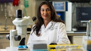
Dr. Padmanee Sharma built a platform at MD Anderson that monitors immune responses across 113 clinical trials in real time, catching how patients respond to immunotherapy while they're still on study. Her team discovered VISTA-positive immune cells that predict who benefits from checkpoint therapy in prostate cancer versus melanoma, validated in mice and now in a clinical trial.
Questions this video answers
Sharma explains that they have restructured clinic visits from 15‑minute to hour‑long appointments to allow thorough consent discussions, including longitudinal biopsies and open‑access data sharing. Patients are generally willing to consent, and the clinic absorbs the altered cost structure.
Sharma notes that healthy donor bone marrow and kidney samples are obtainable via transplant donor programs, but organs like brain or pancreas are difficult. Autopsy programs can provide such tissues, though consent and logistics are challenging due to time constraints after death.
Chapters
Good morning, and I am going to have a very different perspective. I feel when I come to these meetings, I learn a whole other language, and I love learning. It's actually incredible. I feel 20 years of immunology has not prepared me for computational biology, but I'm hoping to get there. So— Thanks.
So I'm going to give the perspective where I am in the clinic, because I see patients in clinic, and so we're dealing from that standpoint.
And I think that the Human Cell Atlas is absolutely necessary, because it's difficult to put all of these pieces together when you're standing in front of the patient, and you need somewhere to start referring back to try and make sense of things as you're taking care of the patients. At least this is my perspective. And maybe we have a little while to get there, but this is certainly the beginning, and we need to make sure that we all invest in this. So, some of the things we considered when we were trying to take this from an immunology perspective again, because I don't know how many of you know, but, you know, for 20 years of immunology, we're sort of laughed at, and then the last few years now we're being taken a little bit more seriously. So, but in order for us to be taken seriously, we actually had to think about things for a long time. And how we were going to go about looking at stuff from the human standpoint. So there are issues to consider, and these were the things we were thinking about: how to actually access all of these cells and samples from living donors, and whether or not we would process them right away, or whether or not they could sit for a while at room temperature or on ice and then be processed, whether or not we could even freeze them, thaw them, and then look at everything in bulk, and whether the freeze-thaw process would have an effect on T-cell subsets, and then whether or not we could use autopsy samples. And I can tell you we did that for a year, and that didn't work out well for the immune cell subsets. So, we actually don't do the autopsies anymore, just because of the issues, again, with ischemia, autolysis, and all of these other issues that affect how the immune response can be evaluated. So, I think these are also similar questions that you will have to deal with as you're looking into building the Human Cell Atlas. I just want to give you some examples, and these are data we've never published because they were sort of just data we used for generating our own SOPs within the immunotherapy platform. So if you take a set of genes and look at the time zero where we actually take the T cells out from the patient and immediately process them from the patient— I'm hoping this is a pointer. But anyway, at time zero when we do this, this is now our standard of where we want to I don't see it pointing.
Oh, okay. So if we look at time 0 here, this is actually right out of the patient and then looking at the—
processing the T cells immediately doing the gene expression analysis, or you can wait 1 hour at room temperature before processing the cells, 1 hour on ice, and you get the picture.
But basically you can see the 0 hour is now where we want to make sure this is what we're comparing everything to, and the longer time it takes you, to actually get this done, you start to see now that you're losing what's immediately taken from the patient.
And this was important to us because, again, we're transporting the samples from clinic.
So I need to know when the blood is drawn in clinic, how much time it takes for the nurse to page the runner, and how much time the runner has to get to clinic and get back to the lab for processing all of this.
And it gives us very different information if we're off for when that actual sample is taken. Similarly, when we do a CyTOF analysis from these same samples, Again, you can see just looking at the red, this is just CD3. Of course, there's 36 markers in the CyTOF panel, but you can actually see over time if I actually process immediately and run the CyTOF analysis versus letting things sit for a while, all of these subsets start to change. And somewhere around 1 hour is when it's the best for immediate.
We looked at all of the clusters in the CyTOF analysis, and you can see I don't need you to go through each of them, but clusters change over time again.
If we're not careful. And so we redefine subsets of cells based on the amount of time that they're sitting there because they lose activation markers and they gain other markers.
And so this is very important if you really want to understand what it is that you're looking at. Are you looking at what's going on in the patient, or are you looking at a matter of what the sample does after it sits for a period of time?
So this is how we created the platform and tried to set up the SOPs based on these kinds of data that we've looked at. And so this is just the immunotherapy platform, which you're trying to build is a lot larger in terms of the Human Cell Atlas. But really, we're here to provide input on the design of the clinical trials based on data that we can obtain. We conduct and interpret all of this immune monitoring data basically in real time, because as I showed you, as we tried to freeze and thaw and do bulk analysis, we don't end up with the same type of data when we do it in real time.
So it's a matter of choosing the appropriate assays so that we can, after every 5 patients, make a decision as to what can be done.
We generate the hypotheses. We actually test them all in the models to make sure we're validating what we think is correct from the data we've generated, and then we integrate the data again in real time to develop the next clinical trial even as the first clinical trial is ongoing. And so when we've set up the immunotherapy platform, it's sort of this vision I had in my head that we would have some scientists and some people around. And now this is the structure of it. As you can see, it's getting bigger and bigger. It's over 100 people at this point, and a lot of it has to do with actually informed consent and sample collection. The scientist part we actually had in place.
It was the whole tracking of samples and de-identification and making sure all the informed consent was done properly so that the regulatory piece is all up here, and this is actually the most of the work. The scientific part is actually down here.
So, so far, there's a number from May 2017, and the platform's been around for 3 years now. So we have about 113 clinical trials across 12 departments with 52 investigator-initiated 49 industry-led studies. We've now consented 2,757 patients, all with appropriate informed consent and clinical data. And it's under an umbrella protocol because this allows us then to do anything we want to the samples.
It's not defined by any of these 113 trials.
It's defined by the umbrella protocol. And so we can even— the umbrella protocol even allows for potential future assays, so we don't have to go back and reconsent patients for any new assays we may develop. As a result of this, we've now analyzed about 27,000 blood samples and about 2,000 fresh tissue samples, and then again about 34,000 archival samples, because from these patients we've went back in time to obtain all of the archival samples available from outside hospitals and at MD Anderson. You're not meant to read this slide. This is just to give you an idea of the amount of work that goes into just collecting the samples.
It's just so that we understand, again, how everything has to go from the time the patient is screened, consented, the blood order is placed in the computer, and then the blood— the patient actually goes to the lab to get the blood, the runner is paged, and then comes into the lab to immediately have the sample processed.
It's an entire workflow, and it took a lot. We're still iterating some parts of it because as new departments join within MD Anderson, then they have to learn the workflow for their particular clinic because everybody's clinic has a different way of working. Same thing—
oh, and I apologize, the slides aren't—
but this is for surgeries and biopsies. We had to restructure all of the ORs, the operating rooms, and all of the interventional radiology suites to do the biopsies are functioning.
Because again, from the OR, I don't want the surgeon leaving the sample on the OR table as he's sewing up the patient, and then the sample's just sitting there until he's done sewing up the patient, and then we get the sample.
Because again, it changes all of our data. Same thing with interventional radiology, the type of needle that is used for the biopsy, whether it's a fine needle versus core needle, whether it's 14-gauge core needle or 18-gauge core needle, makes a big difference in how we can get data out.
And so there are all the SOPs that we've set up in place for making sure that we're getting the samples in sort of the same exact ways every time because now we have a system that works. But when we have a new assay, for example, when we started CyTOF, we needed a different set of SOPs because the assays we'd set at the protocols we'd set up in place before all worked well for flow cytometry or other things, but didn't work so well because we're using different assay now.
So these things are, again, an iterative process. And for us, it's really about doing the clinical trials where we can actually perturb the system. And so we're having a cancer patient in the clinic. We're giving the patient a treatment. So this allows for perturbation of the immune system.
And then we're asking what happens as we perturb the immune system. And so here's a clinical trial example. These red arrows are when we're getting the biopsies and the blood, and the patient is going through multiple different treatments here.
It's prostate cancer patients going through treatment with hormonal therapy as well as immunotherapy, and we're trying to then look at each component and then understand the combination effect. So this is just in one patient.
This is just a CyTOF analysis, again, of that longitudinal biopsies that was done in this one patient over time in the protocol.
And the blue is in the pretreatment samples, the red is post, after hormonal therapy, and the green is after immunotherapy. And this is the immune response we're measuring in the biopsy sample, the three overlaid here.
And just to quickly show you again, this is the merge, but if you look at the pretreatment, you can see that these subsets did not exist. And then all of a sudden they started to exist now in the treatment of hormonal therapy., and here they persist after anti-CTLA-4 immunotherapy.
And this patient is a non-responder.
And so those subsets that we were interested in to understand what they were all turned out to be this novel marker VISTA that we could identify. In the CyTOF, they were all VISTA-positive subsets that we could see. And if we were to look at a responder patient, not to go through all of the data, this patient actually responded very well to therapy. You can now see that this VISTA subset actually disappears over time. And so this patient actually ended up having good response.
And so it was a matter of understanding the VISTA, and it's a novel pathway, and we were trying to look at what it did functionally.
And here you can see VISTA expression. We could look at T cells and macrophages and colocalization of how this protein is expressed. And what we could see is that in the pretreatment samples, which are in the black, triangle versus the post-treatment, which is in the red dots, you can now start to see this increased expression on these macrophages, these CD68 cells. There's some on the T cells as well.
But again, in prostate cancer, we're very interested in this because prostate cancer does not respond as well to immunotherapy as melanoma, for example.
So we actually did a comparison between the prostate cancer samples and the melanoma samples to have a real understanding of whether or not this is something that's specific to just prostate cancer. And again, taking a group of melanoma patients that underwent same treatment with anti-CTLA-4, we could see now that this was definitely something that was different just in the prostate cancer patients compared to melanoma.
And similarly, not only are we running the one assay in terms of the co-localization experiments with multiplex IF, or are we running just the CyTOF, we're also doing gene expression analysis, we're also doing validation in the model systems. And in the gene expression studies, you can see that the M1 gene signature, which is a signature for macrophages that actually help with a tumor rejection, it's much higher in the patients treated with anti-CTLA-4, the melanoma patients, compared to the same treatment that was given to prostate cancer patients. You see more of the M2, which is again the gene signature that goes along with tumor progression, indicating this difference in the tumor microenvironment with this immune cell subset of VISTA-positive cells. And I'm not going to go through all of the data, but again, we were able to show that in the validation setting, you know, in the VISTA knockout mice, you actually have improved antitumor responses. And if you combine an anti-VISTA therapy with an anti-CTLA-4, anti-PD-1, you get much better rejection of these kinds of tumors in a mouse model. And so we designed the clinical trial, actually, that's now ongoing in patients based on this data. So, for us, working with the cancer patients do provide insights that we can gain about what can be done in this particular setting, in the setting of cancer patients and in the setting of an immunotherapy treatment, because we're understanding these immune subsets. But I think for the, the Cell Atlas that you were trying to put together in a much larger way, we need to understand what does the normal immune system look like without cancer, because I'm dealing with patients who've had cancer for many years, so I don't really even understand what these subsets are truly supposed to be like, except this is what they exist now in the cancer setting. So can we identify longitudinal immune responses that become dysfunctional as a result of cancer? Because if we can understand what that is, then you can target that to try and understand now if you can reinvigorate appropriate immune responses in the healthy setting. And then are there immune subsets that existed prior to cancer, and were these eliminated as a result of the cancer? That gives the tumor an advantage.
Because if you understand what those eliminated subsets are, we can figure out ways then to reconstitute them, even with adoptive cell therapy or other immunotherapy strategies.
And then can resident immune subsets in different organs be characterized and compared for similarities and differences? And this would enable hypotheses for why certain tumors preferentially grow in certain metastatic sites. So if you think about it, prostate cancer, for example, always goes to bone. That's its favorite site for metastases, and no one understands why it would do that compared to a melanoma. Which rarely goes to bone. So these are things that I think are still immunologically driven, but we don't have enough understanding of what that immune response looks like even before the cancer develops. So the Human Cell Atlas allows us to start to develop that data and then put it into the context of what we're doing now in the immunotherapy platform. There are many, many other questions we can go into, and I'm sure we'll have discussions throughout this meeting about it. I just wanted to make this brief and give us time to have questions if we have any. And just finish on that point. So thank you very much.
A question? Aviv Regev from MIT and the Broad. I have a question about— patient consent in the experiences you've had, in the challenges around it, and especially around open access to genomic data from patients, which is something that's very important for the Human Cell Atlas. And I think the genomics community has some experience in it, but less so in the patient setting, possibly.
Yeah. So I think it's— I mean, we've been doing it now. You can have open access to the data. And I think a lot of us now are more comfortable with having that conversation with patients. And they're— and all of the patients are willing to consent. What we've done is really restructure the clinics. Instead of 15-minute visits with a patient, we now have an hour-long visit because it's allowed for consent for all these kinds of things, including the longitudinal biopsies. Because in order for a patient to say, yes, you can poke a needle in me multiple times, they need to understand, and it's a longer conversation. And I think most clinics were always set up to have short interactions because then you could bill for more patients. And now with longer interactions, we can actually get more research work done. And the clinic has to absorb a slightly different cost structure. But these are things that I think most people are very much aware of and willing to make those changes, and so I don't think it'll be an issue, especially not from the patient standpoint. They want us to learn.
One more quick question up there. Yes. Stefan Ivanovic, S2 Genomics. I'm trying to piece together a couple of things. One is we talk about how we want things from healthy people, and I see how that works with blood. I'm having a harder time to see how that works with brain or pancreas or things like that. So could you describe how you would see that working for the Human Cell Atlas?
Yeah. Again, you know, so we also have right now healthy donor bone marrow, for example. So if you can imagine, as leukemia patients are getting donors who may give them adoptive cell therapies and for leukemic patient treatments, we actually get the healthy donors to give us samples as well, so, and they do it. And then we also have healthy donors who are being evaluated for other issues, for other, you know, transplant issues that we can get certain tissues from, kidneys for example, and those kinds of things, but not in brain and those kinds of settings. So I agree with you, that's a little difficult, and I do think autopsy programs are good for that sort of thing, to start off with. But again, in our experience when we set up the autopsy program, it's about 6 to 12 hours from the time of the patient passing before you can actually get the family to sign the consent. Because the patient passes, you need to make the phone call, the family member needs to get every other family member in the room to all say their goodbyes, and then you get them to sign the paperwork to say they'll allow for an autopsy. So it's a long process that the body is sitting there and it takes It's a long time before you get any samples out of it. So I do think we're facing those issues. I don't think that means we shouldn't move forward, because there are things you can move forward with. Not everything yet.
More from Joshua







Scientist Stories: Hans Waterham, The value of phenotypic assays in peroxisomal disorders




Scientist Stories: Joel Zlotogora, Genetic carrier screening programs for reproductive purposes