Scientist Stories: Pamela Bjorkman, Neutralizing Antibodies Against Coronaviruses
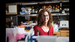
In this recorded lab‑meeting talk, Dr. Pamela Bjorkman (Caltech) reviews structural studies of the SARS‑CoV‑2 spike protein and the neutralizing antibodies that target its receptor‑binding domain (RBD). She explains the up/down conformational dynamics of the RBD, the classification of antibodies into four structural classes (I–IV), and the distinct neutralization mechanisms—class I and II block ACE2 directly, class III bind without blocking, and class IV target conserved, often cryptic epitopes that can still impede ACE2. Cryo‑EM structures reveal unusual features such as antibodies with long CDRH3 loops that are disordered when bound, and antibodies that lock the RBD in the down state (e.g., C144). The talk then shifts to vaccine design, describing a SpyTag/SpyCatcher nanoparticle platform that can display multiple RBDs (homotypic or mosaic) on a 60‑subunit VLP. Mosaic nanoparticles presenting eight different sarbecovirus RBDs elicit neutralizing antibodies against both matched and mismatched strains, including SARS‑CoV‑1, and retain potency against current variants of concern. Advantages of the platform include multimeric antigen display, lyophilization for cold‑chain‑free distribution, and flexibility to incorporate adjuvants, microneedle delivery, or oral formulations. The presentation concludes with ongoing non‑human‑primate studies, collaborations to advance the mosaic vaccine to clinical trials, and future structure‑guided immunogen design aimed at focusing responses on conserved class IV epitopes such as those recognized by S309.
Chapters
Okay, it's 10 o'clock. We'll get started. Hello and thank you to everyone around the world for joining us for this week's lab meeting. My name is Kristin Aboud. I am the science editor at the Human Vaccines Project, and I will be your moderator today. The highly transmissible Delta variant of SARS-CoV-2 continues to be of top concern for public health experts around the world. The variant, which was first identified in India, is now circulating, circulating in at least 85 countries and is responsible for an increasingly significant portion of new virus cases. In countries where COVID has largely been kept under control, including Australia, cases of the Delta variant are rising sharply. In Britain and even in Israel, which has one of the highest COVID vaccination rates in the world, there has been a rise in cases due to the Delta variant, even among those who are fully vaccinated. The rapid spread of this variant is once again making masks a focal point of the COVID response. The World Health Organization is now recommending that even those who are fully vaccinated continue wearing masks indoors and in crowded spaces, while the U.S. Centers for Disease Control and Prevention has yet to change their guidance after loosening their mask recommendations in May for fully vaccinated individuals. Today, we are going to hear about efforts to identify antibodies that will work not just against all the variants of SARS-CoV-2, but more broadly against coronaviruses in general. But before we begin today's presentation, just one note. The information presented today includes some pre-published data that is currently under peer review. At the request of our speaker, we ask that you not reproduce or disseminate the data presented. This session will be recorded and made available on our website and our social media channels for you to review. With that, I am happy to introduce today's speaker, Dr. Pamela Bjorkman from the California Institute of Technology. Dr. Bjorkman is the David Baltimore Professor of Biology and Biological Engineering and a Merck Institute Professor at Caltech. She is a member of multiple prestigious science academies, including the US National Academy of Sciences, the American Academy of Arts and Sciences, the American Philosophical Society, and is an elected fellow of AAAS. She has received numerous awards for her research contributions, including the William B. Coley Award for distinguished research in immunology, the James R. Klineberg Science Award, the Max Planck Research Award, and NIH Director's Pioneer Award, among others, and she was a L'Oréal UNESCO Women in Science Laureate. Her lab studies the structural basis of the host immune response to HIV and other viruses, including SARS-CoV-2. During the presentation, please send me your questions using the Q&A function in Zoom. I will ask our speaker a broad selection of your questions after the presentation, and we will have about 25 minutes for discussion. It is my pleasure to now introduce Dr. Pamela Bjorkman. And Dr. Bjorkman, you can go ahead and share your screen.
Thank you. Okay, well, thank you very much for the introduction, and thank you for inviting me here. I've been enjoying some of these presentations in the past year or so. So let me just get this into Presenter view. I thought what I'd do before I talk about trying to— our vaccine efforts is to talk about our structural biology studies that kind of prompted all of this. So the title of the talk is Neutralizing Antibodies Against Coronaviruses. So I'll just start right in with some obvious things, which is that the purpose of viruses, including coronaviruses, is to get into a host cell, make copies of itself and then release progeny to infect other cells and other hosts. And so the, the point is I'll be concentrating on the spike, which is a trimeric protein on the surface of the coronavirus, and then it binds to the ACE2 receptor, which lines cells in your lungs, for example, and other organs. And so if you have neutralizing antibodies, they are going to be against the spike. And an antibody would look something like that on the left. It's a Y-shaped molecule with two arms called Fabs for fragment antigen binding. And so if it binds to some place on the spike that prevents it from interacting with the ACE2 receptor or somehow prevents entry into the host cell, then none of the subsequent steps happen. So in order to understand how neutralizing antibodies work against the spike, I wanted to remind you of something that was discovered early on for other coronaviruses, and that is that the interaction between the spike trimer and the ACE2 receptor is with the so-called RBD, which means receptor binding domain. So since this is a trimer There are 3 RBDs on each trimer, and these RBDs have the interesting property of being in either an up or a down position. And the RBD has to be up in order to bind to ACE2. So this movie illustrates more what I mean about this. This is a surface representation of the spike trimer, and now what we're going to do is show you the ribbon diagram of the trimer. And point out the RBDs. Now you're seeing one of the RBDs go into an up position, and now it's going to go back down in this computational morph. Now 3 RBDs are up, and now you'll see the— they go— they can fluctuate into up and down positions. And now you'll see that same transition from the top this time. So this is if all 3 RBDs are up. So this spike trimer is conformationally flexible with these two possible, well, there's more than two possible positions, but the RBDs have to be in some sort of up position in order to bind to ACE2. So I wanna just go back in time now. At the beginning of the pandemic, we began studies with our collaborator Michelle Nusenswag who'd been isolating antibodies from convalescent COVID-19 donors. And that's published in their first paper here from Davide. Rohbiani last year. And so what we did, and we meaning Christopher Barnes, who's pictured there at the lower left, we compared recognition of these, of the antibodies in the polyclonal mixture from these convalescent donors to binding to different coronaviruses. And then we looked at if we could find recognition sites that were predominant in the different plasmas. And then we used higher resolution EM electron microscopy techniques to look at single antibodies bound to spike trimers. So the questions we had in mind was, what are the structural correlates of antibody-mediated neutralization? And so I'm going to go— I'm going to just not talk about mapping different epitopes in polyclonal antibodies. That's in the paper that's referenced there at the lower left. But what I'm going to talk about instead are structural correlates of neutralization for the monoclonal antibodies that can be isolated from from convalescent donors. So the approach here is single particle cryo-EM to get structures of the FAB component of the antibody bound to the S trimer. And so I'll show you some examples here that Christopher led the structural biology effort. And this is the first cryo-EM structure he solved. The spike trimer in these figures is going to be gray so that the S1 subunit is in light gray and the S2 in dark gray, and then the RBDs are red. And what you're seeing now in green are the variable domains of the antibody. So in single particle cryo-EM, you can resolve different conformations of the same structure. And so in this particular case, there were two states, one with two up RBDs and the other with three up RBDs. And we saw antibodies bound only when the RBDs were in this up position. Now, Christopher's gone on with the help of others in my lab to solve 30 or 35 more such structures. And I'm showing you just ideas about, or I'm showing you examples of what these look like. And I want to point out that there are many other structures from a lot of labs around the world. And so there are a lot of, there's a lot of compilation now of how antibodies recognize the receptor binding domain, which is the part that appears to be targeted by neutralizing antibodies raised both by vaccination and by natural infection. So here's just 4 examples of monoclonal antibodies isolated from COVID-19 donors. So you can see sometimes there are 3 down RBDs for the C144 case. You can have 2 down, 1 up RBD, or 1 down, 2 up RBDs, or 2 down and 1 up RBD. And so the point is there's a lot of variability here and the antibodies follow different rules depending on where they're binding to the RBD. So I want to tell you a little bit more about C144 because that has an interesting mechanism of neutralization. So C144 only binds to down RBDs. That is, we only saw in the cryo-EM structure, there was only one state, and that had all down RBDs, and all three RBDs had an antibody bound. But the antibody was not bound to a single RBD. Instead, it bridged between RBDs. So if you look there in the center, you see, for example, on RBD-B, the antibody spans between RBD-B and RBD-C. And then since this is a symmetric trimer, you see that interaction reproduced 2 more times. So the result is 3 Fabs bound that lock the RBDs in a down position.. And since they're locked down, they cannot go into an up position, which is what's required for ACE2 binding. So the mechanism of neutralization here is interesting. Not only does this antibody actually physically block the ACE2 binding site, it locks the RBDs down so that they can never go into the up position that's required. And from a structural point of view, it's very interesting. The antibody does this with its complementarity determining region 3, CDRH3 that is, which has two hydrophobic hydrophobic residues in a row. There's phenylalanine 100D and tryptophan 100E, and these actually are exposed in solution, and even though they're hydrophobic, and then they bury themselves in a hydrophobic pocket on the adjacent RBD now, and that's part of locking these down into the down position. So from a bunch of these structures, Christopher classified RBD-binding antibodies into 4 general classes, and I have to say that we're simplifying things a lot here. What we did in this paper from the fall of 2020 is to list some of the antibodies that our lab had solved and some that were available from other labs. There are many more available now. But still, you can use this general scheme of classification with keeping in mind that it's a bit oversimplified here. But in general, there's Class I, which binds to only up RBDs, so it cannot access its site when it is down, when the RBD is down. And these block the ACE2 binding site directly. There are class 2 RBDs. These access— sorry, class 2 antibodies. These access the RBD when it's either up or down. C144 is an example of that type of antibody. There's class 3. Oh, and those block the ACE2 binding site. Class 3 do not block the ACE2 binding site. And they, they can bind to either up or down RBDs. And then Class IV antibodies are on sort of this lower part of the RBD. They only access the spike trimer when RBDs are in the up position. And in general, at the time we published these papers, these, these were not strongly neutralizing. They included CR3022, which was actually isolated from a SARS infective patient and cross-reacted with SARS-CoV-2 but was not very potent. And the reason is, as I'll get back to, this class IV epitope is generally not very accessible when the— on the spike trimer. It binds just fine to an isolated RBD, but on the spike trimer, that epitope is not as accessible. But I'll come back to that in considering how to make a potential pan-Sarbecovirus vaccine. So in this paper, we had a table, and I don't expect you to read any of this except that we put a lot of summary information into this. So if you want to look at it later, what we did was list some examples of the Class I through Class IV antibodies and their properties. And I wanted to point out right now something about these Class I antibodies. They block ACE2. They only access their epitopes when the RBD is up. And they are encoded with a particular V gene segment in the heavy chain locus. It's VH3-53 or its close relative VH3-66. And they have to have a short CDRH3. And let me show you why. For example, here what you're seeing is the interaction of, on the left, a particular class I antibody called C105 binding to the RBD part of the spike trimer. And then in the second from left, you're seeing the complementarity-determining regions from the antibody mapped out onto the RBD surface. And then the second from the right shows the epitope of that antibody mapped in colors of dark green and light green onto the RBD surface. And you'll see that that epitope strongly overlaps with the ACE2 epitope. So that's why it's neutralizing. But I just want to point out something that's unusual among antibody-antigen interactions, which is that the CDRH3 is not contributing very much to the epitope, and it's also very short because it sterically doesn't have a lot of space there. And so the epitope instead is dominated by interactions of the complementarity determining regions 1 and 2 from the heavy chain and 1 from the light chain. And, and, uh, this is— this— these VH3-5-3 class of antibodies have also been pointed out and studied by Ian Wilson's lab at Scripps. So there's some of their papers about these.
Okay.
So if you classify these antibodies into these different classifications, of course, you know, we thought we understood that BH3-53 was in these Class I, but C144 that I already showed you actually has a long CDR H3, and it binds to a distinct epitope from the Class I antibodies. And so how does it accommodate that long CDR H3? It doesn't matter because it's binding to a different place, as you can see there on the RBD. But let me say that once we establish this rule for Class I having a short CDR H3, We got a surprise in some studies with Johannes Scheid at the Broad Institute. He'd also been isolating monoclonal antibodies from COVID-19 patients. So one of Johannes's antibodies was something called BG1-22. It's a VH3-53 member, or VH3-63 member in this case. 66, I mean, sorry. And it has actually a long CDRH3. So we assumed that it would bind to a different part of the RBD like C144. So here's the cryo-EM structure that Christopher did, and to our surprise, it bound just like a Class I antibody. That would be— it bound in the same place as C102, and yet it has a long CDRH3. It didn't bind like C144 with the long CDRH3, which binds to the other side of the RBD. So why is this? Well, the reason is actually that it doesn't even use a great deal of its CDRH3. 6 residues of this are disordered, and I'm showing you the beginning and the end of the CDRH3, and the part that's sticking out is just not visible because it's disordered and not contacting the epitope. And that's these residues right here in the long CDRH3 are disordered. So that's why it can bind like Class I antibody but have a long CDRH3. And I find this just very interesting from a structural point of view because this is not the way antibodies generally interact with their antigens. They utilize their CDR3s, especially their CDRH3, to make the majority of the contacts. This is where the variability is concentrated. And the CDRH3 is generally extremely important. If you look through the protein database, you'll find structures of antibodies when they're free, that is, they're not bound to their antigen, they often have disordered CDRH3s, but you do not find disordered CDRH3s bound to an antigen in the PDB, with the exception of a few designed antibodies that are not, that were not isolated from an infected animal or person. So we find this— there's a lot of new surprises with studying these, these new structures structurally. Okay, so one of the important points to ask, especially now, is what happens— what about the antibodies that are raised by vaccination? And so Vinci Wong in Michelle Nusenzweig's lab went through and isolated monoclonal antibodies from individuals who'd been vaccinated with either the Pfizer or the Moderna vaccine And she got those sequences. We made the antibodies and Christopher did more structures. And suffice it to say that they— these are RBD-binding antibodies again. They're strongly neutralizing in vitro and they bind to the same sorts of epitopes. But there's a predominance of these Class I and Class II antibodies. Those are the ones that bind to that block ACE2 binding. So these are strongly neutralizing antibodies, and you can see that we've mapped out some of the positions here that vary amongst the circulating variants of concern and whether or not the antibodies would be in contact with those. Okay, so I want to switch now to talk about— to use that kind of as a background to talk about then how we think— one of the ways we think it might be possible to get to the point of having a pan-Sarbecovirus vaccine. So this is prompted, of course, by the fact that this is the third outbreak or pandemic of— from emerging from animal sources of a coronavirus in the past 20 years. And so, as you're all aware, first there was SARS-CoV-1 in the early 2000s. And then MERS-CoV emerged in 2012, and then SARS-CoV-2, of course. And there are 4 other coronaviruses that are known to infect humans, which emerged from zoonotic sources, and these cause common colds. Okay, so I think it's clear to everyone that it's unlikely this will be the last emergent coronavirus, and just hopefully we won't have another epidemic or pandemic due to an emerging coronavirus, but I think we should be prepared. So what we've been doing in my lab to try to make vaccines against HIV and influenza and Zika virus is making recombinant nanoparticles to which we attach antigens from whatever virus and then try to observe their immune responses. So I want to introduce the system we're using, which was developed by Mark Hoher's lab at Oxford. So starting with the general principle that antigens are more immunogenic when they're presented as multimers, what Hoher's lab did was to make a nanoparticle to which they genetically fused a bacterial protein called SpyCatcher. And so That's illustrated there as the SpyCatcher virus-like particle or VLP platform, and so they've used different types of nanoparticles. One of them has 180 SpyCatchers attached. It's from an icosahedral phage, and then another one, the one we're using for these studies, is a designed nanoparticle that has 60 SpyCatchers attached, and so the interesting thing about SpyCatcher is it forms a covalent bond with any protein to which you have attached a SpyTag, and the SpyTag is 13 residues long, so this is quite simple to just make a series of SpyTagged proteins and then attach them covalently now to the nanoparticle. And this technique, which they have called a plug-and-display technique, it can be very easily adapted to make what we'll call mosaic nanoparticles, and those would be particles that present more than one immunogen on their surface, and so any— if you spy tag two different proteins and mix them in solution, you'll get a random assortment of two different antigens, and you could do three, or you could do— we've actually done up to nine in the lab, but what I'm going to show you are efforts to use 8 at a time. And so these, the, these multicolored things represent what we call mosaic nanoparticles. So Alex Cohen, who was then a graduate student in the lab who's just recently earned his PhD, he did some work that was published earlier in 2021 to use a mosaic nanoparticle approach to make a potential pan-Sarbecovirus vaccine. So What I'm talking about now are SARS-like beta coronaviruses, and I'll show you later which viruses we chose to have RBDs on this particle. So the— this is sort of a summary of what we've done so far, which has been done in immunized mice. We're starting some non-human primate trials using Bioqual, a contract research organization, but those are not finished yet, but I'll just summarize what we've done that's published. So I'll go through this quite fast since it is published. Okay, so he selected RBDs from SARS-like coronaviruses that there's reason to think might have spillover potential. He made mosaic nanoparticles, and then he looked at the neutralizing and binding responses in injected mice And he showed that antibodies can bind and neutralize to many different strains. And I'll explain the potential that this has for being a potentially protective vaccine against a newly emergent coronavirus. So first, I want to explain the idea behind all of this. And this is something we've been thinking about for a long time because we're very interested in avidity effects, that is the ability of a bivalent antibody. So remember, antibodies have two FAB arms, so they can bind to antigens that are tethered to a surface. They can bind with increased strength of binding if they can utilize both of their arms. So here's the idea, and this is theoretical at this point, but we're in the process of trying to look at these in, in a look at this idea in systematic ways. Okay, so let's say you make a mosaic nanoparticle and it's homotypic, that is, it presents only one type of antigen, and in this case the antigen would be an RBD. So now this, this nanoparticle eventually makes its way to a germinal center in a lymph node and it stimulates B cells with B cell receptors that bind tightly to the antigen on this nanoparticle. Now let's say the antigen has a green epitope, which is a conserved epitope, but it's hard to access, let's say for steric reasons. So most of the time what you'll get are red B-cell receptors that bind to this red triangle here, which represents a strain-specific distracting epitope. So you'll get cross-linking between adjacent RBDs through binding to this strain-specific epitope, which is out there and exposed and very easy for the B-cell receptors to interact with. But what if instead what you injected was a mosaic nanoparticle and it has a series of RBDs, each of which has their own strain-specific epitope? And so there'll be more than one red RBD on this particle, but they won't— they're very unlikely to be next to each other, so you won't get avidity effects helping these red B-cell receptors bind to this mosaic nanoparticle. And instead, what we're hoping for is that you would stimulate B cells that have these green receptors that can cross-link between the adjacent RBDs by binding to the conserved region, which is this green circle here. So the idea is you'd be more likely to make antibodies to the conserved portion of the RBD, and these would be cross-reactive then and potentially protective against conserved parts of the RBD of various coronaviruses. Okay, so here's what Alex started with, is that he went to the literature and looked for sarbecoviruses that have the potential to spill over into humans. And mostly he used this paper from Vincent Munster's lab where they were looking at various Sarbecoviruses that had sequenced S proteins. And so what he did was pick virus— he picked RBDs from coronavirus strains, Sarbecovirus strains from different clades. And then he made a homotypic nanoparticle, which has only SARS-CoV-2 RBD on it. That's the red one there at the left. And then he made two forms of mosaic 4-RBD nanoparticles, which each had four different RBDs. So the first one is 4A, and it had those first 4 RBDs listed there, which come— one of them is SARS-CoV-2, the others come from bats isolated in various places. And then he made 4B, which doesn't have SARS-CoV-2, but which has 4 other RBDs from 4 other sarbecoviruses. And then he deliberately left— oh, and then he made a mosaic 8, which is the combination of 4A and 4B.. And then he deliberately left off 4 different RBDs from— one of them is SARS-CoV-1, because we want to now look at the react— the binding and neutralization responses to the strains of coronavirus that do not have an RBD represented on the particle that was injected. And so we call this a mismatch. So, so you can look at the neutralization responses to matched strains, which are anything that was on the particle, or mismatched strains, which are things that were not on the particle. And he then made sure that he included RBDs from clades 1, 2, and 3 that he left out to look at the neutralization responses. And so I've been talking about conserved regions and the regions that vary, and here's where we were taking advantage of the structural biology knowledge that we have from our lab and others. So, if you look at sequence conservation amongst Sarbecovirus RBDs, what you see is that this is the surface of the RBD, and what we've plotted onto this is the parts of the RBD that vary extensively between Sarbecovirus RBDs, and that's the part in orange, and this is a gradient. So as you get more to the purple side, you have less variability. And what you can see is that at the top, we've also outlined the epitopes of the various antibodies in yellow. And so if you look at the top of those RBDs, they're shown from 180 degrees apart, you can see that the Class I and Class— the epitopes for Class I And class II binding anti-RBD antibodies are there at the top and they're in the orange region. Most of their epitope is in that orange region. So these are the parts that vary between different sarbecovirus RBDs. And they're also where the mutations or the substitutions, excuse me, accumulate in the variants of concern. So this is the part of the RBD that just keeps varying to adapt to different circumstances. And you make very strongly neutralizing antibodies, especially by vaccination, to these Class I and Class II epitopes. Many of them do cross-react to the variants of concern, but some do not. But there's also these Class III and Class IV epitopes, which are more at the bottom of the RBD.. And you'll notice that these are more in the purple area, especially the Class IV epitopes. Now, the Class IV epitope that we— when we started off these structural studies, the typical Class IV epitope was this one right here, which is for this SARS antibody that also binds to SARS-CoV-2 called CR3022. It's very, very weakly neutralizing, and it's its epitope is, however, on this purple part. So in general, the Class IV antibodies, even though they would cross-react across sarbecovirus RBDs and they would not be affected by the variants of concern substitutions, they really were not very good. But we and others have now found that you can have Class IV-like antibodies that have an epitope that reaches up more towards this orange part, but still is in the more generally conserved area. And that's what's called this blue epitope right here. This is from a recent structure solved by Claudia Jetti, and also Christopher worked on this as well with Claudia. And so it reaches up towards the orange part, and it actually physically blocks ACE2 binding, even though it doesn't like sit right on the ACE2 binding site. It actually blocks ACE2 binding, we've shown biochemically. So this is the type of antibody we want to raise. And it's very encouraging that this antibody was found in COVID-19 donors by Davide Robbiani early on in the pandemic. And so these antibodies exist. It's just that they aren't as common as the class I and class II antibodies. So let me show you in this movie where this epitope is. So this is now the, The RBDs are in yellow, and you're going to see one of them in the down position, and now we're going to flip it into the up position, and we're now going to plot the variability. And you can see that the class IV epitope is right at the bottom there, and in the context of the spike trimer, that is not very accessible. And so this is now two new structures of Claudia's. Of class IV-like antibodies that bind sort of higher up on the RBD to block ACE2 binding. And so these are the types of antibodies that we would like to get because they wouldn't— I mean, to elicit because they would not be affected by the variants of concern. And also they'd be cross-reactive across sarbecovirus binding sites. Okay, so I just want to summarize then what we published in this paper. And there's a lot of data in the paper, and I'm not going through it individually, but these were from neutralization assays and also binding assays. So the first conclusion was sort of an obvious one, which is we multimerized these RBDs. We had 60 copies. It enhanced immunogenicity compared to just the soluble antigen. This has been shown for everyone who's made some sort of RBD or spike multimer. You get increased immunogenicity. I'm highlighting two papers there. Tan et al. is from Mark Coworth and Alan Townsend at Oxford. They made a homotypic nanoparticle, which is made the same way that we made our homotypic nanoparticle. And then you have Walls et al. from Neil King and David Wiesler, who made a different type of homotypic nanoparticle, and more recently a mosaic for type of nanoparticle. Okay, so, so that's the first conclusion is you multimerize it, the immunogenicity gets better. But the interesting thing for us was thinking about this when we did the direct comparison between the homotypic nanoparticle right here, some schematized right here, and the mosaic nanoparticle is that the mosaic nanoparticle has 1/8 the number of SARS-CoV-2 RBDs, but if you look at the neutralization response against SARS-CoV-2, what we saw was the neutralization was just as good in the animals that were vaccinated with the mosaic particle as with the homotypic particle. So this co-display approach works just as well as the homotypic nanoparticle to get neutralizing antibodies against SARS-CoV-2. Which is fantastic because it means that you could use it potentially as a vaccine right now to protect people against SARS-CoV-2. But in addition, as I'll tell you, it has the possibility of working against other variants. And I have to say that the, the sera that we raised actually neutralizes the variants of concern so far. Okay, the mosaic co-display strategy also had advantages for making neutralizing antibodies against these emerging coronaviruses and the variants of concern. So what we saw was neutralization of the matched strains. That means the RBD from that strain was present on the particle, so that's what we would expect based on the SARS-CoV-2 results. But the encouraging thing was we also saw neutralization of the mismatched, mismatched strain. And the one in particular that I want to highlight is SARS-CoV-1 was not present on the mosaic nanoparticle, and yet we saw neutralization against SARS-CoV-1. We also saw some neutralization induced by the sera from the homotypic nanoparticle in injected animals. It's just that it was stronger in the mosaic nanoparticle injected animals. So what we're doing right now in our non-human primate study with Bioqual is to inject non-human primates with the mosaic nanoparticle and then challenge them with SARS-CoV-2 and look for protection, but also challenge them with SARS-CoV-1 to look for protection against a strain that was not represented on the particle, which then would, would represent the idea of a particular type of sarbecovirus that could come into the human population. And we also saw neutralization against mismatch strains after only a prime, and that was better for the mosaic nanoparticle-injected animals than for the homotypic nanoparticle-injected animals. Now, interestingly, we're working with Oxford, um, and because they have made this homotypic nanoparticle right here with SARS-CoV-2 RBDs using the same SpyCatch or SpyTag technology, and they're trying to get that into human clinical trials. And once they do that, we We're teaming up with them to try to do the manufacturing to get a mosaic 8 type of nanoparticle also into human clinical trials. And the great thing about these nanoparticles is they can be lyophilized. They can be mailed as the powder to any place. We— that's the way we send these for immunization studies is as a powder. They don't need refrigeration even, so that eliminates the cold chain storage problems, and they can be produced very cheaply. And then in theory mailed to any part of the world and brought up in a buffer and then injected. So taken together, we think a mosaic nanoparticle approach is the best way to protect against SARS-CoV-2 right now, to protect against the variants of concern that are emerging and keep emerging right now in humans, and against new potentially emerging zoonotic sarbecoviruses that may come out in the future. And I just want to point out that sometimes people are a bit leery of using RBD-only-based vaccines because it's a smaller protein. It's still several hundred amino acids long, but maybe there aren't enough T-cell epitopes for T-cell responses. And I want to point out that, of course, if you made a mosaic RBD, you'd present more potential T-cell epitopes than in a homotypic RBD nanoparticle. So with that, I'd like to thank the people in my lab who've been working on this, and I've glossed over a lot of what the— just this tremendous amount of work that they've done starting when the pandemic began and Caltech was shut down. Christopher Barnes began these structural studies, and he started a new job at Stanford actually yesterday. So he is no longer in my lab as of yesterday. It's terrible for us but wonderful for him. So if anyone wants to join a great lab as a graduate student or postdoc, contact Christopher. And Christopher led, worked with a lot of people, both in my lab and in other labs at Caltech, to solve these 30 or 40 cryo-EM structures and analyze them and do a lot of work. On coronavirus. Prior to that, he was working on HIV. And Alex Cohen recently got his PhD, but he was a graduate student when he did the mosaic nanoparticle project that I've talked about. He's continuing to finish up some of these things, and he has done— worked on this for influenza virus as well. We've gotten COVID-19 plasma samples and also monoclonal antibodies from both From Rockefeller, in Michelle Nussenzweig's lab, Davide Robiani led the initial efforts, Minqi Wang did some of the cloning of the vaccine samples, and then at the Broad Institute, Johannes Scheib. So with that, I'd like to thank you for your attention and open this up to any questions.
Great, thank you so much, Dr. Bjorkman. That was really fascinating. I really appreciate that, and we have a bunch of questions coming in, so I will jump right in. And maybe we can start with a couple of the structural questions, including what the function— if you could describe the function of the down RBD conformation. And is this a first step toward furin cleavage, or does it also happen in furin-cleaved spike molecules? And just why does the virus want this conformation?
Ah, well, I don't know why. Wants it, I guess you could say it evolved to have it, I suspect to hide potential epitopes, right? So if it can, there's cryo-electron tomography structures from 4, at least 4 labs now, papers that came out in 2020 of intact variants looked at by cryo-electron tomography, so 3DEM. And these show spikes on a virus. Now, it probably, you know, they're embedded in ice and so on, so we hope they represent what happens on the actual virus, but the RBDs then are in up and down conformations on the virus, and furin would have— these are viruses that could have the furin site cleaved on most of the spikes, I suppose. The resolution isn't good enough to see whether or not that's true, but in any case, I think it must be to actually hide potentially strongly neutralizing epitopes until you need to have them exposed. But this is a fascinating question. I, I'm really interested in the dynamics of what's going on. We're trying to approach that by various spectroscopy techniques to look at what happens to the soluble trimers in solution to try and, you know, nail down what's going on with this apparent equilibrium.
Right, right. So we have a couple questions on the class IV neutralizing antibodies. And is— how— what is the mechanism for how class IV antibodies are neutralizing the virus? Does it change the conformation after binding? And is the region bound there functionally or structurally constrained?
Um, the— okay, so I'll, I'll address the how do they neutralize. I really don't know. That's another excellent question. I also don't really know how Class III antibodies neutralize. It's not by blocking ACE2 directly, except for some of the Class IVs that reach high enough to block ACE2. So then we know how they neutralize. The other ones they don't neutralize that well. But when you look at— we had a structure of one of these C118 bound to the spike trimer by cryo-EM, and it's in that kind of inaccessible part. And I didn't show you that, but it pushed— it apparently pushed the trimer structure such that the RBDs were more separated. So, and I've seen that in examples of that in other cryo-EM structures of these types of antibodies. They tend to— the spike trimer structure apparently— excuse me— is flexible enough that things can reach down in there, but it might be a bit hard. I really don't know, like, why is that part more conserved? It's— it certainly has interactions that need to be preserved to keep the trimer together in the down positions. That could be part of it. There's also something going on that really interests me, which is why you see the substitutions in the variants of concern and also among sarbecoviruses all clustered at the top of the RBD. Like it just— and then if you, if you grow— Paul Bienaszczak's lab at Rockefeller University and other labs are doing this too. You grow viruses in the presence of a monoclonal antibody or plasma, and then you sequence all the mutations that arise to allow the virus, the spike, to mutate away from the antibodies in that. And then you ask what they are, those substitutions, and they're clustering in those class 1 and class 2 epitopes. And they include mutations at position 484, and they include mutations at position 417. And these are the places that, if you recognize the numbers, are being mutated in the variants of concern. Right.
And so there's something going on there.
Yeah, that is very interesting. It could be— it could be at the level of the you know, the codon usage and things like that, or just mRNA, you know, stability. The mRNA, the virus has to be— has to keep certain structures in order to associate with the N protein. And there's all this stuff going on that— yeah, I don't know what the answer is.
Yeah, we have a question on whether there's structural information on the neutralizing antibodies against the N-terminal domain.
Yes, not from our lab. There are— well, we have one. Christopher has one, but it hasn't been published yet. There are structures from David Beisler's lab of antibodies against the N-terminal domain. And in general— oh, I didn't show it in this talk, but when you— we have a movie of where the variants of concern map out on the spike protein. And what I concentrated on today was RBD because that's most of our work involves RBD antibodies. There are mutations in the N-terminal domain, as people probably know, and the variants of concern. And they're not just mutations, sometimes they're deletions. And they tend to be in parts of the N-terminal domain that are disordered in everybody's structures. So when, when we show that movie, one of the things you can see is we just circle that region because We don't know what it looks like anyway, right? And so, so there's some kind of something going on with the N-terminal domain.
Yeah, yeah. So we have two questions asking if you could comment on the impact of glycosylation on the S protein and its role in neutralization.
Yeah, neutralization. So this is something near and dear to our hearts because for HIV there's a massive amount of The HIV envelope is very heavily glycosylated, and the antibodies have to evolve within years in the infected person to reach through this huge glycan shield. And the antibodies often interact with the glycans. They've evolved to actually not only tolerate them, but actually interact with them to reach their protein epitope.. And now you ask, what is— how are the glycans involved in these antibodies against SARS-CoV-2 RBD or the rest of the spike trimer? Yeah, it's not— there's no— I think we saw like touching a glycan once. So, this is not a story for the SARS-CoV-2 antibodies is the role of glycosylation. Yes, there are If you looked at— we always map the glycans because they're there. And these are fully glycosylated spike trimers that we have used for the cryo-EM structures. And the glycans are those things in teal. Yeah. And you can see there's a glycan right there. That might be, you know, every once in a while we see something going on possibly with that glycan. There's another one, you can see it right there. And this one, you know, might have been the one I was thinking about, I've forgotten now, that has a slight interaction with the glycan. Okay. But it's not the same story at all.
Right, right. So maybe we can shift to a couple of questions then about the nanoparticles. We have a question on whether the, uh, SpyCatcher nanoparticles are administered in some sort of adjuvanted formulation.
Yeah, right, we used Adavax in the mice, and we're using just conventional alum in the non-human primates, and you can actually inject these without adjuvant, and they're— if you up the protein concentration, they are immunogenic anyway, but they get even more immunogenic when you put them in an adjuvant. And we're actually working together with Alan Townsend and Mark Coworth. We're concerned about the possible adjuvant because it raises the price per dose, right? The cost of the adjuvant could increase if, if the cost of the particle right now is down very low, like a couple of dollars, maybe per dose, but you could raise it, but you could double it or triple it or factor of 5 by using an expensive adjuvant. And it's just not necessary because these are very immunogenic, and maybe it would be cheaper to just inject them without the adjuvant. So these are some of the things we're, we're looking at in our new non— in our, um, well, not right now in non-human primates, that's not going to happen because even if we had the funding, non-human primates are very scarce. So we're doing it in small animal models to see exactly what we need. And we're working with Pete Patillo at a company who has attached adjuvants chemically to proteins that actually are TLR antagonists so that you stimulate innate immune responses and you move the particles to different locations. They get more taken up by dendritic cells and things like that. And so we're trying to see what would happen if we covalently attached the adjuvant. And then maybe, maybe the vaccine could be delivered orally. That's the hope. Wow. Yeah, but this is very long-term stuff.
Right. We do have a question on whether it might be possible to deliver the nanoparticles via microneedles.
Yeah, yeah, that's the other thing. We were very interested in trying to do that, and we got— we're working with Wellcome Leap, the funding organization, and they are also funding people who are working with the microneedles. And so the idea is for us to team up and giving them these powdered particles and, and see if they could be delivered with the microneedles. Right.
And we had a question on whether you thought the nanoparticle-based vaccine could be used in previously infected or vaccinated individuals.
Sure, yeah. Everything I've— I mean, I'm not a medical doctor, but everything I've seen is that People who've been previously infected make fantastic antibody responses after— if they get a vaccine. People are mixing the vaccines, as you know. Some people are getting a third dose and nothing— it all looks very good so far, I would say. Right.
So we have a question specifically on the antibody S309, which can neutralize both SARS-CoV-1 and 2. And do you have any plans to design structure-based immunogens?
Yes, yes. So what we're trying to do is, is focus the— so these are in— we're doing these first in mice. Alex has designed a series of RBDs, and we're working with Arup Chakraborty at MIT actually, who's done a design to focus the immune response away from Class I and Class II and towards Class 4. And in particular, Arup has made some designs that he thinks might, you know, might allow you— oops, sorry, not cooperating here. So, okay, so Arup has— we don't want these antibodies down here because they they're cross-reactive, but they're very weak. We want these, and Narup has made some— he's calculated some ways that you could use structure-based design to possibly induce those. And yes, S309 cross-reacts also, and it's— we haven't gotten to that yet, but we've— we're trying to do these because they bind to a more conserved area. And S309 would be, it's like over here, but as you can see, parts of it are in this orange part that we're trying to avoid. So for right now, we're concentrating on the class 4 epitope.
Right. So maybe we have time for one more question, um, and that would be, could spike or chimeric immune epitope-based mosaic nanoparticle vaccines be preferable over the RBD-based nanoparticles?
Yeah, yeah, it's quite possible. Um, keep in mind that the RBDs, if you make the spike, there's accessibility issues, right? Because, because the RBDs are not always going to be up. We've certainly made those, and we've— we're starting to look at what antibodies are raised And this is all— I didn't have a chance to talk about it, but Magnus Hoffmann, who's listed here somewhere— well, this isn't quite fair. Magnus is doing— yeah, Magnus has a whole other mosaic nanoparticle approach going on that I didn't have time to talk about that does involve intact spike. And he thinks with that he might be able to make a, not just a pan-Sarbecovirus vaccine, but a more of a pan-coronavirus vaccine, and that would involve intact spike.
Wow, well, that's incredibly exciting, I must say. So thank you so much, Dr. Bjorkman, for that excellent presentation, and it's really fascinating work. So we really appreciate you're taking the time to share it with our audience and for answering questions. Well, thank you. Thank you. I'd also like to thank all the attendees for participating in today's webinar and for submitting such great questions for our speaker. We're always so fortunate to have such an engaged audience for these lab meetings. And I'd like to invite you to join us 2 weeks from today on July 15th for the next Global COVID Lab meeting. Our speaker that day will be Dr. Carl Dieffenbach from the Division of AIDS at the U.S. National Institute for Allergy and Infectious Diseases. His presentation will focus on vaccines for HIV and SARS-CoV-2: How can we build upon our successes and failures? And if you're interested in more research on COVID-19, please sign up for the HVP COVID Report, a biweekly newsletter that provides insights from experts around the globe and highlights the latest scientific articles and data. Finally, please visit our website and follow us on LinkedIn, where we will upload a recording of today's webinar with Dr. Bjorkman. And with that, I'll say thank you again so much for participating today. Stay safe, and we hope that you'll join us again on the next Global COVID Lab meeting.
Thank you.
More from Joshua







Scientist Stories: Hans Waterham, The value of phenotypic assays in peroxisomal disorders




Scientist Stories: Joel Zlotogora, Genetic carrier screening programs for reproductive purposes