Scientist Stories: Saar Gill, CAR macrophage therapies at the limits
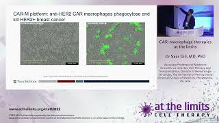
In this talk, Saar Gill (co‑founder of Charisma Therapeutics) outlines the rationale, engineering, preclinical validation, and first‑in‑human experience of chimeric antigen receptor (CAR)‑modified macrophages (CAR‑M) targeting HER2‑positive solid cancers. He begins by highlighting the unique challenges of adoptive cell therapy in solid tumors—poor trafficking, hostile tumor microenvironment (TME), metabolic stress, and antigen heterogeneity— and points out that myeloid cells, especially tumor‑associated macrophages (TAMs), dominate the immune infiltrate across many carcinomas. Using data from a recent Cell paper on tumor ecosystems, he shows that M2‑like macrophages correlate with poor prognosis, while M1‑like phenotypes are favorable. Gill then describes his laboratory’s work engineering human monocytes/macrophages with a chimeric adenoviral vector (Ad5‑F35) to express an anti‑HER2 CAR. The vector simultaneously induces a strong pro‑inflammatory (M1) phenotype. In vitro assays demonstrate CAR‑M specific cytokine release (TNF‑α), phagocytosis, and killing of HER2‑positive tumor cells, visualized by live‑cell imaging. He outlines the GMP manufacturing workflow: G‑CSF mobilization, apheresis, CD14+ selection, GM‑CSF polarization, Ad5‑F35 transduction, cryopreservation, and intravenous infusion. A multi‑center, open‑label, single‑arm Phase 1 trial (fast‑track FDA) enrolled nine HER2‑positive solid tumor patients (breast, esophageal, cholangiocarcinoma, ovarian, salivary). Dose escalation (10‑30‑60% of a 5 billion‑cell target) was well tolerated with only mild fever and low IL‑6; no CRS, neurotoxicity, or lymphodepletion was required. Pharmacokinetic monitoring showed rapid disappearance of CAR‑M from blood, but tumor biopsies before and after treatment revealed (1) increased M1 macrophage signatures, (2) modest detection of CAR transcripts in the tumor, and (3) a rise in CD8 effector‑memory T cells and clonal expansion of TCRs, suggesting secondary adaptive activation. Preclinical mouse models combining CAR‑M with anti‑PD‑1 showed superior survival and marked infiltration of T cells, dendritic cells, and NK cells, supporting combination strategies. Gill concludes by discussing platform flexibility—alternative vectors (lentivirus, mRNA), CAR intracellular domains, and potential to modulate stromal components—and emphasizes that CAR‑M may overcome some solid‑tumor barriers, especially when paired with checkpoint blockade.
Chapters
It's a pleasure to introduce our next speaker, who is an Australian-trained— and hopefully was born and raised, but he lives now in the United States. And he will focus on a different type of cell for immunotherapy, macrophages. And I think I'm looking forward to listening to your talk. Thank you. Thank you very much for the invitation to speak. I do have some disclosures. Most importantly, the work that I describe here today was ultimately licensed to a company that I co-founded, Charisma Therapeutics, though as the rules of capitalism would dictate, I own very little of it by now. So the challenges that I want to talk about with respect to adoptive cellular therapy of solid tumors, you've already heard a little bit about from Dr. Brenner-Pradier. Predominantly. Those are the issues of trafficking and penetration of the tumor, homing, extravasation, penetration, survival within the microenvironment with its associated immunosuppression, its mediators of exhaustion, energy, its unpleasant metabolic challenges that are thrown up against the invading immune cells. And then also, just as importantly, target antigen heterogeneity, which again Dr. Brenner mentioned and the fact— so targeted antigen heterogeneity in solid tumors and the discovery increasingly of the frequency to which we see antigen-negative relapses even in hematologic malignancies. This figure was published quite a long time ago and in fact the reference is from a review of it, but I wanted to start from a high-level overview of what exists, what immune cells exist within the tumor microenvironment. And of course we all focus on T cells largely, that's where most of us did our sort of immunology training, but actually across several different tumor types— and I'll show you a more updated data slide in a moment— the predominant immune effectors or potential effector cell is not the T cell, certainly not CD8 T cells, but rather myeloid cells such as macrophages, tumor-associated macrophages, or myeloid-derived suppressor cells. This really beautiful and rather complicated paper was published in Cell last year and speaks to Cellular Ecosystems in Human Carcinomas. And for those of you who haven't read the paper, the point really is that while there is great heterogeneity amongst hundreds of— millions of people with cancer and a variety of different types of solid cancers, the question that I think this paper sought to address was, can you collapse that heterogeneity into something more manageable by focusing on the immune component or stromal components of the microenvironment? And the answer was yes. This group identified by 10 different cellular ecosystems whereby this great heterogeneity of solid tumors could be collapsed into. And to summarize just a few of the figures from that paper, and I'll show you in a second why I'm talking about this, you will see that across the board in all 16 different carcinomas on the left side of the slide there, if you go from left to right and you look at the colored bar at the bottom, of course epithelial cells and then stromal cells in red and yellow respectively are quite common.. But then again, as I said before, from the older data, you see again that it is largely myeloid cells that infiltrate a variety of different— of these different— 16 different carcinomas, and then across several different— 9 or 10 different cell states. Then if you look over at the right side of the slide, you see the prognostic associations of various— in the various carcinomas, various cell states. And if you focus for the moment on the monocyte and macrophage group there, you can see that in this particular figure, they're able to show you what types of what macrophages are associated with poor prognosis and what are associated with a relatively good prognosis. And then finally, with respect to that paper, which is not my work, I would say that we have known for a long time that there is an M— so-called M1 and so-called M2 phenotype to macrophages, pro-inflammatory and anti-inflammatory, or pro-inflammatory and immunosuppressive respectively. And while the cognoscenti in the field don't like that M1-M2 dichotomy because it's really a spectrum, Nonetheless, for the rest of us, I think it is important to try and simplify things. And so if you follow that M1 versus M2 phenotype, you see largely that in a variety of different solid cancers, increased infiltration with these anti-inflammatory cells is associated with a poor prognosis. So let's go back to one of the first questions, which is what are the challenges to adoptive cellular therapy in solid tumors? And one of them, as we said, was infiltration. So on the left is a very reductionist, very artificial experiment that we did whereby we took human macrophages, labeled them with an infrared dye, injected them into mice. The mice had a tumor that was located in a specific site, specifically subcutaneously in the flank. And then we took out— we sacked the mice, took out the tumor, which you can see on the top left. We took out the lungs, the liver, the spleen. And you can see— I'm sorry, the labeling fell off, but the tumor is the top left one. The macrophages after intravenous administration do traffic into the tumor. Why? Because they follow a chemokine gradient. One of the best characterized but not the only one is the CCL2-CCR2 axis. And on the right side— and so that was our work, but on the right side is from Courtney Crane's lab from Seattle showing— here we have luciferase-labeled macrophages transferred into mice that have a tumor in the flank. You can see at the bottom right, macrophages first go to the lungs. And are abundantly present there, and then over day 1, 2, 7, 14, and 24, gradually locate to the tumor. So in at least 2 of these, not to mention some really early human data from Regensburg, from Germany, showing that macrophages that are transferred intravenously can go to the tumor. So what can macrophages do? Since we think, most of us think about T cells and NK cells as sort of killer cells, macrophages can, of course, phagocytose, if you look over on the left side. Upon phagocytosis, or at least upon encounter of their target antigen, they can produce cytokines and orchestrate an immune response. They are professional antigen-presenting cells, and therefore at least have the possibility, and as I'll show you in the rest of this presentation, do actually promote a T-cell immune response, which conceivably could mediate or at least answer some of the issues that you heard about before with respect to solid tumor adoptive cell therapy, which is antigen heterogeneity, and actually the lack of a very good cell surface antigen to mark solid tumors. And so this work was started in my lab by my first grad student, who is a prodigy, a human prodigy, not a machine prodigy for Miltonie, who then became— who then actually left when he graduated from my lab, went to run the science in the company Charisma Therapeutics. And what I'll show you today, even though there are several different ways of getting transgenes into human monocytes and macrophages, and even though you can work with monocytes or macrophages, and even though you can put CARs, cytokines, enzymes, hormones, peptides, a variety of different things, today I'll really mostly talk about macrophages, CARs, and the use of a chimeric adenoviral vector, Ad5-F35, that was originally described by Andrew Lieber from Fred Hutch. And so what Mike initially did for his PhD project was to develop a way of genetically modifying human macrophages, which really was quite difficult and took a year of his PhD just to work out how to do it. But once he did, what we ended up with are very strongly pro-inflammatory macrophages, M1 macrophages if you like, that express the CAR at a very high level. So UTD untranslated is in red, CAR macrophages is in blue. CD11b is on the y-axis, CAR, in this instance anti-HER2 CAR is on the x-axis. And we could show, although I'm not showing you this data, we could show that the CAR macrophages continue to express that chimeric antigen receptor for several weeks, up to between 2 and 4 weeks in vitro and also in vivo upon transfer into xenografted mice. And when you do a whole lot of flow cytometry or RNA sequencing or ATAC sequencing, Not showing you that data because it's published. We basically found, perhaps not surprisingly, that these macrophages— human macrophages that had been exposed to an adenoviral vector to deliver the CAR, we also kind of got two bangs for our buck because we also got this very strong pro-inflammatory phenotype, perhaps not surprisingly because the macrophages are responding to the entry of an infection essentially by a DNA virus, and upregulate a whole lot of co-stimulatory molecules as well as the antigen-presenting representing machinery. So in vitro, when you take your CAR macrophage in blue and expose it to a tumor cell in yellow, we can then measure cytokine production. Here is TNF on the left, and you can see control, so non-CAR-bearing or anti-HER2 CAR-bearing macrophages, untransduced delta zeta, meaning no signaling domain, or CAR macrophages. You can see that only in response to cells that express HER2 but not in response to cells that do not express HER2 do we get TNF production by the macrophages. And similarly, phagocytosis can be quantified both by flow and by microscopy in the middle figure there. And then killing also is shown here. The red are the CAR macrophages eating, killing, and eating a SKOV-3, which is a HER2-expressing ovarian cancer cell line. So hopefully this will work. Yes, this video shows you CAR macrophages in red encountering, chasing down, and eating, as you'll see serially, green HER2-expressing cells. And you can see, for example, that guy in the middle is sort of busy trying to eat two cells at the same time, and there's a few other ones there as well. Now, on the next slide, I'm showing you on the left is control, on the right is anti-HER2 CAR macrophages. In this instance, we have control macrophages on the left, as I mentioned, the specific macrophages on the right. And we've labeled our HER2-expressing cells, tumor cells, with a pH-sensitive dye such that when the cells are internalized into the macrophages, the pH drops in the phagolysosome, they should fluoresce green, I think it is. And so you'll see in a second that we have a lot of green and reduction of our tumor cells on the right but not on the left. And the reason, again, is that the CAR macrophages specifically encounter phagocytose and start to break down tumor cells. And to show— and I think the time bar is instructive. That process that I showed you takes about a day. So that's all immunocompetent systems and is something that is being carried on further in order to determine how else we can improve upon the system. But while all this was being done— and to be honest, the reason I founded Charisma Therapeutics is in order to get enough money to do a clinical trial so we can really know whether this this has any legs. And so the way this is done is patients with any HER2 overexpressing solid tumor, not necessarily breast cancer, undergo G-CSF mobilization. You think of G-CSF as increasing neutrophils and stem cells, but it also does about threefold increase circulating monocytes. Those— they undergo apheresis as standard. They go CD14-positive selection using a Miltenyi system. Then those cells, those CD14 monocytes undergo polarization to become macrophages upon exposure to GM-CSF for 5 days. And on the 5th day, we add the Ad5-F35 vector to confer expression of the HER2 CAR. And those cells can be easily frozen and reinfused. And the trial is open at 5 sites. I think I'll show you some details on that in a moment. Multi-center, open-label, single-arm study. As I mentioned, any patient with HER2 overexpressing, but it has to be HER2 overexpressing. 3+ or 2+ confirmed by FISH. Group 1, which has just been completed, is an intrapatient dose escalation up to 5 billion CAR macrophages split over 3 infusions. 10% of the dose, then 30% of the dose, then 60% of the dose if we can make that many cells over 1 week. And then Group 2 is to be a single infusion of that total manufacturable dose, and of course, It's done intravenously. And I guess— and I would also add that the product received FDA fast-track designation in September of 2021. So, here are 7 of the 9 patients treated in the first— that I can talk about because this was presented publicly at ASCO recently by my colleague Kim Rice Binder from Penn. The 7 patients, median age of 64, mostly female. All white, largely sort of good performance status, HER2 overexpression, tumor types: 2 breast cancer, 2 esophageal cancer, 1 cholangiocarcinoma over 1 ovarian cancer, and 1 salivary carcinoma, with an extensive range of prior therapies, not surprising for this type of patient population, some of which also had anti-HER2 targeted therapies, and then some of them had prior radiation. Now, one of the key questions, one of the key concerns and fears for me, to be honest, when we started this was all— essentially all our work before the trial was with healthy donor monocytes. And I was concerned that in patients with solid tumors, a lot of the CD14 circulating cells would be MDSCs or about to be MDSCs and that we wouldn't make good quality CAR macrophages. In fact, we did. And I think that speaks to the vector, the adenoviral vector. So you can see that we had high viability of cells with a high purity of CAR expression, high purity of CD14 and high CAR expression. You can see that all the batches, with some variability, so these are, each line is an individual patient product which was taken in vitro and shown whether it can kill HER2 overexpressing cell line, the same cell line, not autologous cancer cells, of course. Some variability between them, and here on this plot in the middle, Lower is better. Lower is more potent because of faster tumor killing. And then if we look at an M1 signature on the right side of the slide, you can see that we started off from fairly M2-type cells, not surprisingly, but at the end of manufacturing, we ended up with M1 pro-inflammatory cells. The cells— the infusion was well tolerated. We put in hundreds of millions of activated macrophages which had been exposed to a DNA virus, and actually there was It was relatively well tolerated. I would add, and I think this is really important, there was no lymphodepletion given. Nothing else was done to the patients, which I think is kind of important to keep in mind when I show you some of the correlative data in a second. No dose-limiting AEs, no need for modification, no severe CRS, although there was some CRS, some fever, and I'll show you some of that in a second. No neurotoxicity. Top is temperature in Celsius and bottom is interleukin-6, tiny amounts of interleukin-6 being produced. Each line is an individual patient. Temperature of 38 is shown at the top. You can see that only patient number 7 had really any sort of fever but no hypotension. And really, that's all I have to tell you. It was quite well tolerated. What about detectability of these cells? We know from CAR T cells that they expand in vivo.. But macrophages do not expand. They cannot proliferate. They're terminally differentiated cells. So we don't really expect to see an increase over time, certainly not exponential as one would see with T cells. And we didn't. In fact, we barely detected any macrophages in the blood. Here is the— I think this was done with ddPCR. So the PCR, quantitative PCR of the product. Again, each line is an individual patient. You can see after each infusion a rise in the number of CAR macrophages and then very quick drop-off with no persistence in those cells. Having said that, one of the strengths of this study is that each and every one of the patients had a pretreatment biopsy and then an early post-treatment biopsy in the same site and then a late post-treatment biopsy of the same site. And if that necessitated another endoscopy and a biopsy, that's exactly what the patient had. So we do have, I think, good quality data that actually showed that we could detect a few. I'm not gonna say it was a lot, but we could detect a few what we call CTO508, the HER2 CARs in the tumor. And we also did single-cell RNA sequencing in all of these patients in all of these time points, and some of these data I can show you now. We saw an increased fraction of M1 macrophages. If you compare the screening, this is subject number 1, number 2, and number 4, and you can see percent of fraction of macrophages with an M1 phenotype in the biopsy. Screening is in the top in the bluish, day 8, which is early time point in the orange, and then week 4, which is the late time point biopsy. We can see generally an increase in the percentage of macrophages in the tumor having administered the cells intravenously that are of an M1 phenotype. And then those cells, patient number 1, an example of patient number 1's phenotype of their macrophages is shown on the right where you can see that there is a significant increase and the size of the circle represents the percent of the cells with that particular transcript upregulated and the color represents expression. So red overexpression but log 2, so not a lot of expression. Largely the chemokines, cytokines, and a little bit in the way of pro-inflammatory cytokines using single-cell RNA sequencing in the tumor microenvironment. What I think is more interesting and more promising is that there is an increase in the T cells in the tumor microenvironment— it's ominous— in the tumor microenvironment of the patients. So subject number 1, number 2, and number 4 are shown. We have screening on the left and then week 4 for each of the patients, so before treatment and then after treatment. And no, I'll remind you, nothing else— no lymphodepletion, no chemotherapy, no T cell just some macrophages administered intravenously. And we see an increase in the frequency of leukocytes that are T cells. If you look, for example, at the color orange, you see that in subject 1, 2, and 4, there's relatively an increase in the CD8 effector memory cells in the tumor microenvironment. Not a great deal of proliferating cells, you can see in the yellow, but I would say that the most sort of outstanding increases in the effector CD8 T cells, as well as in some patients exhausted T cells, which will become very relevant in a second. So single-cell RNA sequencing also allows you to do VDJ sequencing. So look at the— use essentially the identity of the TCR as a barcode to try and tell you, number one, are those T cells that you're detecting in the blood also present in the tumor? Microenvironment, and number 2, what are the characteristics of those T cells, which is something that we've started trying to do here. Subject number 4, this is the TCR diversity screening on the x-axis, and this is in the blood, and then day 15 in the blood on the y-axis. And wherever it's orange, you can see that there is enrichment in T cells at the later time point after infusion compared to at baseline. So something is happening. It could be response to nothing. It could be just 2 weeks later it just changed. It could be something else that happened to the patient. It could be the macrophage infusion. It could be the fact that the macrophages were previously infected with an adenovirus, although we can talk about that later. And then T cell clone expansion in the peripheral blood in these 6 patients is shown. Total expanded is in the sort of colored, whereas the newly detected and expanded is in the sort of lighter colors. And basically what this shows is that the T— this is a TCR repertoire analysis which tells us that there is an expansion of T cell clones in the blood. So far the blood. The next, I think, slide is more interesting, the microenvironment of the subjects post-infusion. So the next slide is we see the same plot as before with the profile of the TCRs day 15 after infusion on the y-axis, screening on the x-axis. And here we're showing you the TCR repertoire in the same patient both in the blood as well as in the microenvironment at different time points. And you can see, for example, subject 4, that there is a vast increase— well, it's relatively because look at the Y-axis— but there is a relative vast increase in the T-cell clonality in the tumor at 2 weeks after infusion. And what can we tell about those T cells in the microenvironment of that exemplary patient number 4? Well, you can see that the most prevalent T cell clone in the week 4 tumor microenvironment of participant number 1, you can see it's increased screening day 15, week 4. You can see week 4 has had an increase in the frequency of that tumor. And here is some example of the phenotype of that clone. It has perforin, it has NKG7, LAG-3, granzymes, eomysoderm, and some increase in LAG-3, as I said, and TIM-3. And so what we think in this exemplary case— and we're chewing over the rest of the results— is that the most expanded clone in the tumor microenvironment of this patient after infusion of macrophages is a CD8 T cell clone, which is a cytotoxic T cell with some suggestion of exhaustion. So where to next? Those of you listening intently will notice that I haven't told you what the rest responses were, because there weren't really great responses. The best responses were stable disease. So why and where to next? So the immunocompetent model allows us to start thinking about combination therapies, which I think is where we will have to go. So this is a similar model to what I showed you before— immunocompetent mice with the HER2-expressing tumor getting the HER2-specific CARs, but also with or without the addition of an anti-PD-1 blocking antibody. And if you look at the different groups, the, I guess, lime green group is the one where we combine CAR macrophages with the PD-1 inhibitor, and that is indeed the one in which we have better survival of the mice. And that is the next iteration of the clinical trial. And if you look over on the right, we have evidence of profound TME activation. Each panel is a sort of replicate, essentially. So we have a control macrophage. Phages, we have our just PD-1 in this non-PD-1 responsive tumor. We have CAR macrophages and we have CAR macrophages combined with PD-1. And this multicolor staining shows you that, of course, we have our stroma, we have our epithelial cells, but we have an influx of T cells, dendritic cells, natural killer cells when we do this combination of CAR macrophages with PD-1 inhibition. And then the second-to-last slide shows us the potential platform of CAR macrophages macrophages, I think of it as just the same as what we've learned that we can— how we can manipulate T cells. Well, we can manipulate macrophages in the same way, potentially giving slightly different outcomes. So you can think of it in 3 different bands, if you like, 3 different axes. Transgene delivery. Well, you can use lentivirus. It just doesn't work very well. It needs a little bit of additional sort of hoops to jump through, but you can. Just isn't as good as Ad5. You can use non-viral approaches such as mRNA, including mRNA delivered by nanoparticles. You can— or electroporation of mRNA. So the next axis is of sort of interesting stuff one can do is, well, what about CAR design? All of the stuff I showed you is actually what one would call first-generation CAR in the T cell world. Why? Because it's not quite clear what the appropriate co-stimulatory molecules are for macrophages. So we— so you could certainly add on different co-stimulatory molecules or you can use different signaling molecules. And the ones we use actually is CD3 zeta, which has no business being in macrophages, but it's close enough to the Fc gamma receptor in its intracellular domain that it actually works just the same. We've done head-to-head comparisons. And then finally, in terms of antigen-specific antitumor signaling, well, I've shown you evidence of phagocytosis in vitro. It's incredibly difficult to show active phagocytosis in vivo. I've shown you evidence of TME activation in vitro, in mice, and in humans as well. Antigen presentation, I think maybe, you know, I think certainly in vitro we can show antigen presentation. I think that you might agree with me that there is some sort of rough evidence of some T cell activation and proliferation, so perhaps there's antigen presentation. And then something that we haven't explored in detail but might be particularly important in the solid tumor is the ability of these macrophages to modulate not just the cellular but also the acellular, the stromal components of the tumor microenvironment. So in conclusion, I've shown you that human macrophages can be readily engineered using a range of vectors and are focused on Ad5-F35, which has desirable properties. CAR macrophages may be suited to the treatment of solid cancers. There is direct killing via phagocytosis there is stimulation of the adaptive immune system, and that I do think that combination with blockade of inhibitory receptors— and I'm really not someone who throws pembrolizumab against the wall and hope it sticks— actually, I think in this case it might actually make sense. It may not be PD-1 inhibition, but another checkpoint blockade certainly to be explored. And then I want to thank the people in my lab who worked on this particular project, and then my colleagues at Penn, as well as colleagues at several other institutions that are running the study right now, including City of Hope, MD Anderson, Sarah Cannon, University of North Carolina, and to the patients and their families who participated in the study. Thank you.
More from Joshua







Scientist Stories: Hans Waterham, The value of phenotypic assays in peroxisomal disorders




Scientist Stories: Joel Zlotogora, Genetic carrier screening programs for reproductive purposes