Scientist Stories: Scott Fraser, Multiplex Imaging of Cells in their Natural Contexts
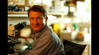
Scott Fraser describes multiplex imaging technologies that capture live cellular dynamics in intact tissues. His lab uses Hybridization Chain Reaction to label up to 250 different molecules simultaneously, two-photon microscopy to image entire embryos in minutes, and label-free methods that detect metabolic states through intrinsic fluorescence without adding dyes.
Chapters
Okay, looks like we will make it through the day on time. It's kind of fitting that we end the day with Scott Fraser, who's the Elizabeth Garrett Chair of Convergent Biosciences here and the Director of Science Initiatives. He's also Provost of Biological Science and Biomedical Engineering here at USC. You know, Scott has a long history background in a lot of the types of accomplishments we've seen with a lot of our speakers all day with— he's a fellow of the European Academy of Sciences and AAAS and National Academy of Inventors. But even more particularly and germane, I think, is his background with interdisciplinary science and translational science, and particularly in the field of medical imaging. So Scott, it's great to have you.
Well, thank you very much for having me. It's a real pleasure to be here. And what I want to try to share with you is some of the multiplex and multimodal imaging techniques that we've been deploying for my sort of day job. My day job is to look at things like how forming organs take shape and the multiple influences into them. And my sort of weekend job, together with some of the people in the lab, is to apply some of these tools over into things like draining lymph nodes, and also to try to see if there's a way to take these imaging tools that we've optimized for experimental animals with encoded genetic markers and carry them into humans, which obviously wouldn't have those markers in place. And so if we're lucky, we'll make it through all of it. I just want to say that I've been very lucky to have an amazing group of collaborators, both outside of the university and inside of the university, and some funding agencies that have been willing to fund us without giggling too much in order for us to go after sort of our day job, which is to try to understand the systems biology of how embryos take shape. Embryos are a great model system for many different systems, even if you don't care about embryos per se, because they deploy so much of their genome in such a small package so quickly. But the problem is that most people study them by doing endpoint analyses, looking at different stages in development and guessing what might have come in between. And I would like to argue that that's always going to lead us astray. If we look at static gene expressions, even things that we think we understand, like the forming heart, where we know some of the cells that form these cell groups that end up making sort of a horseshoe and end up assembling into a forming heart, we think we know how that patterned set of genes in a patterned group of cells would behave, but since this is from fixed tissues, we're sort of stuck. So what we've been doing is deploying in both mammalian systems and other higher vertebrate systems like transgenic quail, being able to make— oh, let's see, I just hit the wrong button— being able to make transgenic quail by, in this case, using lentiviruses, deploying them because they're such a convenient imaging We can take the embryos and put them out in a culture chamber, put them on a microscope, and since we have tissue-specific reporter genes, we can now follow it over time. And now we can ask just how accurate that map of gene expression that should tell us what the cell behaviors were. So I've just labeled a few of the different cell populations here, and what we should have seen is a horseshoe group of cells that were pre-embryonic. Preconfigured to flow forward and make ventricles and atria. And, in fact, that's not at all what happens. The cells— the only boundary that they really seem to obey is right-left, and even that gets violated. So it's clear that what's going on here is that we have evidence that the quail don't read the primary literature. We've gone on to show that neither do zebrafish or mice, and we suspect suspect humans based on some of the birth defects that we've seen. So what we're trying to do then is to make tools that let us carry this analysis into embodied cells that we follow dynamically. And we want to be able to ask enough questions about those cells so that the cellular dynamics and the variance from cell to cell or animal to animal is not noise, but that's really the signal. And that's where it comes in that we have to have these multiplex tools. So I'm going to try to share a couple scenarios with you today, and there's no way to go through all of them that we've been developing, so I'm going to give you just sort of a snippet from each of the columns, so a little bit of a tasting menu of what we're up to. Now many people here have thought about quantitative gene expression this way. Very few of us think about quantitative gene expression by looking at spatial transcriptomics, and that's because most of the tools are really flawed. This is the same gene at the same stage in the same strain of mouse and in the same paper, and it didn't bother the authors that those are so dramatically different. So if I want to ask quantitative questions that I can from a transcriptome and validate it, I'm in trouble. So years ago, when I was still at Caltech, we started using the DNA origami and other tricks that the colleagues there were developing to develop a quantitative in situ and multiplex hybridization technique that would let us take reagents like these hairpins that'll stay stable in solution. They'll stay stable in solution, but they'll self-assemble when they see a target signal. The target signal in this case is an antisense oligonucleotide, and when it sees that, what happens is that this is complementary to that toehold and can branch migrate up. That opens up the first hairpin and exposes a second sequence, which is complementary to that toehold and that stem loop. And so what happens is it explosively grows to make a 200-mer. And once it's made that 200-mer, since it's sequence-specific, we can easily multiplex this by putting multiple dyes on different hairpin pairs. If I take all of them and run them in a gel, they run as a very small molecular weight species, even if I add blender extract of human tissue or zebrafish. But if I add the initiator for one of them, the blue one, it disappears from here and it assembles as a 200-mer. Or the green one, you can see it disappears here and appears up there. So to make it into an in situ hybridization technique, we put those antisense probes onto the initiators and use that to then tag individual RNAs or individual antibodies and then get an amplification of per initiator about 200 dyes. The good thing about this is that the active species is very small, it's very sequence-specific, and it penetrates into prep— into preparations in a quantitative fashion. It also gives us a linear gain, so the things that are lost in a normal in situ, like the nascent transcripts inside the nucleus, because they're orders of magnitude less intense than what's in the tissue as a whole, In conventional NC2s, they disappear. In HCR, they show up quantitatively just as they should. And so the good news is that it works nicely, it's stable, it doesn't care how long you've developed it for. And in fact, this one spent 2 months in the refrigerator and gave us an answer within 5% of the freshly imaged one. Long Cai at Caltech has been pursuing carrying this to higher degree of multiplexing. He's up to 249-plex right now. So that's the good news. And the bad news that comes back to my lab then is that 5 gigavoxels of embryo here means that it takes a long time to do the images. So that's 10 hours' worth of confocal imaging. And, in fact, if we want to carry it to the single message level where Long Cai does and where we do for many of our experiments, it means we have to image about 100-fold lower, and 1,000 hours per specimen clearly isn't going to do it. So the other problem is the colors. I'll just quickly say the way we're getting around the speed is by adopting two-photon microscopy and a light sheet geometry. So we bring in a two-photon laser beam from the side. In this case, I'm shining in blue light so you can see it in this macroscopic thing. Here's the objective that collects the light. It turns that 10 hours into just a few minutes, and it lets us, even with single-cell and even single-protein resolution— in this case, we're tagging the junctional complex in a beating heart— follow those moving cells down to subcellular resolution. Okay, what about the increased number of colors? Now, if I wanted to do it even slower than the 10 hours, I would use CyTOF, but I don't want to do it even slower. I want to do it faster. So what we need to do is come up with a way to deal with this significant overlap between the fluorochromes. And luckily, when I was at Caltech and working a bit at JPL, I was able to steal the technology they use for remote sensing. Sensing technology called linear unmixing to take a spectral cube, so X and Y and the third dimension is the color, and by collecting a spectrum for each pixel from a confocal microscope, for example, we're able to get enough information to make this a well-posed question. So even labels like GFP and fluorescein that overlap dramatically, if we take even just 8 spectral bands in this, we're able to separate them out, in this case the GFP from the fluorescein. There's a little bit of noise propagation, and that noise propagation is a problem because we're always limited by the worst channel. It's sort of like an academic department. The noisiest channel dominates. And so what we needed to do is to come up with a different way to get away from that source of noise. And luckily, Enrico Gratton at UC Irvine had worked out a way for lifetime imaging, and we applied this over to multispectral imaging. So if we take a spectra and take the Fourier transform of it, that spectrum of 32 bands becomes that dot. This red one becomes that dot, a 50/50 mix becomes a dot halfway in between. So all the inverse mathematical problems that were haunting us becomes a forward problem. It means it's both much better performing and much faster. So again, since these specimens can be a gigavoxel of imaging data, if I have to solve an inverse matrix problem for each of the points, what ends up happening is that it takes time and it propagates from the noisiest channel to the least noisy channel, so this The blood vessel label shows up in between the blood vessels as well as in the blood vessels with the normal technique. Using the Fourier domain technique, it's dramatically faster, as fast as collecting the image, and the crosstalk disappears. So what this allows us to do is to take intravital labels like fluorescent proteins or like autofluorescent signal or structural signals. And to unmix them on the fly. In this case, instead of using 5 different lasers to image 5 different dyes, this is using 2 different lasers to image those different dyes. Even though the dyes overlap tremendously, it's easy to separate them out. You can look through the highly autofluorescent RPE in this, in this live animal and see the individual capillaries inside its eye, for example. So this technique ends up really changing the throughput of our imaging. It also changes the capabilities a lot. Okay, we're also one of the test sites for the CyTOF, and we've been, of course, wanting to carry the multiplex imaging all the way to that. One of our problems— well, I think everybody here knows the story of the CyTOF and using metal dyes instead of using fluorochromes, both in a flow approach and in an imaging approach by cutting divots out of the specimen and having those be analyzed. Now, the beauty of that is that it lets you do 36-plex, but I've already told you that we're up to 200-plex with the Muse and HCR, so maybe it's a problem or maybe it's a way of going. What we found is as we started to add the metal-chelating polymers or the nanoparticles that we were using to carry in the isotopes that the CyTOF sensed, it started to become the tail wagging the dog. That is that the label perturbed the energetics of the hairpins. So we had to go over to a different approach in which we used the hairpins with an extended branch off the side that once the amplification polymer forms Those hang off to the side, and then we can secondarily bring in a metal-chelating particle or a metal nanoparticle or a quantum dot or a fluorophore, and we can tag these onto several different things, antisense probes and onto antibodies. Just showing one of the samples that I don't feel guilty giving to physicists, this is fly embryos looking at striped gene expression. Expression. It also gives them a nice defined pattern. What we've been able to do is to be able to show that we can get quantitative capturing of both the RNA and the protein level. In this case, it's a tagged antibody that's amplified up. We've been able to use quantum dots, which makes it about 200-fold brighter, and— actually, I think I got those backwards. This one's 200-fold brighter, this one's 150-fold brighter, the quantum dots. And, of course, the CyTOF works, too, but it's quite a bit slower and harder to do in 3D. One of the nice things about this technology is that it gets close to and exceeds the capabilities of HCR. By having that two-step labeling technique, what we get is both greater signal— this is HCR at its normal intensity in muse. If I histogram equalize them and then I blow them up, you can see that it's a lot easier to do— to do quantitative recognition of the individual message dots within these fixed cells against the background that's in the nucleus than it is with HCR. And so this is allowing us to do these higher-order, sort of, seqFISH, MERFISH, and in situ hybridization multiplexes. That let us carry into the hundreds of targets at a time. Now, that sort of technology has let us ask questions in fixed cells with the HCR and the Muse, and in live cells with the hyperspectral phaser, and others in the lab have been trying to apply that to problems that more people here care about. For example, you know, can we see the cells in the germinal center and traffic— and follow their trafficking as the immune response is being sculpted. And so what we've been able to do, working together with Masahiro Kitano, a very, very good senior researcher in the lab, is be able to collect the movies, like many people have, of both the dendritic cells and the T cells as they're— as the response is first unimmunized, 6, 10, 20, 46 hours. But what we've been able to do is really take a quantitative census and of course then be able to back this up and be able to look through in 3D in these two-photon light sheet images and two-photon laser scanning images. And even in this lighting, I hope you can see the juxtaposition of the blue and the green cells. That's only a part of the problem. What we really want to know is which of the cells that are trafficking from out in the periphery into the lymph node. And by making transgenic animals for KIK-GR, we can use ultraviolet light to activate peripherally those cells that are distant to the lymph nodes and then watch as they traffic to the lymph nodes. Because we can just use infrared light— or sorry, ultraviolet light or two-photon infrared light to activate these cells anywhere we want in the animal and then image because the conversion only takes less than a couple minutes. So that's what we did. Shine the light out in the periphery, wait for a while, be able to see them come in. Hopefully the graphics gods will smile on me.— and we can see if the— okay, so here we— oh, maybe they're not going to smile on me. It was going so— ah, now it's going to work. Okay, so then we can take movies of the cells following both the resident cells, the cells that have trafficked in from outside, and now be able to compare them both spatially and temporally. And as we do that, what we find is a noticeable difference between those cells that— the dendritic cells that have moved into the node. We see a difference in the behavior of these cells. They move dramatically differently. They change their energetics, and that matters for the next thing I'm going to tell you about. And that means that we can make some predictive suggestions based on these invasive experiments with transgenic animals. And what we're hoping to do is to carry this on further, as you can see, most of the interactions are taking place with the cells that have migrated in from the periphery. Very few of them are happening with the cells that didn't come in from the periphery in mounting that response. So I'm going to— yes, how am I doing on time? I'm close, okay. Another couple minutes. So let me just say that what I'm trying to show you is that we've got ways of doing spatial genomics, we can increase our palette, and we can follow cells dynamically, but we have a problem because dramatically few of the patients or subjects that you might care about actually come in with GFP, except maybe they were one of your postdocs. So, what we want to do is take advantage of some of what you saw here, and that is that a lot of these The labels are autofluorescent signals that are intrinsic to the molecule that's there. And in fact, we can see in— by intrinsic signal, the NADH bound and free. We can see retinoic acid and retinol, folic acid, a bunch of other things. It turns out, though, that as you try to image down deep into imperfect optical tissues, the lifetime of the fluorescence —label, that is how long it stays excited before it gives back its light, is a better assay. And I was fortunate to have two people in my lab, one who thought that the world was lifetime and the other that thought the world was color. And so they finally agreed to bury the hatchet and work together. They're now making joint instruments that take the phaser of the fluorescence lifetime in which different labels show up different places in the phasor plot of the lifetime, the hyperspectral, where it shows up different places in the phasor plot for the wavelength, built it to— into an imaging box that can go through an image conduit down a bronchoscope or other endoscope. We're still in the experimental animal cases, but so far we can see tumor stem cells, We can see— we can watch cells as they respond to drugs. We can see stem cells in their endogenous locations. And in this case, it's going into an animal's airway. You can see both structural things. The green that you can see back here that I hope looks filamentous is the collagen on the far side of the tissue. The granularity here are individual cells, and you can see the oxphosphorus is glycolytic as as well as some of the other labels in this in live tissue. What we're hoping to do is, if we can get it through the IRB, is start using this in human subjects. We're at well below any sort of challenge. We're putting photons in and photons out, and we're not applying any exogenous labels. So we're hoping we're going to be able to read the mind of the cells and follow as they respond to drugs, and they respond to immunization. So I'm going to stop there and just say that adding to these exogenous label things, the spectral FLIM approach is allowing us now to look in cells, in organs, and in spheroids, and we hope now in intact people, and follow stem cells and their responses to their environment and to exogenous agents like drugs. And I'll stop there and thank the many foundations and funding agencies that have made this possible. Thanks for your attention.
Yes?
Are you able to connect the autofluorescence imaging that you get from the spectral imaging with gene expression? Because we know a lot about which Yes, and so that's exactly where we're going. Some of the autofluorescent signals are maintained after the cells are fixed. We can then take the live image, fix them, and then do the whole mount HCR and other assays and use the autofluorescent signals that are preserved in the fixed state to re-register, and we're just starting to do that. And if I didn't say along the way somewhere that that's the missing bit, that is still the missing bit. We now need to compare the spatial transcriptome work with the spatial metabolic probing that we're doing. Other questions? Okay. I'm around if anybody has any other questions. Thank you.
Well, I only have about 80 slides, so this shouldn't take long.
But this is—
this has been really an unbelievable day. We saw incredible science. I talked to Gary as he was, he had to go out to another meeting and he was enthralled with everything he saw. So I just want to thank a number of people. One, the events team here at USC. Let's give them a round of applause. They did an unbelievable job. The person I work for at the Vaccines Project, our friend Sandy Glass over here, who got us all here. All the winners again of the Michelson Prizes. And all the excellent speakers over the course of the day. So it's still beautiful outside, and if Steve K. is here, he could tell us You know, where do we get the beer? And with that, I'll close the meeting.
More from Joshua







Scientist Stories: Hans Waterham, The value of phenotypic assays in peroxisomal disorders




Scientist Stories: Joel Zlotogora, Genetic carrier screening programs for reproductive purposes