Scientist Stories: Sidi Chen, Gene Editing in Oncology and Immuno-Oncology
In this seminar, Dr. Sidi Chen presents her laboratory’s strategy for building functional cancer genome atlases by performing in vivo CRISPR screens directly in mouse organs. She first outlines the motivation: the explosion of patient tumor sequencing data demands experimental validation of which mutations drive cancer and how they interact. The lab’s workflow involves delivering guide‑RNA libraries via viral vectors (AAV, lentivirus) to specific tissues (brain, liver, lung) to induce mutagenesis, then sequencing tumors to map driver and passenger mutations. Using this approach, they generated a functional mutanome for glioblastoma, identified key driver combinations (e.g., NF1 + MLH3, PTEN), and extended the method to hepatocellular carcinoma and KRAS‑driven lung cancer, employing sensitized backgrounds (MYC, p53 loss, PI3K activation) to uncover secondary modifiers such as KDM6B and RAP20A. A later screen for immune‑escape highlighted PARK1A, whose loss reshapes the extracellular matrix and recruits myeloid‑derived suppressor cells, illustrating how a single gene can remodel the tumor microenvironment. To dissect complex genetic interactions in metastasis, the lab leveraged the Cpf1 enzyme’s ability to process crRNA arrays, constructing a high‑throughput double‑knockout library covering thousands of gene‑pair combinations. In vivo metastasis assays revealed both monoclonal and polyclonal spread patterns and identified synergistic pairs (e.g., DRAC1‑KMT2C) that accelerate metastatic outgrowth. Finally, Dr. Chen introduced the KIKO platform, which uses Cpf1 and AAV to simultaneously knock‑in CAR constructs (e.g., CD22 CAR) and knock‑out genes such as PD‑1 in primary T cells. The system achieves high editing efficiencies (≈80% knockout, 45% knock‑in) and produces CAR‑T cells with reduced exhaustion markers compared to Cas9‑based methods. She concludes with future plans to scale functional atlases to other cancer hallmarks, integrate single‑cell perturb‑seq, and develop more precise immunotherapy engineering.
Chapters
And once we figure out how to change the setup. All right. Did he show you how to change?
Oh, absolutely. Good to see you. Do you know how to change this? Oh yeah, you're all set.
Perfect. Thanks, wherever, whoever's— they're sneaky in the back. So, um, City Chan is an assistant professor in genetics. He's, uh, one of the leaders and first people to have done in vivo CRISPR screens, and is an expert on a large variety of different genomic approaches from CRISPR also into CAR-Ts and things like that. And we'll let Sidi give his talk. Thanks so much.
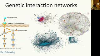
Thank you. Thank you, Marcus, for the kind introduction, and thank you everyone for coming to my talk. Today I would like to describe to you some of the basic research in my lab, briefly some of the old stories, and primarily on the two new stories that we published this year. So in brief, we would like to introduce you the next generation tumor modeling and genetic screening, and then the genetic interaction in cancer, and how do we use gene editing for immune engineering. So why is tumor modeling important? Because we have a large data set now, cancer genomics from patients, thousands and thousands of them, and keep accumulating from the hospital. And this will become a routine, or in the very near future, I believe it's going to become routine to take patient biopsy and sequence them and figure out what are the mutations. And we have accumulating cancer types with mutations, copy number, transcriptome, and phenome, and the epigenome data, and even single-cell transcriptomics. So, but with this giant and massive dataset, we know there are some AI algorithms there, but how can we definitively prove some of those mutations are actually necessary or sufficient to drive diseases? And because there are so many genes, how do we know which of the 2 genes or 3 genes interact together to drive the phenotype of cancer? And exactly what do these new mutations do, for example, Two of the beautiful cases that the last speaker just spoke about, those are novel mutations no one knows— like, no one knows what they do before. And if a patient have those mutations, or if a new patient coming in and have the same mutation, do we know what they do? And how do they drive the tumor progression in the native tumor microenvironment under immune pressure? And with that, we would like to introduce a next-generation tool for us to be able to map the genome to the phenome for cancer. Mutations. Like, how about we just do everything together in the animals and then prove that these are the mutations that are driving cancer and those are the ones that are not? And with that, we were hoping to achieve a clinically actionable map in the future. This is my vision. And two of the approaches we're trying to address these questions are, one is the functional mapping of cancer genome drivers by directly taking cancer patients' mutations, for example, the ones being sequenced, and then we just generate the exact same mutation in the animal and see how those drive cancer. And the other approach is to use unbiased genome discovery, meaning we scan the entire genome and see what would happen, and then we'll see if those are the mutations driving cancer, and if those are the mutations that are also mutated in patients. And one thing we did is we invented the direct in vivo CRISPR screen, or autophagy genetic screen where we would take the virus such as AV or lentivirus or other viral vectors, and we directly mutagenize the right organs in vivo. This can be the brain, the lung, the liver, and the pancreas and other organs. And then when those organs containing the cells of origin were mutated, then you see tumors growing because you hit the tumor suppressors or oncogenes, for example, the KRAS, P53 in the lung that will drive the cancer in the lung, and then NF1, PTEN that will drive cancer in the brain. And then all we need to do is to capture sequence all of the tumors, including the big ones and small ones in the pool, and then we'll figure out what are the drivers, what are the passengers. And the first case we did this is glioblastoma, and this is also the first case done by TCGA in 2008, and then later on 2013 and 2016. This is a very deadly cancer and a lot of mutations. And then what we try to do is we take these mutations, and then we synthesize the guide RNA library. We put them into a viral vector, and then we deliver the viral vector into lateral ventricle or the hippocampus of the brain. And then what we did is we followed survival through imaging and histology and pathology analysis. And these were led by two of my students, one of my postdocs, and that was published a couple of years ago. But the take-home message is that if you have a normal brain and that got hit by a pool virus that is capable of mutagenesis, and that virus is going to generate tumor, and that's close to 100% of the time penetrance. All the animals would fall off, and of course during a long time frame, and then with different penetrance. And these mice would develop glioblastoma, which is mimicking the clinical feature. But this is complex pool. There are a lot of mutations in the brain. Like one of these brain tumors can contain dozens or hundreds of mutations. And then what we did is we capture sequence all the targeted loci that's cut by the guide RNA. And those can contain insertions, deletions, splicing site mutations, just like what you sequence from the patient. But we are doing a simple experiment, the computer in nature, and we are able to capture the entire mutanome in the glioblastoma that's driving directly the tumor growth and progression in these mice. And then with that, we are able to one-shot generate a functional atlas of GBM. And then we compare this functional atlas in animals and to the TCGA data again, and then to our Yale Glioma Center or the GBM patient data. And then we see there's a significant correlation suggesting the functional ones are still the functional ones.. But there are a few outliers that we'd like to point out. For example, the beta-2-microglobulin, which is highly mutated in the animals, but not so highly mutated in the patient tumor, for example, in the Yale patient cohort. But there are sporadic case reporting suggesting HLA is also mutated in the glioblastoma. So that will give us additional confidence. But how many of those are sufficient to drive a glioblastoma? And what we did is, in fact, taking some of the co-mutated ones, and we just knock out 2 genes or 3 genes and see if they are sufficient to drive glioblastoma. And then, as you can see, if we knock out NF1 and MLH3, 2 genes are sufficient to give you a glioblastoma, even though that's only 40%. And then likewise, if you knock out PTEN, that's 100% penetrance. And beta-2-microglobulin and NF1, that's 4 out of 5, this is 80% penetrance. So a small number of genes are sufficient, but of course more genes will drive the cancer being more aggressive. Long story short, we similarly mapped the functional atlas in the hepatocellular carcinoma using similar approach, but with a different viral vector called AAV8 carrying the TBG promoter, which is specifically hepatocyte. And what we did is we deliver intravenously into the tail vein of the animal, and that would mostly home to the liver, and then the TBG promoter can drive the liver specific expression. And then the MRI imaging shows that the mice develop liver cancer, and those are GFP positive because they were driven by the gene editing, and that also turned on GFP. And the mice die, and the histology shows that this mimics the liver cancer, and then we generate a map of the liver cancer by sequencing the entire liver, 5 lobes of each animal, and then we map out the liver cancer. Functional mutanome. So we're scaling this up to the entire genome, but now using different genetic background. For example, all those previous experiments we did were baseline, meaning unsensitized. What we are doing now is using the Myc oncogene to sensitize the tumorigenesis. So looking for secondary mutations that are making the tumors to grow on top of Myc. And then we also have a P53 knockout mutagenized, sensitized, and the PI3K activation sensitized. And basically what we we need to do is repeat experiment with a genome-scale library, because we have sensitizing mutations. So in that way, we can pick up a lot of things that are more subtle. Like we don't have to have the driver that's actually needed to drive tumor, but we can look for modifiers, such as in this experiment we did for liver cancer. And what we did is we have the MYC prime, P53 prime, and PI3K prime tumor types. And then we sequence those mice, and then in the whole genome scale, we identify a few interesting secondary modifiers like KDM6B, GTF2E2, and a few other genes that we know less about, and we actually characterizing this. And the same approach can be done using lung cancer model where the tumor were primed by a KRAS mutation. And what we did is using AAV9 and use catheter to deliver the virus into the lung, And what we saw is the actual tumor formation in the lung. And these are GFP green nodules. And likewise, we sequence those lung to see what mutations are there. And again, we found the RAP20A, U2AF, ROW, and SMOOthen. And those mutations are actually the secondary modifier of KRAS in driving lung cancer oncogenesis. So with that, we'd like to quickly wrap up the story for the functional genome atlas mapping for precision oncology using germs. And we would like to make these germs available and then use them as purative personal avatars, because in the future you can take a patient's mutation rather than the entire cohort's mutation. For example, a patient has 5 mutations, you can just make those models in the animals and test what drugs are most resistant in these models and what drugs are most sensitive. And that can potentially give some information to the clinicians to to consider. And then more recently, we completed a sensitized screen for cancer immune escape in vivo. What we did is we use a liver cancer model with the P53-driven oncogenesis, and then we throw in the BRI library, and then we found that this BRI library mutagenized these cancer cells pretty quickly and then make them more aggressive in growing in various type of host. And what we sequenced later on is that we sequenced the guys and we found the ones that have been enriched. And then one of the genes that we are interested in following is the Park1a, which is less well characterized. And what makes us more intrigued about this gene is that it not only makes the tumor being able— being capable of growing out from the nude mice, but also makes the mutants capable of growing out in B6 mice. And while even the APC or TSC2 or like CSNK1A, those strong tumor suppressors, they couldn't make the MIG-15V cell line to grow in B6. So that's 1 out of the 4 comparison out of the 20,000 in the gene. And then what we did is mapped back because the PARC1A is upstream of PKA, so we hypothesized it must change some type of the proteome or the phosphorylation signaling. And our mass spec data shows us that the Apoc1a hits IWS1 and LIMCH1, and IWS1 is interacting with SETD2 in regulating gene expression. So long story short, we then look at the gene expression by RNA-seq, and what we found is dramatic change in gene expression by just losing one gene, which is PARK1A. And the most striking signature are two. One is extracellular matrices, and the other is inflammatory responses, suggesting that one change in the tumor cells changed the ability of the cancer cells to modulate the tumor microenvironment. And that's exactly true. We also did single-cell RNA sequencing and looked for the tumor-infiltrating immune cells. And what we found is actually the myeloid population and the TAM population being changed most significantly. And then we used flow to validate some of those findings. And we honed into two populations, mMDSCs and pMMDSCs being most dramatically changed upon loss of PROC1A in the tumor cells. And that would give— our hypothesis is that that changed the secreted signature, secreted proteins, and extracellular matrices that are altering the signaling between the tumor recruitment of the myeloid populations, such as mMDSCs. So I'd like to slightly switch gear from oncogenesis to metastasis, because metastases are the ones that kill patients rather than the primary tumor, where we have very good surgeons down here who can take out virtually all the tumor you can see. But metastasis make a tumor become a systemic disease and disable surgery or radiation options. And metastases can be rapidly evolving. But we know genetic interaction networks govern metastasis, because the search for single gene controlling metastasis is successful, but not quite as what we expected. But the genetic interaction can be highly complicated, rather than the canonical view of the single pathway signaling of PI3K-AKT-MTOR or RAS-RAF-MERK-ERK. We believe— as a systems geneticist, the interactions in our genome look like this. It's a big hairy ball. So how can we tease this out? We need better tools. So what we later on trying to leverage is called a Cpf1 enzyme. And that enzyme has a great advantage, which is first invented by Ben Zech, my colleague at Feng Zhang's lab. And he found that the Cpf1 can process array itself, so that we leverage this to generate high-throughput library for double knockout screens. And we have— by the way, we first test the single gene pairs, PTEN and NF1, and we swap the positions of NF1, PTEN, and PTEN, NF1, and both work very well. So then we decided the position probably doesn't matter. So, and then we went to the metastasis genomics, which is the patient's metastases being sequenced by the Michigan Hospital. And we also took the literature-curated metastasis genes, and then we make a metastasis driver candidate gene set. And we computationally calculated all the possible gene pairs of all those genes, and then we designed 4 crRNAs for each gene. And then our synthesized array would target 16 combinations for 225 gene pairs. That's 5,000 arrays. And we also, for single genes, we have 208 single knockouts for the 26 genes, plus the neutral controls, which target two null sites, which means they don't create mutagenesis. So we have the double neutral, single knockout, and double knockout all in the same array. And what we did is we synthesized an array of 11,934 features that have the double knockout, single knockout, NTC, NTC, neutral, double neutral. And what we did is we put this into the antiviral library, and then we use this library to double mutagenize the cancer cells that are CPF1 positive that we introduced. So these double mutant cell pools were injected into the animal, and then we look for how the metastasis happened and which of the double mutants are enriched in the metastatic clones. And because this is high throughput, we can at one shot sequence read out everything together, including a plasmid the cell pools and the primary tumors and the metastases at all of the 4 lobes, as well as any other metastasis we can detect. So as you can see, each dot would represent one gene pair. If they're blue, this means this gene pair is a double knockout. If they're green, this means it's a single knockout. If they're red, then it's double neutral. So the double neutral, the red dots, represent the baseline of how a random double mutagenesis would behave, how a random cell would behave. And then the green dots are how the single gene would behave, and the blue dots are how the double knockout would behave. And if we look at each of the different lobes, we found two different patterns. One is the monoclonal spread, where your primary tumor have quite a few different clones, but only one dominant clone can be able to spread into all the four lung lobes.. But the second type, which is called the polyclonal spread, where each of the metastases is dominated by different genetic compositions, like different double knockouts or single knockouts. And then we have to rely on a lot of statistics to look for how each of those double knockouts compare to the single knockout. And for example, how does NF2 single knockout compare to the NF2 CHM72 double knockout? And then we also fall back on the TCGA pan-cancer and the Michigan metastasis patient mutation data. And then what we found is quite a few interesting gene pairs like DRAC1-KMT2C that not only found in our screen but also found to be mutated in the metastases of the patients. So likewise, we construct the metastasis interaction map, which is indicated the strength of how two genes interact in terms of driving metastasis in vivo. But how are these interactions really driving metastasis? Are these true? And we decided to just test ourselves again by making just one gene pair's knockout, double knockout. So we knock out NF2, CHIM72, and the NF2-CHIM72 pair in both positions. So either as a CHIM72-NF2 or NF2-CHIM72., and we are able to generate those mutations efficiently in one shot. And what we found is that those double mutants do not change the ability of cells to grow, but it did change the cell's ability to migrate transwell. And as well as the most important one is the in vivo validation, meaning knocking out both genes indeed drive metastasis much faster than knocking out single gene alone. So in the patient, if the patient's mutations have those two, then it would be a poor signature for survival. So, and then at the final several minutes of the talk, I would like to turn ourselves to the current immunotherapy regimen. And the first experiment we did for trying to tackle immunotherapy is that we would like to tackle T cells because T cells are the powerful killer of cancer cells. But how can we engineer T cells so that we can modify these and turn them into our flavor to fight cancer cells? And we decide to leverage the Cpf1 system and develop a viral plus non-viral combination platform that's really flexible for us to be— for us to highly efficiently engineer T cells. We call the KIKO T-cell for knock-in and knock-out simultaneously. And then, for example, we first tested the AAV-CPF1's ability to knock out two genes simultaneously using a single array. So this is single array knocking out TRAC and knocking out PD-1, and we're able to use the one single AAV to generate mutation frequency at close to 80%. And how about we create a knock-in and a knockout? So in this case, we keep the array of TRAC and PD-1 double, and then— but we also provided a donor that introduces the C22 CAR-T driven by EFS promoter and then flanked by the TRAC locus homology arms. So when Cpf1's array opens the genomic region of TRAC,, and this would slide in via homologous recombination. So in one infection, we're able to achieve 45% of the knock-in. And of course, and when we look, all these knock-in— most of these knock-ins have PD-1 knockout. So it's a one-step process of creating a CAR with the second gene being knocked out. And what are the characteristics? So these cells, in fact, can grow like crazy. And even though our initial efficiency was like 40%,. And as we keep culturing those, because the mutant cells lose TCR, the CAR-T cells are able to stimulate themselves and have a growth advantage. So by day 9, which is a week and a half, this reached close to 75%. And then in almost all of these cells, they are PD-1 mutant. This is a 90 to 95% efficiency of PD-1 knockout. And how about we try to knock in two constructs, like for example, CAR plus something else, or two CAR. And we tried to use Cas9 to begin with, but those gave us a very poor efficiency, like a few percent. And then we turned into the Cpf1 system, which allowed us to generate pretty efficiently at, for example, at 25% of the double knock-in. So all of these T cells have the CAR19, CAR22, meaning C19, C22 specific, double-specific CAR.. And those can keep growing themselves as well. Like within 2 weeks, this can grow to 60% of the double knock-in. And as we keep culturing them, this grows to 80% or more. We didn't just show the data here. And we tried the other guide RNAs as well, and we confirmed this is independent of the guide RNA, so suggesting this is a platform-specific effect. And what we later on, using the flow and other assays to characterize these cells, we found that The CAR, the KIKO CAR have lower exhaustion compared to Cas9 CAR. And we'll just go straight to the statistics where the Cas9 CAR have quite a bit of PE1 antigen expression and to some degree LAG-3, and the KIKO CAR can reduce this to reduce level of PE1 to almost zero because it knocks it out, even though the Cas9 also knocks it out, but it's not as complete as this. And then also TGF was brought down to about threefold, and also LAG-3 was brought down twofold for these CAR-Ts. And this is happening without compromising or without sacrificing interferon gamma and TNF alpha production, which are the two key cytokines for these T cells. So in summary, I would like to wrap up what we talked about today, is that we have the ways of high-throughput mapping of truly functional drivers from the native cells in the native tumor microenvironment in immunocompetent mice. And that can be done in in vivo screen manner for us to discover therapeutically relevant target in the future. And we can do this in both coding and non-coding elements, and we can knock out two genes at the same time to study the genetic interactions. And then finally, a new approach of engineering T cells so that we can build better and then potentially more efficacious and safer CAR-Ts in the future. So that's what we hope to achieve in the future or can be done in the future is that we can extend the FGCGA or functional cancer genome atlas concept to other hallmarks of cancer. For example, not just transformation metastasis, but more importantly, drug resistance and immune invasion, and then possibly metabolic programming. And we can achieve this at a single-cell level, and now we're trying to do the perturb-seq where we can actually perturb and single-cell sequence them. And finally, we were hoping to identify key drivers for cancer immunity and have better therapeutic targeting. And so, and finally, I'd like to thank the lab and the funding, a lot of colleagues including Marcus who is sitting in the audience, and and other colleagues in my department, and your attention.
Thank you very much. Well, that was a lot.
So, questions from the audience?
I can come up with one if folks are thinking for a little bit.
Just shout out, I'll repeat it. Metastasis study, which you had— you were seeing one mouse, then you call it mouse 1. Yeah, it was a single clone, right? Yeah. Is that clone a major clone within the tumor, or was that a minor clone that was just more metastatically oriented than other clones?
Great, yeah, that's a great question. It is actually a dominant clone, right? Yeah, well, it's like 40%, and then this is the one that spread to all 5 lobes.
Yeah, so in the— now these, the other mouse, they had a baseline mutation that you were looking at, for example, that one, right? One, right? Are those other mutations Do those tend to grow more aggressive tumors and that's why they spread, or are they just two unrelated things? One would be a driver mutation that caused a lot of growth but not necessarily cause a lot of metastasis.
That's a great question. In general, the pattern is the more aggressive you grow, the more likely it metastasizes. This is true from data back from the '80s with Hellman and Hyman's a famous paper on breast cancer. And later on, we did the whole genome scale screen, also confirming the pattern. But of course, there are outliers, like the genes that didn't make the cancer grow fast, but still can metastasize very fast. And we have those outliers as well.
Yeah.
Right. Yeah.
I guess a sort of related question. This is more in your synergistic— well, actually, you're screened for looking at escapers from immune evasion things, and you had NF2, TSE1, a number of those hits are clearly tumor suppressors, and there might be a confounding effect of the tumor growing out a little bit faster, which is one of the reasons possibly for overrepresentation. I don't know if you have thoughts about that at all.
Right, and that's a great question, and in fact, that's why we, validate twice. We first see if it outgrows the nude mice, and then we later on see if it outgrows the B6 mice, which has everything, and then we narrow down to one. And Diane, thanks so much. That went really fast. Yeah, what was your rationale for choosing the AAV vectors in terms of which cells they infect, the size of the insert you can put in, etc.? It's a gene therapy-friendly vector, and it has few immune responses. And also, we use AAV9 and 6, and we compare, and we later on figure out AAV6 is a lot better for infecting T cells. And also, if we put the DNA in, that could cause the innate immune responses for the T cell. But if you use AAV as a carrier, you can dampen that effect. So quite a few different things, and you can use as a gene donor and a carrier for expressing guide RNA, plus all those two other features. But of course, the downside is that it's harder to make, so that's a cost issue. Yeah.
In the earlier part of the talk too, you had beta-2-microglobulin coming up as a cancer hit. I think that was a little surprising.
Any thoughts about that? So I think the reason we hit B2M is likely due to immune pressure, because the immune cells keep surveying the tumor microenvironment. And if we do that in the immunodeficient mice, we don't see it.
But if we do it in the fully immunocompetent, like Cas9 transgenic, and that we saw B2M. And this is mostly the— was that the glioblastoma model? And so something to keep in mind too, and this is also for other folks who might be thinking about this, things like NUDU RAG mice actually still have NK cells, and in other people's screens, beta-2m tends to drop out rather than be enhanced because of NK activity. And I don't really know offhand what NK activity is in, in the CNS, but that may be low endogenously for reasons that folks don't understand.
Any other questions? I'm curious if you can comment about the choice for your CAR T cells. Knocking out PD-1? Is that to try and enhance efficacy in solid tumors? Your construct looked like it was directed at blood cancers. And are you concerned at all that that might increase risk of autoimmunity? Great question. And actually, the choice of PD-1 is just for a proof of principle. And also, we know the T cells can become exhausted in vivo. And that can help decreasing the T cell exhaustion potentially. We haven't done a lot of in vivo work to see how they behave in vivo yet, but given that the technology can basically knock in and knock out virtually any gene of your choice, we can pick other genes.
Yeah. Super. Any other questions? All right. Why do you think the Cpf1 has a higher knockout efficiency than Case 9?
They need a thinner— Oh, this is not a knockout. This is a knock-in, and the efficiency in our hands is better if you're trying to knock in two rather than one. We compare the single gene knock-in, we don't see any difference in the knock-in efficiency. And but if we do multiple, meaning make it complicated, then this seems to be the better strategy.
Yes. Alrighty, any other questions?
All right, well, thanks very much, Sidi. Thank you. And Xavi. Thank you.
More from Joshua







Scientist Stories: Hans Waterham, The value of phenotypic assays in peroxisomal disorders




Scientist Stories: Joel Zlotogora, Genetic carrier screening programs for reproductive purposes