Scientist Stories: Sinem Saka, A DNA Toolbox for Spatial Biology from Imaging to Sequencing
Sinem Saka describes DNA tools for spatial biology, including SABER for signal amplification and LightSeq, which combines imaging with spatially resolved transcriptomics. LightSeq uses light-directed barcoding to map cell types in mouse retina and identify rare dopaminergic amacrine cells without destroying tissue.
Chapters
Welcome everyone. Hope everybody had a nice summer break. So today it's a great pleasure to introduce Sinem Saka, who will give this Embryogenelia Bioimaging Seminar. So Sinem originally studied molecular biology and genetics in Ankara in Turkey, where she finished in 2008, and she then went on to do her doctoral studies at the University in Göttingen here in Germany, where she worked on nanoscale protein organisation in cellular membranes. And for this she used already novel super-resolution and multimodal imaging approaches. And after that, she then moved to the US to work at the Harvard Medical School and the Wyss Institute. And there she developed the highly multiplexed imaging methods that I think still form sort of the basis of what she wants to talk about today. And she then luckily joined EMBL in December 2020, where she is now leading her own group in the Genome Biology Unit. And she keeps now developing and applying new labelling tools for spatial multi-omics and other methods to investigate the functional state and the spatial and molecular organisation of cells across scales. So thanks a lot, Sinem, for agreeing to talk in our seminar today, and I'm really looking forward to your presentation.
Please.
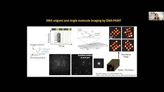
Thank you, Robert, and thank you for the invitation. It's great to be here. So it's going to be mostly imaging-focused, but I'll talk a little bit about unpublished stuff that goes more into the omics direction too. So it could be a nice bridge, I hope, between different disciplines too. So yes, as Robert mentioned, I've been in EMBL for like over 1.5 years now. Sorry. Yeah, let's switch. Yeah. And we are really interested in investigating molecular organization in context. So as opposed to focusing on just one cell or molecule of interest, and labeling and observing it in physical or contextual isolation. We want to really make use of imaging so that we can investigate it within context and learn who else it hangs out with, what's happening around, and how its state interplays with its surroundings. So with that type of mindset, we are really broadly interested in connecting these local subcellular organizational features and how their heterogeneity impacts the cellular state. Of course, this first domain, things like spatial organization, cell-cell interactions, even protein translocations or moonlighting activities, as well as other concepts like cellular condensates or aggregates, are mostly observable by imaging-based methods, whereas this deep cellular state profiling, omics type of technologies including genomics, epigenomics, transcriptomics, etc., has been the way to go. But there is a bit of a discrepancy between these, mostly because earlier wave of omics technologies have been really dissociative and where the spatial context was lost. And with this new wave of spatialomics, things are coming back together a little bit, and we are really interested in connecting these domains also across scales. So previously, I've been working on developing and applying new methods for labeling and detection of biological targets to be able to address the limitations in the imaging domain and study particularly nanoscale spatial organization. So it was more revolving around super-resolution microscopy and things like correlative mass and fluorescence in imaging and probe development. More recently, we've been particularly repurposing DNA as an engineering tool to improve the resolution, multiplexing, and sensitivity of microscopy for spatial biology applications via DNA-based imaging for single-molecule localization microscopy like DNA-Paint and DNA exchange imaging for multiplexing. And I'll be focusing a little bit on these approaches because initially, I mean, our thinking is that we can really empower some of the main limitations in spatial biology context that DNA toolbox. And we've been developing or contributing to a number of methods that really address the limitations that are fundamental to fluorescence microscopy. For example, the resolution through DNA paint type of approaches for single molecule localization, and also combining some of our labeling tools with expansion microscopy to go below the diffraction limits. For multiplexing, we have been coming up with this DNA exchange imaging concept, which I'll talk a little bit more about. And the main part would be the sensitivity improvements with a method that we again used DNA engineering for improving signal-to-noise, and how we can start to connect or integrate imaging with sequencing of cells and tissues through a new approach we've been working on called LightSeq. So just as a recap, of course, there has been many applications that showed use of DNA, particularly in the beginning for super-resolution imaging. The main concept here is that in conventional fluorescence microscopy, a target molecule is labeled with a fluorophore either directly or via probes like TIS or antibodies. But there are a limited number of fluorophores that can be applied at the same time. And to circumvent the spectral overlap, we can instead uncouple the labeling from detection by using DNA-based reversible probes. So in this case, we barcode the target either directly or via probes with short DNA sequences that will be used as docking sites for complementary fluorescence strands, which are called images in this case. So The good thing is there, you know, this hybridization-based detection is really controllable because we understand the factors around this hybridization. So we can really modulate that and reverse this when we need to. And by creating orthogonal barcodes on a library of targets, we can actually one by one or multiple at a time by using different spectral channels detect many different targets and multiplex this targets in this fashion. So this is the general concept for what we call DNA exchange imaging and has been also used in different contexts. I think the first, it came out around as an extension of the DNA paint approach that was developed by Ralph Finkbein and colleagues. And in this case, the same approach is being used, but the imager and docking site interaction is designed to be very transient. So This binding happens and then dissociation happens very often spontaneously. So this gives this blinking behavior where things bind and unbind with predictable binding times. So every time something binds, you can detect a peak, and then when it unbinds, the signal is lost in the background. And this way you can do, of course, single molecule reconstruction by doing iterative imaging. And one could get fairly high resolution with, like, there's some implementations of down to 5 nanometer precision with these type of approaches. This has been utilized also for in situ imaging afterwards by us and by many other groups as well. This exchange plane principle, this was one of the earlier implementations of that shown on DNA origami, has been utilized again on the DNA origami forest and then have been moved more onto cellular imaging. Of course, in this case, it's just like origami structures that show different numbers have been labeled with different sequences and one could in the same sample image all of these sequentially as well. When you now want to actually use it for in situ applications, one could actually change this engineering and make the binding a bit more stable. That would be diffraction limited, but it will be really useful for doing other types of applications beyond super-resolution. In this case, we again multiplex with simultaneous application of probes and sequential application of imaging of fluorophores. Some of the earlier demonstrations were done on cultured mouse hippocampal neurons in this fashion with primary, secondary antibody complexes using stable binding of these probes as much as possible. It's again reversible, but then it lasts a lot longer by engineering the DNA strands to be much longer in hybridization or by modulating the buffer conditions accordingly. So this allows us to apply, you know, all probes like antibodies in one step and then do fast exchange cycles because these small oligonucleotides are very fast in diffusing and unbinding and being washed. And that really helps with multiplexing applications. However, when things come to tissues, of course, there are other issues like autofluorescence fluorescence and scattering. And the variation expression level and epitope access could be actually very different. And proteins, you know, have a very broad range of distributions. It could be just a few copies per cell to millions of copies. And the detection sensitivity is a bit of a problem. So we thought that there is actually quite a need for DNA-mediated signal amplification, particularly to support multiplexing as well. And one way to do that would be, you know, if you were— had to have your target molecule, if you were able to put a concatamer of DNA strands, we would be able to bring a lot of images, and this would create an in situ signal amplification approach. So of course, one question there is how do you create this kind of concatamer in a reproducible and also controllable fashion? So for this, we have been utilizing a new approach which we call SABER. The basic idea comes from the primer exchange reaction, which is a reaction engineered for programmable single-stranded DNA synthesis. So in this case, we utilize this short primers. These are usually 9 to 10 nucleotides, and it's complementary to this overhang of a hairpin molecule, and it can bind here. Here, what you see in red is what we call a stopper moiety. So it could be as simple as a C nucleotide, but then you don't include G nucleotides in the reaction mixture. So there is nothing to add by the polymerase in there. It could also be some modified RNA residue or other things that doesn't allow the polymerase to go forward. So This type of reaction is autonomous and isothermal, so happens in one temperature by itself. I mean, it's just a small animation. So we have the primer binding and a polymerase. This is a strand spacing polymerase, starts to extend it using the templates, and there is like self-dissociation through branch migration process. And this process repeats in this tube over and over at the same temperature, and every time adds one more unit sequence of the primer in there. So you can use it for also creating complex structures, but initially we've been using this for creating long concatamers, which we can use as amplifiers later on. So this way we can create, you know, this amplifier concatamer that we use for in-situ signal amplification. In this approach, we call signal amplification by exchange reaction, or in short, SABER. I mean, the neat thing about this is that we can really create concatamers of predefined sizes because the reaction is really easy to control. One could change the hairpin concentration, polymerase concentration, magnesium concentration in the buffer, or incubation time at one temperature. So it's like a really easy version of a PCR, essentially, where you don't need to do temperature cycling. And you can visualize the products on a gel if you have a fluorophore on the primer to see what kind of lengths you are getting. So, you know, after a few type— a few trials on the reaction, you can really identify the parameters to know at what condition you get what type of length. And this gives you a bit of a control over the signal amplification later on as well. So that's the basic idea. And we have been designing to enable high multiplexing about 50 orthogonal amplifiers. And so far, we have tested 30 of them and validated in situ detection without crosstalk between them for multiplexing applications. So I mean, the way it works is very simple then. We have DNA-barcoded antibodies on the targets. And then we incubate the sample with these in vitro-generated concatamers. We could even do this in branched fashion. So we can even do secondary concatamers if needed and then bring in the fluorescent imagers to be able to detect targets, multiple channels at once, and remove them, add them again, add the new ones again to detect more targets. Exactly. We have done proof of principle experiments in mouse retina tissue. These are quick 40-micron-thick sections from fixed frozen samples. Mouse retina tissue is great for these benchmarking experiments because it really has stereotypical organization and the cell types are defined, so we know where to expect what type of marker. It was producing very well without crosstalk the layered organization of of the mouse retina. This is a 10-color demonstration with multiplexing in linear fashion. And sorry, so this I already showed. We can also do the branching to amplify the signal even further overall, and this can be done on the same sample. So we can do linear or branching applications on the same sample to improve the signal and to tune the signal levels that was— that could be different across different targets. For example, this is an This is a demonstration on human tonsil sections. These are FFPE, so formalin-fixed paraffin-embedded samples, which are classical for medical archival. And some of these proteins like IgA, IgM are very highly expressed, so they can be detected with like linear amplification using fairly short exposure times. But some of them are a bit lower level like the CD8 or Ki-67, which we can now do branching to bring them to the same level of signal. So similar imaging parameters can be used for all of these in multiple channels. One could then do very fast imaging of large tissue areas, this centimeter-scale whole tissue imaging, for example. This same approach is also applicable with RNA targets or DNA targets as well in the SABER-FISH approach. In this case, again, this is a demonstration on retina tissue from 7 different targets across the retinal layers where we can amplify the signal. It really helps to be able to get both specificity and with using low probe numbers because we do still tile, but we get additional amplification from all of these concatamers as well. And we could also do DNA FISH. This is an example from— I'm sorry, it's metaphase chromosomes where a 17-color walk was performed on one arm of the X chromosomes. One additional direction, of course, is to go under the diffraction limit. We can go— we can combine the approach with expansion microscopy. In this case, they are kind of complementary to each other too, because the expansion also dilutes the signal a little bit since now your signal per pixel is going down. But we can bring in some additional amplification through use of SABER. And also you don't have to reapply antibodies every round, which is a pain for these very large samples. And instead you can just switch the images. So in this case, we are actually using the classical expansion microscopy approach from Ed Boyden's group. But we are using staining with DNA-conjugated antibodies And our concatamers are modified with acrydite-modified primers. So, the primers themselves have the acrydite group, which can be cross-linked to the gel. And then when they expand it, they stay on their respective locations. So, this gives, of course, the opportunity to be able to separate and look at structures beyond diffraction, such as these pre- and post-synapses in culture, and also applicable in tissue experiments as well with expansion. So, and it has the additional de-crowding effect as well that the expansion offers too. So as a summary, DNA nanotechnology toolset I presented really allows for multiplex amplification of proteins and nucleic acids in cells and tissues and has a workflow that goes from, you know, designing primers, barcodes, probes, and preparing the concatamers with quality control in vitro and applying them in situ for multiplex detection. And it's like really a DIY system where you can combine different things together up to the amplification level based on the needs without doing in situ enzymatic reactions. And it's really, you know, compatible with standard equipment and even can support high-resolution imaging. If it comes in discussion, we can also look at it, but it seems that at least in our measurements, the— for diffraction-limited imaging, the linear and one round of branching is not creating very big artifacts in terms of the resolution and still staying in a diffraction-limited regime as well. And mainly because we are using linear concatamers rather than very big DNA nanoballs that are several microns potentially long. So, that is kind of the imaging-based tools that we are trying to use for looking things more in context now with different applications varying from, you know, cancer and neuropathologies into the cellular organization. But we have been more recently trying to really connect these type of imaging-based approaches with sequencing-based approaches. Because even if you do these many more cycles of imaging, it's— you still would spend a lot of time to get to the transcriptome level or the whole proteome level. So it's at some point great to be able to get real transcriptomic profiling information, for example, from the cells we image. So for this, we have been really working on merging these two domains from, you know, going from imaging of the samples and maybe adding the sequencing information on top for the same cell, of course, this is a little tricky and there has been a lot of technical limitations. So our newest approach is trying to go into like this challenge where we are using light-directed in situ barcoding of biomolecules in fixed cells and tissues for spatial index sequencing. And this is the work that I've done at the end of my postdoc and has like very big contributions from a lot of members of Peng Yin's group and Kostya Sapko's group, particularly Jocelyn, Kexin, Yining, Liu, and, and So the envisioned approach for this is, you know, we can image with our favorite microscope the cells of, like, you know, the tissues and cells and find interesting areas and then define these ROIs, regions of interest, for in situ barcoding with iterative barcoding rounds so that later on we can actually extract the barcoded reads from only these areas or only these subsets of cells and sequence them with a bulk next-generation sequencer. Sequencing to know what are the different transcriptomic profiles for each of these locations. And ideally, in this process, we don't want to be dissociating the cells or disrupting the samples. So we can even go back after sequencing and do follow-up assays on the same sample, like more multiplex imaging and so on. So to be able to enable this type of an approach, we had to come up with a number of innovations and then apply them together so that we could really go from imaging to sequencing. The first one of these is the fast photocross-linking for a light-controlled in situ barcode incorporation. So this is what gives us a spatial selectivity. The second one is a novel one-step DNA stitching reaction, which we call cross-junction synthesis, which helps us to convert spatial barcodes to sequenceable reads or indices. And third one is like the workflow that allows a non-destructive extraction of transcriptomic information from fixed cells. So we can go from transcriptome to NGS without destroying the sample. And I'll go through each of these to explain a little bit how these steps work. So starting with the spatial selectivity, we actually are using— sorry— like a photolithography-inspired approach. Which, I mean, photolithography is a patterning process in which a photosensitive polymer is selectively exposed to light through a mask, and it creates, like, the opportunity to create different patterns. We're not really using a real mask in this case, but we are using photomasks to expose certain areas to light, which is then used for a photocrosslinking chemistry. So the nucleoside chemistry that we are using for this is called tricyanovinylcarbosol, CNVK in short. This has been developed quite a number of years ago by a Japanese group. And this modification really allows interstrand cross-linking in hybridized DNA strands, as shown here, with pyrimidine residues. And it's very sensitive to thymidines. So when it's opposed to a thymidine residue, it can cross-link under UV illumination. This is 365 nanometer level in 1 to 10 seconds to almost completion. Very high efficiency. And then you have a covalent bond formed between those two strands. So this is the trick we wanted to employ for bringing in spatial barcodes for in situ samples. And we have also utilized actually the same chemistry before for super-resolution labeling of samples, combining like DNA paint with the CMV-K chemistry and controlled photocross-linking as well. So it was one level higher, really high-resolution labeling. But in this case, we we want to bring it back to tissues for doing spatial indexing. And the way we also do it in a more high-throughput way is that we use pattern elimination to be able to identify or expose the areas of interest. So we created DNA barcode strands that are modified with the CNMeK moiety. And this is just like an example for doing a surface barcoding experiment for testing things out. So in this case, let's say this is our docking sequence sequence, and we apply the CNV-K initially in the absence of UV, and it hybridizes to its target sequence. And then in only certain areas, we can expose it to UV and cross-link, and the rest will not be cross-linked. So when we do a stringent wash, we would remove everything else but just leave the cross-linked strands still bound. This is how we typically bring in the spatial indices onto these locations, in, you know, in a nutshell. So we can— this was an in vitro demonstration, and we use a digital micromirror device, or in short, a DMD, to be able to do it in high-throughput fashion. In this case, it has a lot of little mirrors that can be turned on and off, that which creates a customizable photomask. So you can go from any image, like our image here, to a DNA photomask that will help with macro-patterning of DNA. So in this case, we had just a surface of DNA alone where we brought in fluorescent oligos at the desired locations using a black and white mask. We could, of course, do it in multiple colors as well by doing sequential rounds of bringing in different fluorophores, different targets. Like, this is a Penrose triangle that was divided into 3 different regions where it's sequentially barcoded with one oligo. Then you wash, you bring in another oligo with another fluorophore optionally, and so on. So one could also do that in multiplex fashion for identification or labeling of multiple ROIs. So these are just some of the surface experiments, but we can also do this type of photopatterning in situ. I mean, with a DMD, you also have a lot of options to do, you know, checkerboard patterns, micron-scale dot arrays. These are just like fun experiments we have tried initially, or do custom cell selection based on fluorophores, or, you know, go for really manually drawn ROIs for regions of interest in tissue. So these are the possibilities of using the in situ photopatterning approach for different purposes. And we saw that it's feasible to apply it also in tissue samples directly. And next, we wanted to figure out how we can utilize this, like, cross-linked spatial index to be able to actually read in a sequencing reaction, because they are still on— like, they have the still cross-link, and we need to put them into one strand to be able to read it out by sequencing afterwards. For this, we came up with a cross-function synthesis, and this has some inspiration from the primer exchange reaction I've previously talked about. So the way this works is that if you think of this as the, like, a docking site that we have in situ, and this is our cnV-K barcode, if we now create— use this as a template using a DNA polymerase, so this would be the primer, and now we copied this out, this part would be essentially acting as a bit of a stopper because the polymerase normally would not be able to jump through this. But we have designed some internal complementarity here to aid for this process. So when the polymerase or the new strand reaches out here, it cannot really extend normally, but the strand-displacing polymerase could help a jump or a hop here to happen through internal complementarity. So we have this, like, let's say P domain that is normally coming across the P* domain. But because we have another P* domain here, by just DNA movement and unbinding and rebinding, it could actually unbind from here and find another template strand here and continue the synthesis. And in this way, we can essentially now take the transcriptomic information from one side and connected into one strand with the spatial barcode that we brought in. And so this creates the whole template in an enzymatic fashion after the spatial barcode incorporation. So I hope that was clear, but this is how we create the template for the sequencing later on afterwards. Now I want to put all of these together in a workflow so we can really go from one step to the other. So the idea is that, you know, we have fixed permeabilized samples on a slide or in a coverslip, and we first do an in situ reverse transcription reaction, which converts all the RNAs to cDNAs using random primers. So these are 5' and 3' primers, which incorporate internally to all the different RNAs and then used to convert them to cDNAs. Later on, we also do process called A-tailing. So this is like an enzyme that brings a lot of A nucleotides like we have in the poly(A) tails of mRNAs without any template. So this creates another site on the 3' end of the cDNA. In 5', we have our docking site of the primary— our docking site for spatial barcodes. And in 3', we have another handle that's made of poly(A) tails. So then we can start the process for iterative rounds of light-directed barcoding and bring in all the different barcodes for different regions. Of course, one could do actually a lot of imaging in between for different biomarkers, etc., to define those regions. And afterwards, after we are done with barcoding different areas, we do an RNase H extraction. So this enzyme specifically cleaves the RNA in RNA-DNA hybrids. So only for the ones that we created cDNA RNAs, we can cleave off RNA to release all the cDNAs without destroying anything in the sample. So it's very mild for anything other than RNA. And then we move on to doing the cross-junction synthesis because we have all of these junctions that we now convert to readout strands. Then we perform more classical PCR amplification and the next-generation sequencing library prep to prepare samples for RNA sequencing afterwards. Afterwards. So that's like the whole workflow and how different parts come together. I mean, the first experiments we have done were regarding visual pooling of cultured cells. So we co-cultured mouse cells with human cells. In this case, the human cells were expressing GFP. And we can really use that as a marker to label different cell populations. So the human cells got one of the barcodes, the mouse cells got the other barcode. We don't have to, but we used fluorophore-labeled barcode strands just to be able to validate we were labeling the right population in situ. And you can do this for like fairly few number of cells. So in this case, we have done 25 cells of each from like an almost 6mm by 6mm chamber with a lot of other cells. So it was just a small population of samples we actually used. And we were able to map the reads correctly to human or to mouse, but like more than 90% accuracy. That was not bad, but we actually have improved this accuracy quite a bit afterwards, after this first experiments, because one of the reasons why we discovered that there was a bit of a barcode mismatch was just like the background binding of the fluorophores. With more blocking, we actually even improved that in future experiments. But this first pilot experiments gave us a lot of confidence that the whole workflow is feasible to do and one could actually get to the level of whole transcriptome information to find back where they came from and assign the regions back to the in situ cells as well. It also gave us a number of different transcripts that are enriched for different cell types. I mean, in this case, we are not just using the poly(A)-tail but internal priming. So we don't only have mRNAs, but also things like microRNAs, long non-coding RNAs. So a lot of different varieties varieties, which can be really transcriptome-wide in this case. So, you know, that's also an additional advantage of the approach. Then we move on to try— we moved on to tissue samples to do more regional level of transcriptomics. We utilize the retinal cells because we had a lot of experience with them, and there is quite some data on single-cell levels, cell types in the retina. So we had something to benchmark against. And in this case, we have labeled 3 different layers, functional layers in the retina. So the, this 3 layers go with different barcodes. One of them is really enriched in rod and cone cells. This is labeled with the pink. The other one, the BCL layer, is bipolar and Müller cells, mostly in horizontal cells. And then the ganglion cell layer, the green one here, is the, that's really enriched in ganglion cells. So with the different cell types, we were able to really go back from the sequencing results back to the original and make sure that we got the right area. So this is again, more of the benchmarking of the methods as well. This is how it looks after we incorporate different fluorophores to all of these different regions. And we were able to, after the sequencing, able to get 3,400 differentially enriched genes across all of these different layers. So had a pretty good coverage for the different layers. I mean, I will probably not go into all of the details of the target replicates, but it was really confirming the right type of cell types that come from each of these layers. So we had actually pretty good accuracy. We also compared our results across replicates, and it seemed that we had a very good, like, principal component analysis results that show that they were quite reproducible. And more importantly, we have also compared our results to a single-cell sequencing method called DropSeq, where they looked at retinas from the same age of mice, and we were able to capture over 98% of the genes previously covered by DropSeq. This is only focusing on the genes that are actually captured in DropSeq, which are mostly mRNAs due to poly(A)-tail priming. But we have additional sequences as well due to non-coding RNAs that we capture additionally. The sensitivity, that's of course an important aspect, and it's actually quite low in most spatial omics methods. So we we are in principle on par with most of the other spatial omics methods. In this case, we are measuring the sensitivity as opposed to single-molecule FISH. And we are very similar to DropSeq in this case for sensitivity, if you look at the number of expected transcripts from smFISH and what we observe in LightSeq or DropSeq experiments. So of course, smFISH type of methods, which are targeted, would be a lot better in capturing all the copies of a transcript. But our sampling seems to be not so far from doing single-cell level sequencing with drop-seq type of approaches. So that still was quite encouraging. And we also validated that we can do other assays like immunofluorescence after the sequencing of the sample. So this was all the barcodes we incorporated. And we revisited the sample, same sample, same section after sequencing. And it actually antibody stains for some of the different markers. Markers like VSX2, which we saw enriched in only one of the layers. And the sample was still intact for protein imaging. So, that's a possibility as well to go back and validate some of the findings even in the same tissue section. And lastly, we actually applied the approach now for some new insights and getting down to single-cell subtype resolution. So, the idea was to find cells that are really rare, which is a big limitation in single-cell RNA sequencing because you have to dissociate the sample and find those very few cells in a big population and sequence those. But typically it's not an easy task to do. And there is a particular subtype we focused on in this case that's called dopaminergic amacrine cells, in short, DUCs. And these are particularly challenging because they are less than 0.01% of retinal cells. I mean, we only have 4 to 8 cells per section. The cells are rather large. You actually see here the cell bodies. In this case, we have 1, 2, 3, 4, 5, 6 cells, but they have very long processes that are intertwined quite a bit. And because they are among all the other types of omicron cells, they are a bit of a pain to really isolate and then capture in RNA sequencing reactions. And since it's known that this population is dopaminergic, we at least know some biomarkers to find them by imaging. In this case, we are using a tyrosine hydroxylase that's part of the dopamine pathway as a positive marker by immunostaining to identify these cells. So, in this implementation, we have used the stain to find them first in the tissue sections, and then Now we have introduced the barcodes to only the cell bodies of these cells. Then we have also looked at— this is the tyrosine hydroxylase-labeled amacrine cell with the barcode. We have also labeled all the nearby cells in green here. These are mixed population of amacrine cells. This is the target cell group. To be able to get the backgrounds of the general amacrine cell population so we can find the cells that are transcripts that are differentially enriched in just the dopaminergic ones. We have two different barcodes now we are comparing afterwards as well. In this approach afterwards, we moved on to the sequencing from, in one tissue section we only get as few as 4, maximum 8 of these cells. But we were still able to recover 36 transcripts that are differentially enriched for dopaminergic ones in comparison to all the other mixed microglial cell populations. We followed up with doing RNA-FISH using SABER-FISH approach, validation of all the significant targets here so we can make sure that indeed we detected targets that are specific to dopaminergic cells. Among these were, of course, a lot of dopamine pathway genes, which are more like positive controls. But we've also seen interesting targets that have not been observed before because it's very hard to study these cell subtypes, rare cell types. Subtype. So one of them was a voltage-gated calcium sodium channel, normally fairly expressed in neuronal populations. It's more of a cardiac form, but we can see that it was expressed in this cell type. And the other one was a STRAS6, a vitamin A transporter. This is implicated in retinal disease, but so far potential phenotypes related to ducks were unknown because it wasn't known that this is very enriched in dopaminergic population. So these are interesting biological sites to follow up further. But we were very happy to see that the method could be actually used for pooling material from S2/S4 cells from the whole tissue section. So yeah, so this is kind of the whole population and all the ones with star are— have a high enrichment, and those were the ones we have validated with, with SABERFISH approach. Of course, we cannot do the FISH afterwards in the same sample because we already removed the RNA, but we have the confirmation experiments in new animals, new sections as well. I mean, the stereotypic RNA helps with those validation— stereotypic organization of retina helps with those type of comparisons. These are the new markers we have identified. We have also done antibody staining for those in the same section to validate as well. Of interest. So some of the quantitative aspects of these results for those of you who also are a bit more into the sequencing side of things. So we— the UMI, like, UMIs essentially are unique molecular identifiers. They give us an idea of the unique transcripts we are getting. And we were able to get essentially for, you know, if we define the unit area for now as a 10-micron by 10-micron area, because this is a definition that has been given in other spatial transcriptomic methods like the slide-seq, maybe you might have heard of it, or the DBiT-seq type of approaches. And we were able to get UMIs or transcripts from this type of area up in the range of 8,000, like our 6,000 to 10,000. These are all the different replicates. And if you look at the cell size and kind of calculate the transcripts per result, it's again in the 6,000 to 10,000 range. So this many transcripts that we are able to actually read, unique transcripts we're able to read. This is after removing all the, let's say, let's contaminated species like ribosomal RNAs. These are the more informative transcripts. And we are on par with some of these other approaches, both commercially available or other developed approaches for spatial omics, which have been also getting results around 1,000 to 5,000. Per this type of unit area. These results are quite variable because the tissue preparations or tissue types really affect them. But we think that's actually great that we are on par with other approaches or even better in terms of our recovery from the same sample without dissociation. So going forward, of course, we want to integrate some of these multiplexing approaches with the LightSeq to be able to really leverage both the imaging side and the sequencing capability. And for example, we can really do multiparametric imaging with DNA barcoded antibodies or morphology markers beforehand for really good phenotypic characterization and subcellular level imaging, identify different phenotypes or cells of interest based on that, also utilizing machine learning type of approaches, and then define the ROIs like this based on the classification patients and follow with the gene expression to really understand how the phenotype is linked to gene expression. And we can validate these with secondary assays. We haven't tried doing mass spectrometry, but in principle, we believe that's also a follow-up assay we could do afterwards and trying to run some tests on that now. So some of the, like, really good advantages of this approach is that we are really concentrating the sequencing reads only for the cells or areas of of interest. And that actually saves a lot. You don't have to sequence, you know, whole tissue sections and so on if you are interested in only one subtype or one particular phenotype. So that's actually one good, very affordable side of the method. And it's compatible with different microscopy modalities. So you can do your imaging on whatever approach you want to use or whatever microscope you want to use, and then do the barcoding. One thing, of course, a limitation, things are really a bit more onto the 2D level because in 3D 3D, it's hard to really pinpoint the exact location without having light exposure at the bottom or top plate layers. But in 2D samples, you know, at the level of 20 microns, 10 to 20 microns, it would be really applicable. And the approach is quite accessible and affordable for people to be able to do in their labs, just using maybe like a scanner or a DMD on top of an existing microscope. So there are a lot of potential applications. You know, we've already shown you can mix profiles from mixed samples like multiple cell lines, perturbation, or screens. We have shown the capturing of rare or hard-to-isolate cell transcriptomes, and I think that this would be quite valuable for certain follow-up assays. And one could really capture interacting cells, and this is something we are actively working on. And this could also be going into the in situ micro- microbiome. Because we have the full sequencing capacity, we can find the locations of microbes and get their sequences. And in general, identifying spatial heterogeneity and neighborhoods in cell subtypes, and even for creating reference or validation maps for multimodal data. Was there a question?
Oh, okay.
Sorry. So, and in the near future, we want to bring it to Sorry, we want to bring it to even maybe subcellular levels. So we have done this regional sequencing, and we haven't quite done like one cell sequencing, but you know, now we are very close to that. And we could also use photomasks for finer patterning and going down to subcellular levels. We know our chemistry would be able to do that, and it's more of optimizing the conditions and photomasking for for this. So this is like a small trial where we were trying to barcode small subnuclear regions as well in the same cells using actually like confocal microscope and 405-nanometer illumination. So that's also possible, and one doesn't always have to go to UV, although the chemistry is a lot more sensitive in UV. And we are trying to connect the barcoded transcriptomes with protein markers as well to have a multi-omic view. So with that, I want to kind of conclude. Of course, as I said, This was very nice collaborative work with Joshua Elkington, Yuning Liu, and Emma West from Pank's lab and Konstantin Sapko's lab. And my group actively, actually Valerie, Yuxuan, and Jennifer Caron are actually working on developing this further and bringing its new capabilities. We are working on right now. And we also have, if you know anyone who might be interested, potential postdoc fellowships available. Available for this on Connected Methods too. Thank you for your interest, and I'm happy to answer any questions.
Great. Yeah, thank you so much, Sinan. Really interesting and fascinating. So do we have any questions from the audience? If so, then please just raise your hands or simply unmute yourself. Yeah, Mark. Please.
Sure. So I was wondering, in terms of regions of interest, it looks like you were doing this on a tissue scale and ROIs would be on the scale of nuclei. Have you ever thought of going further into maybe looking at space within the nucleus, if there's any asymmetry, say, within the nucleus for transcription?
Yeah, so this is something we are actively trying to do, going to subcellular levels. Indeed, I mean, we have some applications now focusing on, let's say, neuronal compartments, but also we want to extend this to subnuclear regions indeed. So it is possible. We are trying to at the moment characterize the limits, you know, how many cells we would need to pull to get enough sensitivity. The main consideration there is, of course, the labeling is quite specific, but if there is any background stickiness and so on, all of these will contribute to the background. So, and in a way, background scales with the amount of cells you actually have in your whole sample. So it seems like going for smaller chambers and so on is really helping to also not lose too much material during pipetting and so on. So we are optimizing some of these technical parameters, but it seems it would be definitely feasible. We are just trying to see the limits of how many cells would be, uh, too low or would be enough at the moment. But yeah, that would be super interesting to follow that up.
Great, thank you.
Uh, Boaz, please.
Hey, hey, great talk. Um, I have a few questions. I'll start with, with, um, the SABR antibodies with expansion look amazing. Uh, is there a way to get these things? Do we have to make them? How easy it is? How many times does it work?
Yeah, those are good questions. So currently We don't have a provider that sells you everything, unfortunately, but I think they are easy to make. We do DNA conjugation to the antibodies ourselves. There are very cheap DIY methods, but there are also commercial kits available to do these that we have characterized. We actually— I will show this. We have two websites to help people a little bit with if they have run into trouble. So this would be like for ImmunoSABRE and SABRE FISH. So we have some advice on how to, how to do these things, but we have a lot of people that use help desk that come up with their own applications and reach out for advice. So we are trying to support the potential users as much as possible, but there are a lot of kits and I can give specific advice on what might be, what has worked best for us to do the reagent generation. The concatamer generation itself is actually really easy, and we have very detailed protocols and the supplements of the papers, and we have a lot of people who successfully reproduce the, you know, the concatamer generation and so on without any problems as well.
So just a quick question on those. Have you characterized the, like, the length distribution so we can see them in the— right, as different stripes, but within a stripe, like how much variability do you expect there to have?
There is certainly some variability because it's an enzymatic process and kinetic process. I mean, if you were to really boost the signal, we would also see more truncated forms, but majority of the like extended products is at a certain regime. And we have seen that it is— that regime is actually a lot tighter compared to some of the other methods. So that's actually nice. We are We're also trying for certain applications where quantification might be more critical, like have tried some approaches to enrich particular lengths as well, you know, from like cutting it down from the gel or purifying certain lengths. So those could be done. We don't routinely do that for all applications, but that's possible to do for things that are really sensitive. Overall, it changes a lot from sequence to sequence, the efficiency of that. Because it's a kinetic process. But we have in the paper, you can find in the supplement actually, like a validation for different types. So we have— we are showing the full gel and you can see the distribution. And we have characterized the best-looking sequences for high-fidelity applications as well.
Got it, thanks.
Good. Do we have any other questions? If not, maybe I have another one more technically, and it actually goes back to some of the discussions we already had seen, and especially on the spatial barcoding, you know, for the photocrosslinking, right? I mean, you're using this one photon illumination and the digital micromirror for this. So have you already looked into whether you could also do this in a two-photon process?
Yeah, this is something that I definitely want to follow up with you as well. I mean, I've been looking if there is any information on the chemistry and whether it's reactive to, like, to photolumination. So far, I think that has not been studied at all. So this is something we have to characterize. I mean, so far we have tried a few different parameters for there, but we didn't see good activation or good cross-linking. But we haven't been very comprehensive. And so I think this is something we need to do a bit more of a screening of different parameters to identify what works. I think it's could be feasible, but we don't have any results on that yet. And this is something we definitely want to invest a bit more energy to see if that's a possibility, because a 3D application would be, would be quite interesting.
Yeah, exactly. Yeah. And otherwise, do you like— what is sort of the limitations and how, you know, deep you can go into a certain tissue? Um, so the tissues can be for—
I mean, it's— so basically it could be fairly thick in terms of the light penetration, but the main problem is that of course you would be labeling everything like a column. Unless you have the two-photon. So we haven't tried very thick tissues. The other limitation, of course, in doing things in thick tissues is, of course, the reagent penetration and collecting RNAs and so on.
That's what I was referring to.
Yeah, so that's— we haven't gone very thick. The retina tissues we have used were, I think, at the 10 to 20 micron range. So that has been as thick as we have gone with the approach for FFPE samples and things like that. Usually the range is more in the 5-10 micron. I think for those applications, you know, the 2D capability is pretty good because we are in the one, almost like one cell thickness. But we haven't gone too far based on some of our previous experiences with like SABER penetration and so on. One could probably go thicker in terms of penetration of reagents, but we haven't tried extracting RNA like, or cDNA from much thicker samples so far. So hard to comment on that.
Okay.
Well, thank you. And I'm looking forward to what's coming next in this direction. Akanksha, please. I hope I pronounced your name correctly.
I can ask another question in the meanwhile. Have you compared the ImmunoSABER to normal primary and secondary? I know in the paper, but just in terms of like quantifying?
Yeah, if you can wait a second, I can pull a slide for that. Let me— yes, we did. So maybe I can show the data rather than— so this is, sorry, yeah, so this is kind of the application on the FFPE samples. Like these are tonsils, and we are looking at like a particular region here in the germinal centers. All of the dots are copy. And the red is a Ki-67, a proliferating cell marker that we have labeled with, you know, like an antibody that has a DNA barcode. For the unamplified case, we have this no concatamer but only like one imager binding site. I'm sorry, I think I wanted to show the secondary antibody fluorophore too. So with the— I mean, this would vary a little bit from sequence to sequence, but this particular case, we have quantified things side by side. This is kind of the linear amplification around 6-fold, and the, the secondary antibody fluorophore was giving around 8-fold. Again, this would be variable again depending on the secondary antibody and how many fluorophores each one has, but it seems like the linear one is giving comparable amplification overall in our experience to the secondary antibody-secondary primary complex. And if we go with branching, we go way above that. So in this case, with one round of branching, we were able 50-fold. And then we can even do an iteration, one more round. And this brings us to like almost 188-fold. So you can also just use the amplification itself without— for like really rare targets without multiplexing. And, you know, push things down up to like 200-fold. I'm looking for another slide. Maybe we have like a high zoom versions of this to to show, but maybe I don't have it here, to show that— I'm sorry, I might not have it here— but to show that we have still very good morphology after these. So this marker is a nuclear marker, and even after like 200-fold amplification, we can actually see the signal still confined to the right compartments, whereas other amplification methods like TIRAM and signal amplification, which are quite diffused using are starting to go even outside of the nucleus in terms of the label kind of spill out. So this is why we think it might be good for like higher resolution subcellular level. I mean, not super resolution, but high resolution applications too in general. And I couldn't find it now, but we definitely have some applications— sorry, no, I couldn't find it— have some application also on like microtubules and so on to show that we can do dense labelling on these kind of structures. So, the concatameres are not like, you know, creating just point-like spots but allow you to label almost like continuous microtubule structures too.
And in terms of diffusion into thick tissue, how long do you think— how long are you waiting, or have you tried?
Yeah. So, for— you know, we have tried. I've tried myself up to like 100-micrometre tissue sections. These were cleared sections, so it might be different for different types of preparations. So in this case, we went usually, you know, 1 to 2 days of incubation times for the antibodies. In general, my observation is that the concatamer penetration works well at the level of, you know, antibody penetration, because after a certain regime, also the antibody penetration becomes very uneven. So it seems that there is like— it's kind of similar between the penetration of antibodies and the concatamers. But having said that, if the concatamer lengths get too long, we see an adverse effect. So because we can, you know, actually extend things up to 1 to 2 KB, but that seems to be a little bit not as helpful, both used to penetration. And I think things start to have more like secondary structure or inhibiting binding. So typically, we keep more at the 500 nucleotide level, 500, 600 nucleotide level for optimum penetration. Binding.
Got it. Thanks.
And I see now a question on the chat. I was just wondering, normalization across different barcodes, area efficiency of the barcoding process itself creating variability. So are you asking about the LightSeq approach here? Right, LightSeq, right? So indeed, if one uses different areas and so on. This could create variability, but some of these type of variability has been addressed very well in the sequencing field because they have different number of cells in the samples and so on. So there are a lot of statistical normalizations that are done in analysis packages for a different number of cells because in the single-cell sequencing or bulk sequencing, you know, like in the single-cell sequencing, there are a lot of like zero fields and so on. So these type of problems are actually common in that field and have been addressed with statistical normalizations. For example, the tools that we have used is from Wolfgang Huber's lab called the eSeq2 that has a lot of normalizations happening in the background for different sample sizes based on the expression profiles. That's one thing. In terms of the cross-linking efficiency, if the different ROIs are affecting— the different sizes of ROIs are affecting the binding. This is actually something we are quantifying in a bit more detail now because we are also trying to use like a scanner-based, laser scanner-based system as an alternative to DMD. And in this case, of course, the scan times are varying a bit based on the ROI, whereas in the DMD it is probably less of an effect and things are more linear. So, so this is still— we are working on whether there's an effect, and I think it really It depends on how the scanning is done and the synchronization of the laser and ROI definition. So if the power density that the sample was exposed to is relatively, like per pixel, is relatively standard or it's not constant, I don't expect a huge change. But if that is changing due to the assay and ROI size, then there might be some problems. Does that answer your question?
More from Joshua







Scientist Stories: Hans Waterham, The value of phenotypic assays in peroxisomal disorders




Scientist Stories: Joel Zlotogora, Genetic carrier screening programs for reproductive purposes