Scientist Stories: Vamsi Mootha, Mitochondrial Diseases and Function
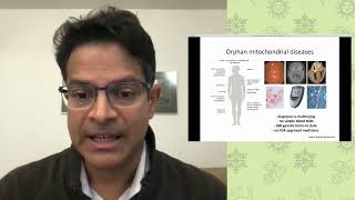
Vamsi Mootha explains how mitochondrial dysfunction causes age-related and rare genetic diseases, and describes his lab's catalog of 1,100 mitochondrial proteins. His key finding: exposing mice and cells to low oxygen (11% instead of normal 21%) suppresses disease symptoms, extends lifespan, and even reversed brain lesions in Leigh syndrome models, suggesting defective mitochondria accumulate toxic oxygen.
Chapters
Hello and welcome to this evening's Science for All Seasons talk. The Science for All Seasons talk give you a chance to explore hot topics in genomics and biology with experts from the Broad Institute. My name is Tom Ulrich, and I'm the Associate Director for Science Communications at the Broad. And it's my pleasure to welcome all of you here this evening and to introduce our speaker, Vamsi Motha. Vamsi is an institute member here at the Broad and is the co-director of our metabolism program. He also holds professorships in systems biology and medicine at Harvard Medical School and molecular biology at Massachusetts General Hospital. Vamsi is going to talk about our mitochondria, which we all know as the powerhouse of the cell. But in his studies of mitochondrial diseases, Vamsi has come to the idea that our mitochondria have another fundamental function that also impacts our cellular and overall health, which he'll discuss tonight. If you have any questions for Vamsi, please ask them using YouTube's chat feature, and we'll do our best to discuss them during the Q&A portion of the evening. And if you're tweeting, please use the hashtag #BroadSFAS. Vamsi, the microphone is yours.
Good evening. Thank you so much, Tom, for that kind introduction. And it's a real pleasure for me to be here today and speak at Science for All Seasons. Over the next hour or so, I'm hoping to take us all on a small voyage into our mitochondria. And this is an organelle that first captured my imagination when I was a medical student more than 25 years ago or so. And the electron micrographs that you see in the background, these are electron micrographs of mitochondria from different organs in our body's tissues. And it's these beautiful images that just initially grabbed my fancy. And as I learned about their evolutionary history, their roles in disease, I was hooked. And this is all that I've been working on since then. And I've had my own laboratory, as Tom said, at MGH and at the Broad for a little bit over 15 years or so. This is the outline of my talk today. I'm going to provide a brief primer on mitochondria. And then I want to introduce you to what happens to mitochondria in a variety of human diseases. And then I want to share with you our approach to mitochondria, and I want to go deep on one story towards the end that I think is focused on disease but is providing new insights into the role of mitochondria. So this is what mitochondria actually look like in living cells. I think most of us are accustomed to those static electron micrographs that we've all seen in our high school biochemistry textbooks, but if you actually image mitochondria mitochondria in living cells using fluorescence microscopy, this is what they look like. So every single one of these red thread-like structures is one mitochondrion. And as you can see, within each of our cells, they're swimming around, they're dividing, and they're fusing. They almost look like tiny microscopic life forms. And that is, of course, because about 2 billion years ago or so, they were free-swimming bacteria. So our mitochondria that look like this today, about 2 billion years ago, were probably free-swimming bacteria, not that different than E. coli. So how did this happen? So there's something called the endosymbiotic event. I'm sure many of you have heard about this in your high school or college biology classes. But what we think happened was that approximately 2 billion years ago or so, there's a close relationship between one cell, an archaeon, and then another bacterium not that different than E. coli. This relationship became very close, and at some point it became permanent. And this cell is actually the common ancestor to humans and plants and slime mold and baker's yeast. And so clearly one of the most important events in the history of life. Now, this is a cartoon of a human cell, and inside of the nucleus, you're going to see chromosomes. And so most of our body's DNA is in the nucleus, and this consists of about 3 billion bases. And we receive these chromosomes and this DNA both from mom and from dad. But our mitochondria are really living fossils. If you look inside of them, they actually still have a tiny circular genome that looks a little bit like a bacterial genome. It's much smaller than it was 2 billion years ago, but it's still a reminder of the bacterial ancestry of this organelle. And this particular genome is passed from mom to offspring, from mom to offspring. Now, as we all know, the mitochondrion is called the powerhouse of the cell. So why is that? Well, the organelle houses a number of different machines. It's super important for metabolism. But inside of these inner membranes of the mitochondrion are the machinery called the respiratory chain. This membrane, this inner membrane, if you splay it out from one individual, that's basically the same surface area as an entire football field. It's the most proteinaceous membrane in the human cell, and it's chock full of these little protein complexes. And what these protein complexes are going to do, it's called the respiratory chain, and they're going to enable what's called oxidative phosphorylation, or OXPHOS. So this is the process by which the food that we eat gets burned with oxygen and then gets converted to ATP.. And just some food for thought. Each one of us in a given day is breathing about 500 liters of oxygen, and we're turning over about 50 kilograms of ATP per day. So that's how active the inner membrane of our mitochondria are every day. Now we have two pathways for making ATP. We can either use glycolysis in the cytosol, and that's going to yield about 2 ATP per glucose molecule.. And that doesn't need mitochondria or oxygen. However, if we choose to use our mitochondria and if we use oxygen, we'll now all of a sudden be able to get about 36 ATP molecules per glucose consumed. So a huge power boost. And this is why the mitochondria is called the powerhouse of the cell, for good reason. Now, I want to introduce a water wheel analogy because I think this might be useful later on as we talk about what happens in mitochondrial disease. So what's happening inside of that inner membrane? And I think it's very analogous to what's happening with a water wheel, and if the water wheel is actually equipped with a turbine. So as water flows from high elevation to a low elevation, the water wheel turns, and that mechanical energy is going to get converted into an electrical energy by the turbine. Now, inside of the mitochondrion, the food and the oxygen They wanna react with each other. And if they do, there's a lot of energy to be harnessed. However, there needs to be a path by which they can react. The mitochondrion provides a vehicle for those two to react with each other. And there are tiny microscopic turbines, the machines that I just showed, that now spin, that will now effectively convert the ADP to the ATP. And this is the cellular energy currency that powers our motors, allows me to think right now. So hopefully this water wheel analogy makes sense, and we'll get back to it a little bit later. So that's a brief primer on mitochondria, and now I want to share with you what happens, um, in, in, in health and in disease. It is now well appreciated that virtually all of us, as we age, we experience a mild decline in mitochondrial function. This is one of the strongest biomarkers of the aging process. Now, what's unclear is whether this is actually driving the aging process or if we have sick tissue and hence sick mitochondria. At the opposite end of the extreme are an extremely large collection of individually rare orphan diseases. These are typically children, but sometimes adults, that are born with birth defects in some part of their mitochondria. This leads to single organ or multisystemic disease. In these diseases, there's little doubt that the mitochondrial defect is causal for the pathology. And then there's some common diseases between these two, like where again we observe mitochondrial dysfunction, but cause or correlation, it's unclear right now. So what's happening with aging? Ian Traunce showed a few decades ago or so that if you biopsy skeletal muscle from individuals of varying ages, we can purify their mitochondria, put them in a test tube, and measure rates of oxygen consumption and ATP production. And what you can see is that this declines as a function of age. This extends in vivo as well. So it's possible to take humans of varying ages, you exercise them, and measure how much oxygen they're consuming while they're exercising as fast as they can. After the age of 30, this particular metric, the VO2 max, declines about 10% per decade in all of us. And so that's the bad news. But the good news is we know that regular exercise can send this trend upwards. I want to introduce that opposite end of the extreme now, these rare orphan mitochondrial diseases, because this is what we have been focusing on historically, both because they represent a great unmet medical need and also because studies Studying them may provide insights into some more common conditions. These are tough diseases. Diagnosing them is really hard because virtually any organ system can be impacted. Some of these patients have one organ system impacted. Others have multiple organ systems impacted. And when a diagnosis is finally arrived at, the patient has gone to more than 7 different specialists before the diagnosis is achieved. We don't have any simple blood tests right now that tell us whether the mitochondrial disease is getting better or worse. There's more than 300 genetic forms to date without a single FDA-approved medicine. So that's sort of the spectrum of mitochondrial dysfunction from the rare into the common. And what's super fascinating is that this field is now gaining widespread interest. A few years ago in The New Yorker, there's actually an article entitled Silicon Valley's Quest to Live Forever. There's a lot of interest in aging.. And in this particular article, the author takes us through a party at a Hollywood party where Goldie Hawn is sitting on a sofa and she asks a question. I have a question about the mitochondria. I've been told about a molecule called glutathione that helps the health of the cell. And like I said, I've been studying mitochondria for more than 25 years. Never in my wildest imagination would I have thought that there'd be an article in The New Yorker that would have Goldie Hawn asking a question, a very good question also, about mitochondria. So now I've provided a primer on mitochondria, and I've introduced you to what happens to mitochondria in health and in disease. And now what I want to do is I want to share with you our lab's approach to this organelle. So we're really trying to take a systematic approach to mitochondria. And I like to use a particular analogy to describe what we're trying to do. I grew up in Southeast Texas where one of my brothers was a real car enthusiast. He would go and buy old cars and then we would work on fixing them up. And the first thing that we would do when he would buy a new old car is we'd go to a store called AutoZone and we'd buy what's called a Chilton manual that specialized for that particular make and model of a car. And as you open up the pages of this booklet, The first few pages have all the parts of the car and their parts numbers. The next few pages have all of the wiring diagrams of the electrical system, the air conditioner, the transmission, and the carburetor. Then the next few chapters are about how to debug the car when it's not starting properly and how to fix it. And to a large extent, this is what we're trying to do, not for a 1965 Mustang, but for the mitochondrion. And so the first step in our lab's efforts was really to try to figure out what all the parts are. Back in 1981, Fred Sanger and colleagues sequenced the mitochondrial genome from humans and reported that it encodes 13 proteins total. I'm sure you can appreciate that when you look at the electron micrographs of mitochondria, they're so glorious, they must consist of more than just 13 proteins. And by necessity, all of the other proteins must be coming from the nuclear genome. This was sequenced in the year 2001 in draft form. And I trained with Eric Lander in genomics. And my goal subsequently has been to try to figure out what all of the components are that come from the nuclear genome that end up in the mitochondrion. And so over the course of our lab's a little bit more than 15-year history, we've worked very closely with the Broad Institute's proteomics platform to build a parts list for this organelle. And this is really work that was initially spearheaded by David Pagliarini, one of the first postdocs in my laboratory, as well as Sarah Calvo, a computational biologist in my laboratory. And using a mix of mass spectrometry, imaging, and computation, we've been able to put together a list of 1,100 proteins that comprises this organelle. And we've continually updated this list And we've made this freely available to the research community. Now, a large number of these proteins have never been studied before, and we would love to know what they do. And so what's beautiful about the new tools of genomics is that we can study all 1,100 of those proteins, get snapshots of all 1,100 of those proteins at once using new tools like RNA-seq and microarrays and proteomics. And so we love doing those types of genomics experiments. And then we develop computer algorithms with which to mine all of that data, as well as what's available in the public domain. And through this experimental and computational approach, we're working out the function of a lot of the circuits of the mitochondrion. We're also very interested in understanding which of those mitochondria circuits are altered in disease. And to understand this, what we've been doing is we've been collecting blood from patients in close collaboration with our clinical colleagues throughout the world. We collect blood from patients and their family members that tend to be unaffected. And then we use next-generation DNA sequencing to try to understand what— which of those protein components are defective. And then we also use something called metabolomics to try to understand all of the biochemistry of these disorders. So in this way, we are identifying the genes and the biochemical pathways that are altered in some of these rare mitochondrial diseases. And this is our long-term goal, precision mitochondrial medicine. Our hope is that from a single tube of blood, we should be able to establish a genetic diagnosis. And from that same tube of blood, by looking at the chemicals, we ought to be able to stage the severity of the disease. Now, if we only have a therapy, then what we can do is we can monitor these chemicals to see if the patient is getting better or worse. And so, what's really missing right now is a novel therapy. And that's what I want to focus on in tonight's talk in the time that remains. So, back in 2014, we completed what I think is one of the most important experiments in my laboratory with generous funding from the Marriott Family Foundation. We used a new genomics method to screen for ways to cope with a broken mitochondria. And this was a screen that was performed by a talented graduate student, Isha Jain, at that time. Now, this screen gave rise to a very counterintuitive idea. It gave rise to the idea that hypoxia, so low oxygen, might actually serve as a therapy.. And this is a bit counterintuitive because we usually think of oxygen as being life-giving. And any of us that have ever gone to the hospital, one of the first things that will happen is that we will be given oxygen. And so this screen actually told us the exact opposite. It was telling us, let's dial down the oxygen when there's mitochondrial disease. I want to share with you some of the work that we did with this idea. And first, I need to just introduce a disease and a disease model in which we've been exploring this idea. The disease in which we've been exploring this idea is called Leigh syndrome. This is a disease that was first described in 1951. It's the most common pediatric mitochondrial disease, and it's associated with a subacute degeneration of the gray matter of the brain. There's more than 80 different genes, either on the nuclear DNA or the mitochondrial DNA, that have been identified, and we don't have a single FDA-approved medicine for Leigh syndrome. A bit more than 10 years ago or so, Richard Palmiter's group in Washington described a mouse model of Leigh syndrome. They knocked out one of those nuclear genes that's important for the functioning of the mitochondrion. And these mice are born fine, but then right around at 2 months of age, they become very sick. They stop moving properly, they'll lose their body weight, and then they'll develop these lesions in their brains by MRI that are resembling the lesions that we see in the humans. So we received these mice and we wanted to see whether this idea of dialing down the oxygen might work. And so I'm going to show you a video of 5 mice. All 5 of these mice have this neurological disease, except 2 of them have been breathing 11% oxygen Now, all of us at sea level are breathing 21% oxygen, but what we're able to do is we're able to dilute the air with nitrogen that two of these mice are breathing. So now they're breathing only 11% oxygen, and they're— all five of them are placed in the cage at the same time for the purposes of the video. But see if you can guess which two mice have been breathing thin air. So, I hope that was obvious. The results were really, really striking. We were overwhelmed by just how powerful the effects of hypoxia were in this mouse model. I want to point out that we did these mouse studies in collaboration with Warren Zappal's laboratory at Mass General Hospital that had experience delivering hypoxia to mouse models. Now, this mouse model will die at 2 months of age from the neurological disease that I just told you about. However, if they're breathing thin air, this is what the survival curve looked like. At the time of publication, you know, there were zero mice that had died from this disease. Post-publication, we carried out the survival curves, and the mice are not fully cured of their disease. They'll still die from their cardiovascular disease at approximately 1 year of age. So a dramatic extension of lifespan from about 2 months to almost a year just by dialing down the oxygen levels. And as you can see from the video, it wasn't just lifespan. But the healthspan was also dramatically improved. They're putting on body weight, they're moving around, they just look a lot more robust. A really natural question is, if low oxygen is good, what happens with high oxygen? We decided to provide the mice with 55% oxygen. This is an O2 level that's often experienced by patients in the hospital, and these mice will die within about 3 to 5 days of exposure to 55% oxygen. So these mice that have mitochondrial disease are very sensitive to high oxygen. Within one week of us publishing this paper, I received telephone calls and emails from clinicians around the country and the world that actually shared with me vignettes of their outpatients that had mitochondrial disease that were actually placed in hyperbaric oxygen. That's a great way of flooding the system with oxygen. And they shared with me anecdotes of even outpatients that became comatose within 24 hours of hyperbaric oxygen and never woke up again. And so these are anecdotes that suggested that hyperbaric oxygen may be detrimental, but when combined with our mouse studies, strongly suggest that high oxygen can be toxic in the setting of mitochondrial disease. Now, we've been working with Fumito Ichinose's laboratory at Mass General Hospital to understand exactly what's happening. His laboratory is able to place probes into the brains of these mice, which lets you measure what the oxygen levels are. This is what the oxygen level should be in the brain of a healthy wild-type mouse. As you can see in the mouse model of mitochondrial disease, there's dramatically elevated levels of oxygen in the brain. Now, we're interpreting this as high unused oxygen in the brain. Remember, all the oxygen that we're breathing, a very large fraction of the O2 that we're breathing, is actually getting consumed in the mitochondrion. And so our inference here is that when the mitochondrion is broken, the oxygen is still being delivered, but it's not being extracted. And as a consequence, it's accumulating. And by simply dialing down the oxygen that's we're breathing in these mice, we can normalize this brain hypoxia. And so the canonical model of mitochondrial disease is based on the canonical idea that it's the powerhouse of the cell. When the mitochondrion is broken, there's less energy, there's less ATP. And this is definitely a component of the pathology. I'm not arguing that. However, I think what's, what's often overlooked And I think what our work is telling us is very important, is that when the mitochondria is broken, there's also high unused oxygen. And that this high unused oxygen can be toxic and it can actually cause pathology. And this is why when we dial down the amount of O2 that's being breathed by these mice, we can actually alleviate their disease. I want to just go back briefly to this water wheel analogy. So again, The water that's flowing is turning this water wheel, it's turning a turbine, and it's producing electrical energy. If the water wheel is blocked for some reason, sure, there's not going to be enough electricity. However, we're also going to see some flooding upstream and even a drought potentially downstream. And I think what our work is telling us is that this flood of oxygen can damage parts of our cell. And this is where a lot of our research is focused right now. We're trying to understand what parts of our body cells, molecules, and pathways can get oxidized by too much oxygen. And in the same way that a copper penny can turn green or an iron nail can rust, that's all because of oxidation. We believe that oxygen itself can damage certain parts, biomolecules, or cells. And this is also contributing to the pathology. So I've shown you that chronic continuous 11% hypoxia is promising in mouse models. Everything that I've shown you so far requires that we place the mouse 24/7 in a box breathing 11% oxygen. Now, translating that into a therapy might be challenging. And so an important question is whether something more practical may work. We've tried intermittent hypoxia in these mice models, and that does not work. So we can't do 12 hours on, 12 hours off. How about something like instead of 11%, which is Pikes Peak or Mont Blanc, how about if we went to Denver instead? So 17%. Unfortunately, that's also not effective. There's also genetic ways and chemical ways that we can trick the cells into thinking that they're hypoxic.. And in the context of a mouse model that I just showed you, tricking the cell into thinking that it's hypoxic is not good enough. So the only thing that's working right now is chronic continuous 11% oxygen. I also wanna emphasize that it has dramatic effects on the brain disease, but it's not fixing the cardiovascular disease. So these are some of the limitations of the approach that I'm sharing with you. And I wanna emphasize, please do not try this at home. This is preclinical research. Hypoxia can be very dangerous. And so we have a lot of preclinical research that we need to do before we can try to move these ideas into patients. Now, a really important question is whether we can reverse any of this disease. Most of these patients will actually present with some of these neurological deficits already present on their MRIs. So a really important question is whether any of that is reversible. An advantage of this mouse model is it has a very stereotyped trajectory. And right around 2 months of age, these mice will now fulfill our hospital's euthanasia criteria. So we can ask, what if we start hypoxia at this point? And when we do, we see that the mice are now regaining their body weight. It's quite dramatic. And we actually call this affectionately, our lab's Lazarus effect on this NDF-S4 mouse model. If we do brain MRIs on this mouse model, what you see is that these lesions that we see by MRI, we can make them disappear after a few weeks of hypoxia therapy. And so we find this to be very, very exciting. And we believe that this is also supported by human anecdotes as well. And Missy Walker and Maria Pilar Miranda in my group have recently written a review article that has shared some cases of reversibility in humans. We believe that the observations we're making in mice also extend to humans as well. So we believe that we have made a really important and what I think is a super exciting discovery, and that is that hypoxia is a suppressor of mitochondrial dysfunction. What I mean by that is hypoxia allows cells and organisms to cope with broken mitochondria. And we're really excited about this discovery. And we have a lot of active work ongoing right now aimed at trying to understand the full mechanism by which this is working, both in cell culture as well as in vivo. We really want to know whether this will generalize to other diseases. As I said earlier, There's about 300 monogenic mitochondrial diseases, and everything I've shown you so far is focused on one of those. And then finally, I've shared with you some of the limitations of being able to translate this into the clinic. So we're working on trying to identify safe, practical, effective means of moving the science forward. What's really exciting is that in newer work that we've done in cell culture, at least, there appear to be hundreds of mitochondrial mutants that show a fitness defect at 21% but can then be suppressed simply by lowering the oxygen tension. So at least in cell culture, it looks like this is going to generalize, maybe not to every one of those mitochondrial diseases, but to a lot of them. There's one disease that we're really excited about, and that's Friedreich's ataxia. This is actually the most common monogenic mitochondrial disease. And it's due to a recessive loss of a gene called frataxin, which was historically considered to be an essential gene. Nobody had ever knocked it out completely. But in human cells, in yeast models, as well as in worm models, we can completely knock out this gene and make the cell, yeast, or worm survive simply by dialing down the oxygen. I want to just highlight this one particular slide from C. elegans worm models. This is what worms ordinarily look like. Each of those squiggly lines is one worm. If we knock out this gene completely in a worm, you don't see any worms because it's lethal. So this is consistent with the idea that this is an essential gene. Now, if we just dial down the oxygen, these worms not only survive, but they will now complete their entire reproductive life cycle as well. And so it's a dramatic effect in worms and in yeast and in human cells. And now we're trying to move this up the preclinical chain, if you will, with the goal of trying to impact this disease. I want to now take a step back and just think about what the broader implications of our work is. When we think about human disease, we often think about the cross-product of genes and environment. And I think what our work is telling us is that when it comes to mitochondrial disease and mitochondrial genetics, the key variable is environmental oxygen. Remember, our genes co-evolve with the environment, and By studying patients, we've uncovered what I think is a really important G by E interaction, genetics by environment interaction. And this particular interaction may actually be related to an even more ancient event. Now, I told you earlier about the endosymbiotic event that took place about 2 billion years ago or so. A really important question is, what was the selective advantage for this archaeon and for this mitochondrion to get together and form a permanent union? One of— there's a number of theories as to what the selective advantage was, and, you know, one of the ones that's commonly cited amongst high school students and college students is the ATP power advantage. The mitochondrion that was engulfed provided a lot of power to the host, and I would emphasize that that probably is true, but the question is, could there be other advantages as well? Now, if we go back a few hundred million years prior to this endosymbiotic event, we have to remember that life on our planet started out in a completely anaerobic state. And then around 2.4 billion years ago or so was what's called the Great Oxygenation Event. And so we went from no oxygen to some oxygen. And again, oxygen is life-giving, but it can be corrosive as well. And this was— it probably caused a lot of problems for anaerobic organisms. And so a possibility is that another advantage of endosymbiosis was the ability of this mitochondrion not only to produce a lot of ATP, but also to consume oxygen and detoxify it. I think our work has important implications for rare mitochondrial diseases. We will instinctively give supplemental oxygen to patients when they come to the hospital, and in the vast majority of cases, this is totally fine. However, if somebody has rare mitochondrial disease and they don't have any signs of hypoxia, I think our work is suggesting that we may need to think twice before giving supplemental oxygen when it's not even indicated. And so our community is now beginning to take notice about this possibility. I think our work has potential therapeutic implications as well. We're doing a lot of preclinical work in mouse models and in worm models that I told you about earlier, but we've also just recently completed a safety study study of hypoxia in the hospital. So my colleagues Lorenzo Barra and Stuart Harris have created hypoxia in a tent at Mass General Hospital. And we've brought in a couple of healthy volunteers that spend about a week or so as the O2 levels are dropped. And so we're gaining experience delivering hypoxia in a hospitalized setting, just trying to understand what the human physiology and metabolic responses are. And so these are still in healthy humans, but hopefully The information from these types of studies or preclinical studies will then allow us to develop therapies for some of these patients. Now, everything that I've told you about today is focused on rare mitochondrial diseases, but might there be broader benefits of hypoxia? Remember, I told you that virtually every single common age-associated disease is associated with a subtle decline in mitochondrial activity.. And so does anything that we've learned have relevance to more common diseases? And so now I'm definitely speculating. And what I wanted is only to share with you this one paper that was actually published by the Indian Army. Now, the Indian Army usually is not in the business of publishing health outcomes research studies, but they made a really interesting observation that they felt compelled to share with the broader community. India in the 1960s had a lot of border disputes with China. And at that time, what India ended up doing was to deploy 150,000 troops at the Indo-China border. Now, it's a vast border. And out of those 150,000 individuals, 20,000 of them were at high elevation, comparable to the levels of oxygen that we're talking about today. The, the other 130,000 were either at the plains or at sea level. And they were stationed there for about 7 years. And over the next 7 years, the clinicians in the army were able to see, you know, how these soldiers were doing. And what's remarkable is that over the 7-year period, the incidence of diabetes, hypertension, ischemic heart disease were all much lower in the individuals living at high altitude. Now, of course, lots of things are different between sea level and high elevation. The temperature is different, UV radiation is different, the clothing is different, the food is different, the activity level is different. But one of the things that is different is the oxygen levels as well. And so it's tempting to speculate that chronic mild hypoxia might actually have some health benefits. Again, this is pure speculation, but the types of hypotheses that are raised by our work. So in closing, I've shared with you a primer on mitochondria. I've told you about mitochondria and human disease. And I've shared with you our approach to this organelle that's led to some new insights, namely the idea that low oxygen can actually buffer defects in the mitochondrion. And that this has, I think, potential implications for understanding the basic biology of the organelle, but also for understanding how to alleviate mitochondrial disease. Now, this is probably the most important slide. I really want to just thank the past and present members of my laboratory, as well as our wonderful collaborators that have helped us with all of the work that I've shared with you today. This has been a lot of teamwork over the more than 15 years that we've had our laboratory. And I really want to do a shout out to Dave Pagliarini and Sarah Calvo. Those are the two that really created that first Mitochondria Inventory that's so widely used today. I also want to do a special shout out to Isha Jain. She was the graduate student that did that initial genetic screen that gave rise to that very initial idea that low oxygen could be beneficial in the setting of mitochondrial disease. And they were all helped by a number of other members of our laboratory. And also, I'm very grateful for our collaborators, most of whom I have mentioned throughout the course of this talk, but including Dr. Warren Zapol at Mass General Hospital, who's been a really wonderful collaborator for hypoxia studies. Gary Rubkin, with whom we're doing some worm studies of hypoxia. And then others that have helped us in our proteomics work, our genetics work, and biomarker studies. These are the generous funding agencies that have invested in our labs. I really want to thank them. And finally, I want to just dedicate this lecture to Dr. David Holtzman, He was a pediatric neurologist that actually saw patients with mitochondrial disease at MGH, and he was also a mitochondriac. He loved this organelle. And as it turns out, he left us exactly 3 years ago today. And so I just want to do a shout out to him because he was a dear friend to our laboratory.
So with that, I'm happy to take any questions that you may have. Thank you so much, Ramzi. That was a fantastic talk. We've had a few questions come in, and one of the ones that I wanted to actually start with was, how did you get interested in studying mitochondria?
What got you fired up about this organelle? It's a great question. And what I would say is that when I was a first or second year medical student, we were in our pathology class. And quite honestly, it was those electron micrographs. We had a tiny, tiny section in our pathology book on mitochondria and maybe just 10 minutes of an introduction to what are called mitochondrial myopathies. And when I saw those images, I learned that they used to be bacteria, that they still have a DNA, and we're just briefly introduced to their elaborate biochemistry, all of those turbines, if you will, I just thought that this is the coolest organelle. And basically from that point onwards, that's all that I've been working on, both as a student, as a fellow trainee, and in our own laboratory.
And then speaking of the bacterial ancestor of the mitochondria, how big do you think that ancestor's genome might have been? I mean, clearly it's lost— the mitochondria lost a lot of genes over time as it became part of other cells. Um, what kinds of genes do you think might have been lost, and how many genes do you think it might have originally had?
Yeah, no, great question. And so, you know, we think that that original bacterium probably had a genome with about 1,000 to 2,000 proteins that it encoded. And then roughly once every million years, one of those proteins was either lost altogether or that DNA was recoded into the nuclear genome. So this is what we call reductive evolution. That original genome that encoded anywhere between 1,000 to 2,000 proteins, it's been whittled down tiny, tiny, tiny, tiny, so that today our mitochondrial genome only encodes 13 proteins. And a whole bunch of those original genes have either been lost altogether because they're just not needed, or they've been transferred to the nuclear genome and then re-imported from the nuclear genome into the organelle. And our mitochondria today have a lot of eukaryotic innovations as well. Our mitochondria can do a lot of things that, that original bacteria couldn't do. So the proteome of the mitochondrion really is a mosaic consisting of some ancient proteins as well as some newly derived proteins as well.
So that actually leads into another question that I wanted to put to you was, what kinds of genes and proteins from the cell, from the, from the nuclear genome, interact with mitochondria and the mitochondrial genome?
Yeah, it's a great, great question. And so that mitochondrial genome it only encodes 13 proteins and all the RNAs that are required for the translation of those 13 proteins. And so the 13 proteins that are encoded by the mitochondrial genome, they're all a part of that respiratory chain, those turbines that I showed earlier. And those 13 have to work with at least 77 other proteins that are coming in from the nuclear genome. In addition, the entire ribosome for the mitochondrion, all the protein components are encoded by the nuclear genome, imported, and all of those proteins interact with the RNAs coming off of the mitochondrial genome so that you have a functioning mitochondrial ribosome, which is completely distinct from the ribosome in the cytosol.
Wow, that's— that sounds quite complicated, uh, more than I can fully grasp. Um, Are there any correlations— and this is from one of our viewers— are there any correlations between mitochondrial DNA mutations and nuclear DNA mutations?
Do you see certain sets of variants or certain sets of mutations that get passed along together? That's a really, really good question. This is what we call epistasis, where a variant in the mtDNA sort of segregates with a variant in the nuclear genome. We don't— we believe that this is happening in humans, but there isn't any really good rigorous data to support that yet. But if you look in yeast, if you look in Drosophila, there's pretty good convincing examples of where a genetic variant in the nuclear genome is matched to a particular mtDNA. And when they're not matched with each other, you get problems. But when you put— if you have either one of them in isolation, you have problems.
But when when they're present together, everything is okay. One of our viewers is asking about the methods that you use to change oxygen concentrations in the laboratory. I mean, I understand that with mice, you were saying, you were telling me before that you basically pump oxygen or pump nitrogen into the cages so that it sort of dilutes the air a little bit. But in the laboratory, so in an incubator in a lab, what method do you use to get the oxygen concentration lower?
Yeah, actually, whether it's cell culture or worms or mice or even humans at MGH, it's the same strategy. We have nitrogen generators or we have nitrogen tanks, and we're basically diluting that 21% oxygen that's found at sea level. We dilute it with nitrogen to bring it down.
So it's actually the same strategy in all cases. Switching a little bit to something more physiological, you talked a little bit about how mitochondrial function changes with age and how it gets reduced over time. Do the number of mitochondria in our cells change as well, or is it just the function? And do they change with age or with environment, or do they sort of stay, you know, somewhat level over time?
Yeah, great, great questions. And so as we age, both the number of mitochondria decreases and the activity per mitochondrion decreases. So that initial in-tround slide that I showed, that was actually normalized to the amount of mitochondria. You could see the downward slope, but the number of mitochondria goes down as well. So this actually is one of the strongest biomarkers of the aging process itself. And then in terms of the environment, when one goes to high altitude, to low O2 levels for a long time, we do lose some of our mitochondria at high altitude as well.
Interesting. One of our viewers was asking about other methods that the cell might have besides OXPHOS for boosting ATP production. Are there other ways that the cell is capable of doing that or that the mitochondria is capable of doing that?
Yeah, great question. So the classic example that most students are familiar with is what's called glycolysis.. And so that doesn't need the mitochondrion at all. And the glucose will get converted to lactate. And in that process, you'll be able to generate about 2 molecules of ATP per glucose molecule. There's a few other pathways like that. They're broadly called substrate-level phosphorylation, which you have a substrate-level phosphorylation pathway that's resident within our mitochondrion that doesn't need all those turbines and it doesn't need oxygen. So there are a few other backup pathways for making ATP as well. Okay.
Are there any forms of subclinical forms of mitochondrial disease that you've— that you or others have come across that can be triggered by exposure to high oxygen levels over their lifetime?
Yeah, no, this is a great, great question. And so there's actually a disease called a retinopathy of prematurity. And so a few decades ago, what was happening was that when infants were being born premature, they'd be placed in a small incubator to keep them nice and warm and keep their gases controlled. And those preemies would actually be given supplemental oxygen. And a fair number of them were actually developing a retinal disease because of the high oxygen. And then Later on, somebody named Beckman, a very, very famous bioengineer, developed one of the first oxygen meters. And somebody had the hypothesis that maybe the high oxygen that the infants were receiving was actually causing the retinopathy. And once they could measure it, they started to decrease the O2 levels, and now the retinopathy was not forming. And so, there's instances like that where supplemental oxygen can cause pathologies. And if somebody receives really high oxygen for a long time, they can develop pulmonary disease as well, including something called respiratory distress syndrome. So oxygen is a little bit like Goldilocks, as a lot of people say. You definitely don't want too little, but too much can be bad, but you want the right amount.
A couple of our viewers have been asking for a little bit more detail about what counts as a mitochondrial disease. And I know earlier in the talk you sort of listed off some things affect the brain, some things affect the heart, some things affect other organ systems. Like, what are some of the mitochondrial diseases that I know they're generally rare, but which are seen in the clinic in general?
Yeah, so there's debate about this. I mean, this is something that I try to depict in that slide with the entire spectrum of mitochondrial diseases, all the way from aging to rare mitochondrial diseases that are, that are due to a single gene defect. So when I'm talking about those 300 single gene defects, I'm talking about a defect where the mutated gene is encoding a mitochondrial protein. So there's 300 monogenic disorders of the mitochondrial proteome. Okay, now you can classify diseases in a variety of ways. This is what's called nosology, right? You can classify diseases on the basis of the genetics or the biochemistry or on the basis of the clinical presentations. And so usually when I'm talking about a mito disease, I'm usually talking about a disease where the defect in the mitochondrion is really playing a causal role in the pathology. What I'll say is that The diseases that you may be familiar with are diseases like Parkinson's disease. That's a relatively common neurodegenerative disease. I think it impacts about 5% of individuals over the age of 60 or 65. So it's a very, very common neurodegenerative disease, and it is associated with mitochondrial dysfunction. And there's a lot of people out there that will actually argue that Parkinson's is the most common mitochondrial disease. But there's a lot of active debate right now whether or not the defects— you definitely see defects in mitochondria in Parkinson's disease, but whether they're the drivers of the disease or whether they're secondary, that's something that's debated right now. But I would say Parkinson's may be the most common condition that a fair number of the community believe is a mitochondrial disease. And out of those 300 rare mitochondrial diseases, the most common is something called Friedreich's ataxia. And that one impacts about 1 in 50,000 individuals in North America.
One of our viewers had a question about the benefits of exercise. You mentioned in one of your slides how as you're aging, you can sort of, not reverse, but ameliorate the effects of aging on your VO2 max and on your mitochondrial function with exercise. And the question was that, do you think that local hypoxia hypoxia might have some of the beneficial effects of physical activity on mitochondria?
It's a great, great question. You definitely form local hypoxia with exercise and whether that in any way is related to the dramatic effects that we're seeing of low oxygen on these rare mitochondrial diseases. It's a super interesting question. What I can tell you is that I had that one text slide in our mouse models of mitochondrial brain disease. Intermittent hypoxia does not appear to be working. However, for that particular brain disease, but there is a small thread of literature that actually suggests that intermittent hypoxia can be very helpful for spinal cord injury as well. And so whether the benefits of intermittent hypoxia for spinal cord injury, whether the benefits of exercise, which does cause localized hypoxia, are related to the biology that we're focusing on.
It's an open and wonderful, wonderful question. When it comes to the root causes of the mitochondrial diseases that you see and that others see, do you find that their genetic causes tend to be more mutations in the nuclear genome or the mitochondrial genome? Because you said there's a lot of interaction between the two.
Or does it really depend on the individual condition? Condition. So if you take all of these genetic mitochondrial diseases, the vast majority of the disease genes are nuclear, right? Because the nuclear genome just has an outsized genetic impact on the organelle. However, if you take the number of individual patients, most patients actually have mutations in the mitochondrial genome. And the age of onset tends to be very different for the mtDNA diseases versus the nuclear diseases. So these follow a bimodal distribution. And so the ones that are appearing earlier in life, they tend to have mutations of the nuclear genes. And those that present later in life in teens or in early adulthood, those tend to be due to mutations in the mitochondrial DNA.
This one that just came in that caught my eye. So essentially, in your models of in your models, your mouse models of mitochondrial disease, when you put them at different oxygen levels, do you see any relationship with the changes in red blood cells or hemoglobin?
Yeah, so definitely. So what ends up happening is when we place the mice at low oxygen, they will mount a polycythemic response. So they will increase their red blood cell mass. This is what happens to healthy humans when they go to high altitude.. And this is what happens for our mouse models as well. And so that's the normal physiologic response. And so we actually think this is why the intermittent hypoxia may not be working, because when we do something like 12 hours on, 12 hours off, the 12 hours when they're in hypoxia is increasing their hematocrit, the amount of oxygen-carrying capacity. And now they're coming into normoxia, hypoxia, normoxia, hypoxia. And we are speculating that this is giving them a gush of oxygen that's actually toxic.
That's what we think is happening. And in your mouse models, especially the Lazarus mouse model, so the one that you're able to rescue by putting it into a hypoxic environment, do you see differences not just in their activity and their survival, but in their ATP production?
Yeah, great question. We actually have limited means of measuring ATP production capacity in brain tissues in these mouse models. What I can tell you is that we're developing these various blood tests that I told you about using that technology called metabolomics in our mouse models of mitochondrial disease, as well as in our human patients with Lee syndrome or MILAS, we're actually not typically seeing a biochemical or metabolic signature of ATP deficiency. Okay. The biochemical signature is actually related to that buildup of oxygen and NADH, if that makes sense. And so we don't see a strong ATP deficiency biomarker, at least in the blood. In trying to make these measurements in the brain right now is very challenging.
Okay, just a couple more questions. So one of them is, do you think, or have you seen any evidence of antioxidants like vitamin C or vitamin E or selenium or things like that having an effect on mitochondrial function?
I am so glad that somebody asked that question. So antioxidants have consistently failed efficacy studies for Friedreich's ataxia. In our mouse model, we and others have tried very strong antioxidants And this is a really important point, and I didn't get into in my talk, but we actually do think that high unused— we think that a function of mitochondria is to consume oxygen. When they're not functioning properly, the oxygen is accumulating, and that can cause damage. However, it's not the traditional superoxide and hydrogen peroxide type of a free radical type of a damage. What we think is happening is that the dioxygen itself is oxidizing things. And so as an example, if I have an iron nail, if I have oxygen, it's possible to actually oxidize that iron. And when you oxidize iron, you've oxidized iron and you've produced a free radical. And you can throw antioxidants and vitamins on the free radical to make it go away. However, the damage that has happened is we have oxidized the iron. And so we actually think that a lot of the free radicals may be epiphenomenon and that the dioxygen itself is oxidizing precious biomolecules. And that's the pathology.
And if our hypothesis is correct, uh, and we're still testing it, then traditional antioxidant therapies should not work. So can't necessarily reverse anything It might keep more damage from happening, but whatever damage has been done has been done.
Kind of like you can't de-rust a nail. Yes, except, you know, that Lazarus model, what we are seeing, Tom, is we actually see, you know, if we get to the point of no return, if the mouse is really dead, I very much doubt that we're going to be able to reverse that. But at the early stages, when we see the MRI lesions, in the brains, that part in that Lee syndrome mouse model, we're actually able to reverse that. And what may be happening is we're preventing further damage. And now perhaps the low oxygen and the damaged tissue, there may be factors that are now going to help to promote repair. So we're actually working on trying to understand whether the low oxygen is only preventing further damage or if there's an active process of repair as well. Okay.
So last question that I wanted to throw at you, and I want you to sort of like look into your crystal ball a little bit. Further down the line, when new therapies for mitochondrial diseases start to become available, do you imagine that there would be a suite of treatments that could be used across different mitochondrial diseases? Or do you imagine that individual patients would likely need truly personalized therapies based on their own mitochondrial nuclear mutations or whatever pathway happens to be broken in that individual patient?
Yeah, it's a great, great question and something that we think about a lot and deeply. And I think we're going to need to have a mix of approaches. As of today, there are 300 monogenic mitochondrial diseases, and most of these are not just rare, but they're ultra rare. And so I think biotech pharma may go after some of these to try to use gene replacement technologies or gene editing technologies to fix the root genetic cause. However, there's a very long tail of very rare mitochondrial diseases. These literally impact 10 known patients in the world right now. Trying to come up with a, um, a gene therapy or gene repair type of a program for that very long tail is going to be tough. And so we think that we're going to need other therapies that, you know, impact some of the downstream consequences of a breakage in the electron transport chain. And from our laboratory, you know, what we think is happening is there's a flood upstream and there's a drought downstream. And we think that the drought and the flood are contributing to some of the pathology. And so the hope is that the molecules and the approaches that we developed to address those two aspects will actually enjoy application in a number of these different conditions. So I think we're going to need to have multiple approaches. And I can't underscore enough how important biomarkers, really high-quality biomarkers, are going to be so that we can make sure to provide the right therapy to the right patient. And we can also see whether it's working in a rigorous way.
All right. And with that, thank you so much, Framzi, for giving such a wonderful talk this evening. And thank you to all of you for virtually joining us for this evening's Science for All Seasons talk. We hope you enjoyed it. We hope you like what you heard. And if you did, then please consider watching other talks from the series. You can find them all at broadinstitute.org/fsas or on the Broad Institute YouTube channel. A recording of tonight's talk will be posted on the channel at youtube.com/user/broadinstitute. To find out about other livestreamed events and new videos from the Broad Institute, consider subscribing to our YouTube channel and tell your friends and family to subscribe as well. Thank you again for joining us this evening. We hope you found the talk enjoyable and informative, and have a great night.
More from Joshua







Scientist Stories: Hans Waterham, The value of phenotypic assays in peroxisomal disorders




Scientist Stories: Joel Zlotogora, Genetic carrier screening programs for reproductive purposes