Scientist Stories: Xiaojie Zhang, Expect the Unexpected in Cellular Cryo-Electron Tomography
In this seminar, Xiaoxue Sun (introduced by Robert) presents a cross‑scale investigation that began with an interest in biomolecular condensates and culminated in the discovery of a persistent, stress‑reactivated Mumps virus infection in HeLa cells. The talk outlines the biological background of liquid‑liquid phase‑separated condensates, introduces cryogenic transmission electron microscopy (cryo‑TEM) and cryo‑electron tomography (cryo‑ET) as tools for visualising these structures at molecular resolution, and details a comprehensive correlative light‑electron workflow that combines cryo‑confocal microscopy, focused ion‑beam (FIB) milling, and subtomogram averaging. Initial cryo‑ET of stress granules revealed unexpected helical filaments, which were later identified via mass‑spectrometry and immunofluorescence as Mumps virus nucleocapsids. Quantitative PCR, transcriptomics, and phosphoproteomics showed that cellular stress (heat shock) reactivates the virus from a persistent state, leading to coarsening of viral replication factories, a conformational switch of nucleocapsids from a curved to an open helical state, increased phosphorylation of the viral P protein, and enhanced interaction with the viral L polymerase. High‑resolution subtomogram averages (5–6 Å) of the nucleocapsid in both open and closed conformations reveal structural features that modulate RNA accessibility and polymerase binding. The presentation also highlights technical advances: a cryo‑confocal microscope for accurate targeting, automated FIB‑lamella preparation, a streamlined subtomogram‑averaging pipeline (WARP, RELION, EMAN), and machine‑learning‑assisted data annotation. The work demonstrates how integrating cellular cryo‑ET with mass spectrometry and biochemical assays can uncover viral reactivation mechanisms and provides a versatile platform for studying biomolecular condensates and virus‑host interactions at nanometer resolution.
Chapters
Okay, then I guess good morning to our colleagues in the US at Janelia and good afternoon to everyone who joins here in Europe from EMBL. So it's my pleasure to introduce the next speaker of our joint EMBL-Janelia Bioimaging Seminar today, who will be Xiaoxue Sun. So Xiaoxue studied molecular biology in China, first at Jilin University, then at Tsinghua, University, where she did a PhD in structural biology with Professor Haithi Li, where she worked on structural and functional aspects of protein complexes and their role in epigenetics. She then moved to Europe and more specifically to Dresden to do a postdoc with Tony Hyman, where she worked on biomolecular condensates, and this is also where she got in touch with her current supervisor, Julia Mahamid, And so she joined her lab when Julia was setting it up here at EMBL, and now she continues here as a senior postdoc. And today she will tell us probably about her more recent work in cellular cryo-electron tomography to study both biomolecular condensates but also the role in viral replication. So it sounds really interesting, and I'm very much looking forward. Thanks again for joining this seminar series and for presenting. So please take it away.
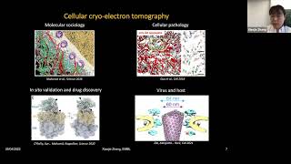
Thanks a lot to Robert for the kind introduction. And I also want to thank the organisers for giving me this opportunity to share our story today. So I'm very excited to share our unexpected discovery about stress-induced Mumps virus reactivation, which was a cross-scale study enabled by cellular cryo-electron tomography. So when I started my postdoc, I was particularly interested in a field called biomolecular condensates. As shown in this video, the C. elegans germline granules indicated by the GFP-labeled key component of the pea granule compartment, they actually behave quite dynamic. They are mesoscale, micrometre-scale assemblies containing mostly RNA and RNA-binding proteins. They show this type of liquid-like behaviours, fuse and fission over time. They are very important for the developmental biology of C. elegans. And this was also shown at a later stage in mammalian cells, the HeLa cell stress granules, when cells respond to heat shock stress, for example. You can see cytoplasmic assembly of the mCherry labelled G3BP1, a microprotein of stress granule. They are also very liquid-like. So, these type of compartments, in contrast to conventional membrane-bound compartments, they actually assemble via a mechanism called liquid-liquid phase separation. And in recent years, there are more types of biomolecular condensates which were recently found, they have multiple functions. They have very different functions ranging from cell signaling to transcriptional regulation in diverse cell types. And what's more interesting is that these components of biomolecular contents are also related to the development of neuron degeneration diseases. For example, in this image, you can see In the patient neuron cells, the stress granule component FAS actually forms cytoplasmic inclusions by immunohistochemistry staining. So, to understand the molecular basis for the assembly and dynamics of biomolecular lysis and how they might be related to disease development would be very interesting. So, What was striking at the beginning of this field was that people found simply by using purified key component of biomolecular condensates in the in vitro system, people can reconstitute these droplet structure, which recapitulates the behaviour of biomolecular condensates inside cell. Normally, these key components contain multimodular domains, and they have disordered features. And later people found, although in other types of amyloid condensates, in the in vitro reconstitution system, they can also observe such droplet-like structure, but they also convert into amyloid-like fibrils, which was speculated to be a link to the disease stage of these proteins. So, I was particularly interested to study the underlying structural basis for these mesoscale assemblies, first in vitro and later, at a later stage, also directly inside cell. So I want to introduce the technique cryogenic transmission electron microscopy, which enabled the study of these biomolecular condensates at high molecular details. So in contrast to light microscopy, as we see in this image, transmission electron microscopy actually use electrons as source and also electron magnets as condenser lens instead of glasses. And then this technique allows a visualization of specimen at molecular details with higher magnification. But apparently, with this technique, we cannot image live cells. We have to fix them either by chemical fixation or flash freezing. So chemical-fixed sample has the advantage of imaging the specimen at room temperature, but the flash-frozen samples, although technically more challenging, actually allows preservation of the high-resolution information of the structures inside the specimen while the specimen was imaged at -185 degrees Celsius. So, in order to image biomolecular condensates, which is actually a relatively thick sample for the electrons to go through, I took the advantage of a branch of the cryo-TEM technique, which is called cryo-electron tomography. So, in this case, I can use the state-of-the-art transmission electron microscope called TEM, but tilt my specimen containing the biomolecular condensates at a range between minus, plus 70 degrees. And by getting the projection views of the biomolecular condensates, we can later computationally reconstruct these projection images into a three-dimensional volume in order to see the molecular resolution details within the biomolecular condensates. So, we have recently written a step-by-step protocol about how to image biomolecular condensates with cryo-ET technique. So, I have imaged multiple types of biomolecular condensates. For example, first, I observed the condensates assembled by stress granule protein G3BP. BP1 in presence of RNA. And we can see in this image, there is indeed detailed feature within the condensates. For example, the cluster-like structures or some sort of beads-on-string structure. And we can even visualise very thin RNA fibres at the periphery of the condensates. However, if we look at another FAS protein condensate, which is also a component of stress granule. Here, this condensate looks very different from the one on the left image, it appears totally amorphous. And we also see fibres originating from the periphery, the boundary of the condensate. So both proteins are stress granule components. And then I wondered what type of structural features is actually the physiological structural basis of stress granules in the cellular context. So, I thought about using a more complicated technique, the cellular cryo-electron tomography, to directly visualise stress granules in their cellular context. So, before going for the experimental workflow of the cellular cryo-electron tomography, I want to first show you the potential applications of this technique. For example, we can actually use cellular cryo-ET to visualise the molecular machineries at the nuclear periphery inside a HeLa cell, or at a disease context, we can visualise how the inclusion bodies aggregates inside cytoplasmic region interacting with proteasomes and introduce dysregulation of the function of proteasomes. In another case, we can also directly see the coupling between the macromolecular machineries, in this case ribosome and the polymerase, directly from intact bacterial cell, which is very potential for development of drugs based on cellular structural biology. And recent years, we also see this technique is very useful in directly revealing the interplay between the virus and the host. For example, when HIV particle, viral particle entering the nuclear pore, which is of similar size in the diameter. So with these potentially useful technique, we probably wonder how the experiment can be done. But there are indeed challenges in cellular cryo-electron tomography in comparison to a simplified cryo-ET of in vitro sample. So, in this case, we are looking at a HeLa cell from the side view. And you can see a HeLa cell is about 5 to 10 micrometres in thickness. This is very thick for the electrons to go through the biological material. So, to enable transmission electron microscopy, we first need to thin the section of the cells down to a very thin, like, blade away most of the cellular material to only preserve a very thin section of the cell in order to let the sample to be electron transparent. And secondly, since I was interested in the structural basis of stress granules, how could I locate stress granules within such a large cellular volume? So we would have the challenge of targeting. And within the next slides, I will show you how these technical challenges were overcome by our development developed, the pipeline developed in our lab. So, first of all, so cryo-electron tomography starts from freezing of the sample, as I mentioned before. So, in this case, we have the cells seeded on the transmission electron microscopy support termed TEM grids. So, we once we induce the formation of stress granules inside these cells monitored by live imaging, we can immediately flash-freeze the grids with the cells seated on top with commercially available devices called Vitrobot or Leica EMTP2. And by scanning electron microscopy, you can visualise how a grid looks like and how the cells are nicely seated at the centre of each square. To enable the next step sample preparation. And as I mentioned, one challenge of the workflow or the technique is to target stress granules in my project. So, when I started, I worked with Leica company to develop a prototype cryoconfocal light microscope. As shown in this image, the main difference between a cryoconfocal light microscope and the room temperature SP8 microscope was actually this cryostage mounted in the optical path, because we need to keep the sample frozen all the time and maintained at a temperature of -180 degrees. So, of course, we also need a transfer toolbox to load our grease with cells frozen into this cryostage. And when zooming in, we can see how this imaging— the sample is loaded onto the cryo stage. Actually, there's continuous cold nitrogen gas going through the cryo stage chamber and keeps the sample at a frozen state and a low temperature. So, when we image the samples at a cryogenic temperature, meaning -185 degrees Celsius, apparently there are more challenges in comparison to the room temperature confocal light microscopy. First of all, as we imagine in this cold nitrogen gas atmosphere, there is potentially water condensation which could potentially be deposited to the surface of the grid especially on the surface of the cell. So, this would be a big obstacle for TEM imaging, which I will show you very soon. And secondly, of course, we always want to get good image quality. But in this case, we also need to take into consideration the time that will be taken to image them, for example, with more averaging or like longer dwell time because we really want to get image done as short a possible time as possible. Because over time, there could also be contaminants accumulated on the surface of the sample. And there could also be drift and vibration during long-time imaging. As shown here, when we examined with tetraspec bees, which emit at multiple channels, multiple fluorescent channels, the strategies, the two strategies scanning between stacks or between frames actually give very different behaviour of the signal. The scanning between stacks, meaning we first image one channel through Z and then different channels. And the second strategy scanning between frames means we first image at a fixed Z height and then different channels and later we change the Z height to get the Z-stack images. So as we see, our final purpose of using cryoconfocal is actually to do correlative light and electron microscopy. So the coordinates of the signal among different channels should be as consistent as possible to give us the location XYZ coordinate information. But apparently, if we use the first strategy scanning between stacks, the coordinates are not so consistent. And if we go for the second strategy, it seems at least we can get more consistent information about the coordinates of the signal. But when we look at the images, these two strategies apparently have different behaviors. The second strategy, although it has the advantage of consistent coordinate information, but apparently there could be more vibration and drift happening during the longer imaging period. So during the experiments, I always need to do image registration after data acquisition. In order to get them better aligned to get more accurate information about the coordinates of the signal. So, these are the YZ view of the fluorescent beads in the maximum intensity projected view. And so, by developing this microscope and technique, it enabled a publication about nuclear pore complex in their cellular context in recent years. So, for my project about stress granules, now we can actually use the cryoconvocal light microscope first to get the navigator view of the TEM grid. And then we zoom in to the individual square to look for the cells. So, here you can see the red signal is the MKII fluorescently labelled stress granule microprotein FUS. So, we can see nicely distributed assembled stress granules inside the cell. And then these green signal is actually the fluorescent beads serving as fiducial markers for the correlative light and electron microscopy pipeline. So, we will talk about their usage on the next slide. And with this sample, we next can think about the challenge of the thickness of the sample. So in order to get a thin section of the cell that is electron transparent, we use the device, the Aquilose focused ion beam scanning electron microscopy, which has dual beam inside the chamber. Allows sample preparation and monitoring of images by SEM at -185 degrees Celsius. So, again, if we look at a HeLa cell, now with this technique, the cryofocused ion beam milling, we can actually really remove the majority of the cell material and in the end only preserve a thin section of the cell. At the site of interest in my project, the location of stress granules. So, with the three-dimensional confocal imaging data acquired from the cryoconfocal light microscopy, now we can correlate the data, the 3D data, to this 2D view, the FIB view. So, by a procedure of 3D to 2D correlation enabled by software called 3D Correlation Toolbox, we can prepare the thin section of the cell containing the stress granule locations. So, the resulting sample looks like this. We have two views from the top, the SEM view, and from the side, the FIB view. So, we can see this thin slab of the cell of a thickness around 200 nanometres now actually contains the location of stress granules. And when we load this thin section of the cell, which we call lamella, onto state-of-the-art transmission electron microscope, now we can visualise the lamella part of the cell at first of all intermediate magnification. So, here you can see the sample is actually heavily covered by these black blobs, which are ice contaminants. So, as I mentioned before, in this cryo-electron tomography pipeline or cryo-TEM technique in general, we want to try to avoid the ice contaminants on the sample as much as possible. But luckily, there are still regions corresponding to my target structure, the stress granules, for acquisition of the tilt series while tilting the sample in the microscope. So, with the tilt series, I can do the reconstruction of the cellular volume, into this tomographic data. And when we go through the Z direction in the XY view of the tomographic volume, we can see conventional cellular organelles such as mitochondria, ER membrane ribosomes, microtubule, and actin filaments. So, we can really try to resolve these molecular structures or cellular organelles to very high resolution. And what was striking to us at that point was we observed a large patch of filamentous structure at the region, presumably to be the stress granules, since we were using the correlative light electron microscopy pipeline. And when zooming in, Here we can see these filaments actually give repetitive features along the helical filaments, along the filaments indicating a helical structure, and the filaments are actually also hollow in the middle. So, since we were targeting stress granules, we were very excited to see these filaments in a large patch. We thought, oh, maybe these filaments are scaffold proteins and structures of the stress granules. So, I next continued to trace these filaments along the central line of this helical structure. I generated equally distant points along this helical structure and cropped subvolumes along these lines.. And then we call them subtomogram particles. And by aligning, computationally aligning and averaging these subtomogram particles, I generated an average structure of these helical filaments. As we see in these different views, indeed, the filaments are helical and it's hollow in the middle. But so my big puzzle was, first of all, what is the identity of these helical filaments? And if we can resolve, how we can resolve them to high resolution so that we can know the identity of them, and also the potential function of these filaments in putatively the stress granule location. So, back then when I worked on this project, we actually didn't have established a pipeline to obtain high-resolution subtomogram averages or structures of these helical filaments. I had to first go through the biochemical isolation of these helical filaments from the cell lysate. And luckily, I was able to enrich these helical filaments 13,000 G pellet. And by negative staining TEM, we can see here these filaments are nicely preserved in this pellet fraction. And further, by optimising the isolation procedure, I performed sucrose gradient centrifugation experiment and was able to better purify and enrich these helical filaments at certain fraction of my centrifugal tubes. And in this case, I was aiming to work with my collaborator, Sindhu Jha, in Savitsky group to perform quantitative mass spectrometry in order to identify the molecules responsible for such helical assemblies. However, over a long time of trials, we weren't able to find any potential candidates that can assemble into these helical structures. We were very disappointed, but I decided to continue to explore more about the property of these helical filaments. And I found by adding an RNA analogue heparin, because initially I thought this protein might be at least RNA-binding proteins, this actually induced morphological change of these helical filaments from a curved state to a relatively straight state, along with other conditions. So, this later enabled acquisition of these tomographic data of the helical filaments. As we see in this slice view along the Z dimension, although the helical filament is still sort of curved in Z. But with this 3-dimensional volume data, I can perform alignment and averaging of these helical structures in order to get a high-resolution structure of the filaments. So, with this, I worked with a master's student in the group, Tim Pfefferster. We were able to resolve the structure of the helical filaments to about 5 to 6 angstrom resolution at the beginning. And from this resolution detail, we could already see— actually, it's also a random encountering of similar structures when we read literature. We had the suspicion that these helical filaments might be related to some new the nucleocapsid structure of viruses. So although the chance was very little, but we still decided to ask our collaborator, Cynthia, to again examine our mass spectrometry data with a list of viral protein that may potentially form such helical structures. So we actually were very surprised to see Indeed, this is a structure assembled from a mumps virus protein, the nucleocapsid protein of mumps virus. So although I was very shocked, but I still decided to at least validate first if this is, you know, indeed a virus protein by immunofluorescence light microscopy. So in this image, you can see with the antibody against the nucleocapsid protein of Mumps virus, we see all the cells in my cell culture actually contain the Mumps virus infection. And later, by transcriptome sequencing, we confirmed again the existence of the viral genome in the cell culture, and also the transcription happening inside these cells. So, I had to revise my cell culture model. So, in this image, from this data, we postulated that our cell culture model is very likely a persistent infection model of the Mumps virus, because in all cells, we can see the existence of their nucleocapsid protein shown by the cyan-coloured granule. And since the cell line I used was also fluorescently labelled on stress marker, stress granule marker protein G3BP1, the magenta colour, we see the cells were not actually under any stress from the viral infection during the cell culture. So, this is indeed a persistent infection cell model, and we suspected the viral replication and the host cell immune response were somehow maintained at a balanced state during the cell culture and experiment. So, you may wonder how the viral life cycle of mouse virus actually looks like. So we can see here in this illustration, the mouse virus contains a minus-stranded RNA genome encapsulated by its nucleocapsid protein, N protein, and this forms this nucleocapsid which serves as template for the viral transcription and replication. And these events were carried out by the RNA-dependent RNA polymerase, the L protein of the virus itself, with assistance from the phosphoprotein of the virus. So these together constitute the core of the viral transcription replication events inside cytoplasmic region, termed viral factories or viral replication factories. This corresponds to the cyan colour structure we see in the immunofluorescence light microscopy images. So, at that time, this was a big puzzle for us, why we initially started to look into the structural basis of stress granules, but it turned out, we found large accumulation of viral nucleocapsids in the cytoplasmic region, which also appear to be sort of phase-separated because they do not really have membrane at the boundary of the entire structure, but they only locally exist. So, this could also serve as a good model for studying the structural basis for biomolecular condensates or compartments inside cell. So, we decided to continue with the project. But now, of course, the purpose was a little bit shifted towards the viral activity, infection, and stress response. So we wondered if our persistent cell viral infection model somehow may provide information for understanding the viral pathogenesis. So when we look at literature, It's very interesting that we found persistent Mumps virus infection was related to chronic diseases. For example, here I'm showing you chronic myositis, which is indicated by the weakness of the muscle groups of patients. And you can see in the muscle biopsy of the patients, there is large accumulation of this mumps virus nucleocapsid structure. And when zooming in, we can see these nucleocapsids also appear to be very straight. So we suspected, or sort of we thought, our persistent mumps virus infection model could initially be a stage of the balance between the viral replication activity and the host immune response. But maybe by accident, the stress response— the stress treatment actually induced reactivation of the Mumps virus activity, which resulted in observation of the large accumulation of Mumps virus nucleocapsids in cytoplasmic region. Because this stress-induced reactivation of viruses have been observed in other types of viruses, for example, human simplex viruses or immunodeficiency viruses, but little was known about the reactivation of RNA viruses, for example, mumps virus, under stress response. So this was very interesting to us, and we also wondered If there is indeed stress-induced reactivation of the viral activity, then what are the underlying molecular mechanisms? So, first of all, we carried out quantification of the viral mRNA and genomic RNA by quantitative PCR experiment. And we found, interestingly, along the time course of stress treatment, there is clearly increase in the viral transcription and replication inside these cells. So indeed, stress can induce the reactivation of these viruses from a persistent infection stage. And then what would be the underlying cellular basis for these reactivation events? When we look at the viral replication factories by immunofluorescence light microscopy, we see along the time course of stress treatment, of course, there is assembly of stress granules, the magenta colour structure, but we also observed change in the viral factories, the cyan colour structures. To be noted, the viral factories in many cases exist in close proximity to stress granules. This in a way explains why in our correlative light and electron microscopy experiment to target the stress granules, accidentally, we observed the existence of Mumps virus replication factories. So, the real change in the viral factories in this stress treatment experiment was that we observed continuous increase in the average size of biofactories, which happens concomitantly with a decrease in their numbers. So when this phenomenon of the biofactory largely resembles the behavior of liquid-like biomolecular condensates in their growth phase. So we can see when the condensates grow, either by a mechanism called coalescence, meaning the condensates fuse with each other, or at a procedure named Ostwald ripening, meaning the bigger condensates grow at the expense of the dissolution of the smaller condensates. By either mechanism, the condensates can grow an increase in the size, along with a decrease in their numbers. So, this phenomenon we call coarsening behaviour of the liquid-like condensates. So, the phenomenon observed for viral factories in our stress treatment experiment is reminiscent again of the coarsening behaviour of biomolecular condensates, indicating the viral factories in our stress stress treatment behave as liquid-like biomolecular condensates. So, we wonder then what was the molecular mechanisms underlying such coagulating behaviour of biofilm bacteria during the stress treatment. So, biomolecular condensates are mostly mediated by RNA or RNA-binding proteins, which have disordered features shown in this illustration. So when we look at the most abundant protein of the Mumps virus, the M protein and the P protein, based on their sequence prediction, we can see the C-terminus of the M protein and many regions across the P protein of the virus actually appear to be disordered. So we wondered if by transfecting these viral genes into the non-infected cells, we could already potentially see the phase separation behaviour of the viral protein. So indeed, when I transvected the viral plasmids into the non-infected cells, even without any stress treatment, the p-protein on its own can form condensates inside cytoplasmic region, and in presence of the viral M-protein, they actually co-localise to form large cytoplasmic assemblies reminiscent of the morphology and the size of the real virofactories in my infected cell model. So, this shows us indeed the viral p-protein can be a driving force for the phase behaviour of virofactories during stress treatment. And my recent data, although not shown here, also indicate the p-protein can behave as liquid, so can assemble into this liquid-like condensates and fuse with each other under stress treatment, even in these non-infected cells. So until now we know, okay, the viral p-protein could potentially drive the phase separation behaviour of biorefactories observed in my stress treatment experiment. But then what is actually the molecular trigger or the switch for such coarsening behaviour of these condensates? So, although our mass spectrometry quantification didn't reveal change in the intracellular the total level of the P protein inside the cell. But interestingly, we observed there is clear increase in the phosphorylation level of the P protein at this site indicated by the green colour. So, this phosphorylation site exists at the interface between the viral P protein and the viral polymerase L protein. And based on our structural analysis, due to the complementary surface charge between these two proteins, the phosphorylation could potentially enhance the interaction between the viral P protein and the polymerase. This would facilitate the viral transcription and replication carried out by the viral polymerase machinery. And indeed, later on, By many biochemical experiments, we validated the viral NPL protein duoform complex. And interestingly, the biopolymers is more enriched into this complex when cells are under stress. And with my collaborator, Sindhu Jha, we further performed solubility quantitative mass spectrometry profiling, which is a technique to reveal the change in protein solubility at proteome level at different treatment conditions. And in this case, we found along the time course of stress treatment, although most of the viral protein didn't change their solubility property, we interestingly found the biopolymers L protein is more enriched into— it's decreasing in its solubility. And in the concept or context of biomolecular condensates, this indicates the biopolymers has higher potential to be enriched into the viral replication factories when cells are under stress. So, from these evidences, we show stress treatment induced change in the viral protein interactions. And then I wondered if we can directly visualise the change in the structural features of viral factories directly inside the cell. So, with this purpose, we now come back to the technique I introduced to you at the beginning, the cellular cryoelectron tomography to directly visualise the biofactory and their change over time of stress treatment. Again, this is intermediate magnification view of a thin section of the cell containing biofactory with stress granule in its close proximity. And here at early time point of stress condition, we can see similar feature of the viral nucleocapsids, the filaments we have seen in previous, and they appear to be highly curved and compact in this cytoplasmic region, which do not have membranes, meaning they are packed in this phase-separated compartment. However, what's most interesting to us was that while I treated the sample, the mumps virus-infected HeLa cell for a longer time, along the stress time course, we observed clear change in the viral factory and their nucleocapsids. So, first of all, we see the viral factory is getting more crowded, In these images, we can see this black density is getting more in the same cellular volume. This was also quantified as in this plot, the black dots of the cell fraction, the volume fraction of the nucleocapsids in viral factory. So, and also we can see these densities attaching to the surface of these nucleocapsids, which are more prominent when the cells are under prolonged stress condition. And the second striking discovery of this experiment was that I observed there is morphological change of the nucleocapsids. These filaments converted from a curved state to a more straight state. And interestingly, this straight conformation of the nucleocapsid is highly reminiscent of that was observed in the disease state muscle biopsy image of the patient. So our study actually indicates a link of the reactivation of monkeypox virus in persistent infection to the chronic diseases. So, as I mentioned before, for this project, I developed a pipeline, a septomogram averaging pipeline, to get a high-resolution structure of these heterofilaments, which was enabled by incorporating a suite of software packages named WARP, RELION, and EM, which were initially developed for high-resolution structure resolution, averaging of the ribosome structure in bacterial cells. And I incorporated this pipeline and modified it for resolving the helical structure to higher resolution. And again, with isolated nucleocapsids, I first optimised this pipeline, starting from segmenting of the filaments and sampling along the helical structure and aligning these subvolumes and averaging them at different binning factor of the images. And in the end, I was able to resolve two conformations of the helical structure which have very distinct features. So this video shows you, first of all, the majority class of the helical structure. You can see it's helical, and here I'm showing you 4 subunits coloured differently, and the RNA is encapsulated at the surface of the helical structure. And then soon enough, if we look at the looming view of the structure, we can better visualise the elements responsible for the assembly of this helical structure. So you can see the C-terminal domain arm region and the N-terminal domain arm region are the key elements for the assembly, and the iron density can be better visualised from the side. And we can see these majority and minority class actually have very different conformation, which resulted in a difference in accessibility of the RNA genome of the virus, and also a flexible region at the C-terminus of the protein. So this is a better visualization of the difference between these two conformations, and the majority class contains a larger helical repeat in comparison to the minority class. So this means the majority class is at a relatively open conformation compared to the minority class, which is at a relatively closed conformation. So from the LUMIN view, we can see this is enabled by the rigidification of a key element, the C-terminal domain arm region of the nucleocapsid protein. And the resulting function is that the open conformation actually allows more space at the surface of the helical structure for the viral genomic RNA to be more accessible to the viral polymerase machinery, and also more space for the C-terminus of the M protein, the nucleocapsid protein, to reach the surface of the helical structure in order to interact with the viral polymerase machinery. So, these two confirmations were obtained at early time points of stress treatment. And with the pipeline developed to resolve such hybrid structure, I was able later to resolve the street nuclear capsids directly observed in the cellular volume. So, with together with a master's student, Saiying Cheng, in our group, we developed, optimised this pipeline to obtain high-resolution structure for cellular material as well. And the resulting structure looks like this. It highly resembles the open conformation of the nucleocapsids from the isolated nucleocapsids at early time point of stress treatment. Of course, we again see the similar feature of these key elements and high accessibility of the biogenomic RNA and the C-terminus region. And what's interesting from the cellular electron density map is that we additionally see extra density at the surface of the nuclear capsids, which was predicted to be the location of either the C-terminus region of the nuclease protein, or the interacting partner protein, the viral p-protein. So this indicates again, the straight conformation of the viral nucleocapsids observed at prolonged stress condition inside cell is supporting a state for viral transcription and replication. So, with this, I want to conclude. We proposed a working model where we discovered the stress-induced transition of the persistent Mumps virus infection from a basal level to an activated state. So, we observed coarsening, or yeah, the coarsening of the viral factory, along with the morphological change of them, especially the viral nucleocapsid. We identified change in the phosphorylation level of the viral p-protein, which potentially stabilizes the viral polymerase together with the conformational change of the viral nucleocapsid and higher accessibility of the viral genomic RNA. At a prolonged stress condition. Altogether, they enable the reactivation of the virus at stress conditions. This was, of course, also orchestrated by the downregulation of the host immune response, which were reviewed from our quantitative mass spectrometry of the host cell factors. So, I want to show you from this study over the years, the efforts from our group now really enables high-resolution subtomogram averaging directly for cell samples at different steps. For example, we can now have a micropatterning technique to better spatially control the seeding of the cells on the TEM grid. And we also have automated focus the IMB meeting for preparation of the sample. And also recently, we developed a practical pipeline for directly imaging the lamella after field meeting. This provides even better, higher precision of the signal, fluorescence signal, for correlation and targeting. And the convolutional neural network and aided data annotation now allows high-throughput data mining in a less time-consuming manner, and the developed pipeline for subtomogram averaging is also now more streamlined for generating high-resolution structures. So with this, I want to show you that we think cellular cryo-electron tomography is really a cross-scale technique that bridges high-resolution structural details all the way to molecular events and organelle interactions inside a cell, even at tissue level when we incorporate even more complicated cryo-FIG technique. So, to integrate— when we integrate cellular cryo-electron tomography with mass spectrometry and other techniques, now we can really resolve the cellular molecular and structural mechanisms underlying basic biological processes. So, with this, I want to thank the entire Mohamed Group for support in this project, especially my mentors, Yuria Mohamed, for all this advice and support, and people involved in this project, the funding resource, and especially my collaborator, Misha, and Sindhuja, for all the mass spectrometry quantification and my previous supervisor, now my collaborator, Tony Hyman in Max Planck Institute, for insights in the cell biology aspect and all the support from facility people and useful discussion from the external user experts. And I want to thank you for your attention. And I'm happy to take questions.
Sorry for the long talk. Oh, absolutely fine. Thanks a lot, Xiaoxue. Really great talk with a very impressive array of technologies that you presented, going all the way from the cellular scale to really the nanoscale. So I think we can open the Q&A. So if you have any questions, please raise your hand, and if possible, ask them in person. Yeah, Brian, please go ahead.
Oh, that was absolutely fantastic. Absolutely fantastic, mind-blowing talk. Congratulations. Thank you. I have a question about these stress fibrils, right? You're forming these fibrils. Do you think these fibrils are then reconverted to make the capsid envelope? Or is this just a byproduct of making too much when you're virally making all these things? Do you think there's another process where you make, where where you disassemble these fibrils? Yeah, I hope I get your question correctly. So the filaments I'm showing, or the helical structure in my slides, are actually the templates of the virus. It's a nucleocapsid, meaning we have the nucleocapsid protein as a scaffold, and then the virogenomic RNA sort of wrapped around at the surface, right? Each filament actually corresponds to one genome of the virus. So I think, you know, for example, during the transcription replication procedure, of course, this serves as a template, and then synthesis of new nucleocapsids. So you might see sort of assembly, but I'm not sure if there is actually disassembly. Of such type of structure.
Okay, yeah, thanks. Any further questions? If not, I had a question towards this of the correlative aspects, right? Because you use fluorescence microscopy to really target your tiny structures in really large volumes. So I was quite impressed, you were saying the lamella that you sort of like cutting away only 200 nanometers thick, and still you are accurately sort of finding, finding your target structure with fluorescence microscopy. So can you talk a bit more on how you get this high registration accuracy, given that with a confocal microscope still your axial resolution is fairly poor in comparison, right?
So yes, so in the real experiment we additionally introduce another channel corresponding to the fluorescent beads, right? So in that case— oh, sorry, other than fluorescent beads, we can have a different channel of the internal structure, for example, staining of the lipid droplets. So that will actually serve as our internal reference, first of all, because as you mentioned, due to the diffraction limit, right, we cannot actually have accuracy at, you know, 200 nanometres with confocal, right? So, actually we use the internal reference, such as lipid droplet structure, to improve the accuracy for correlation, because we can see both the fluorescence signal of lipid droplet and the structural feature of lipid droplet, because we know they have membrane, right? You can easily distinguish lipid droplet in TEM data.
But then have you sort of quantified the accuracy? I mean, in sort of—
Yes, not my study per se, but in the paper I briefly mentioned at the end, the FAP automation paper recently published from our group, Sarah, now a postdoc in our group, actually quantified the accuracy. So if we have like this such internal reference, it's relatively accurate.
Okay, yeah, thanks. I'm waiting for the raised hands. In the meantime, sort of a more general question that I maybe didn't fully get this. So when you were saying that, you know, the Mumps virus particles are sort of like, are in your HeLa cell culture, so is it only the case for your HeLa cell culture? You think it's a more general phenomenon, right, that it's already—
Yeah, this is a very interesting question. Initially, we really thought it's somehow randomly introduced into my cell culture. But when we later talked about this story to the general community, we found it was actually identified many times in other labs. So, we don't know, you know, HeLa cell is generally used cell culture model across the world. But during these 50 to 70 years, we don't know what was introduced sometimes randomly, right?
So then likely it got introduced already earlier, I guess.
We guess so, yes.
Okay, good. Really interesting, very unexpected. Yeah. Okay, any further questions from the audience?
I'm also happy to take questions in case you want to communicate in emails or something. Yeah.
More from Joshua







Scientist Stories: Hans Waterham, The value of phenotypic assays in peroxisomal disorders




Scientist Stories: Joel Zlotogora, Genetic carrier screening programs for reproductive purposes