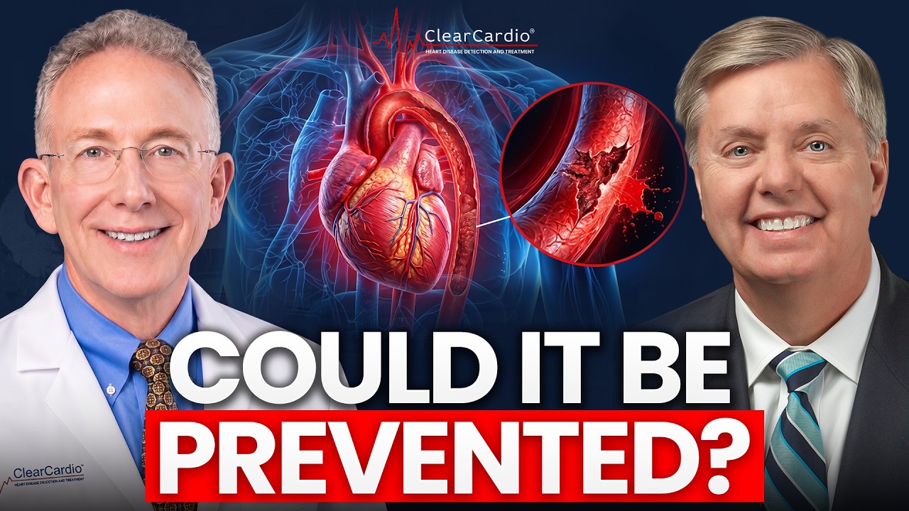
Look How DANGEROUS PLAQUE Looks Inside Your Heart Arteries

Dr. John Osborne uses AI cardiac imaging to show patient Jamie Gruber's plaque buildup: 26.4 mm³ in the LAD artery, mostly soft plaque. Total plaque volume, not visible blockages, predicts heart attacks. Major trials found stents don't improve survival over diet, exercise, and medication.
Chapters
They do not treat the disease. It does not detect early disease. It can't detect early disease. Use AI to see things that would otherwise be invisible. So as it turns out, it is not the blockages that we have been looking for and fixing mechanically for decades. That is not what predicts heart attacks or mortality. What actually does predict mortality, cardiovascular mortality, deaths from strokes and heart attacks, and heart attacks is actually total whole heart plaque volume. This is Dr. John Osborne, Chief Medical Officer of Cleer Cardio. I'm here with Jamie Gruber, who flew in all the way from—
Punta Cana, Dominican Republic.
All the way from Dominican Republic to get his scan. We're going to actually go through the whole process, look at all the pictures. He got scanned this morning.
I'm not a big hospital guy, but I mean, it was 10, 15 minutes. I was shocked at how quickly I was done. I didn't know what to do with the rest of my day. So, yeah, so I came here and excited to see—
We try to under-promise and over-deliver.
Very simple, very easy. It was great, honestly.
Fantastic. So glad you had a great experience. And so in the background, we did the scan, we get this incredible multi-gigabyte dataset. It's a massive dataset. It actually takes a DVD to store all the data on it. And all that data obtained from your heart is obtained in about 1 second. Everything else is putting in the IV, position you, putting you in the CT scanner. Another thing I'll ask you about, because this is a common question, right? So we use a CAT scanner, a CT scanner, right? So, but I think sometimes people can confuse easily a CAT scanner versus an MRI. So an MRI is a whole different imaging device. If you've ever— more typical with that, it's with a, with a CT scanner, CAT scanner, whole different technology. It's a donut, but it's open. You got plenty of room. It has another advantage of it beyond the speed and the resolution of this machine for our purposes. It also has the biggest bore, that is the biggest hole. So just less. More room for you and less issues with cholesterol. So I'm glad, glad you did well with that.
No issues. Yeah.
All right. So, you know, it, it is interesting that even people who are in great health can have terrible heart disease, which you don't have.
Okay. That's a good reveal.
I'll take that. Uh, but also too, and here's the, the magical, incredible breakthrough of adding AI to cardiac CT. So I've been doing cardiac CT for 25 years. Uh, and in the last few years, we now can layer on top of that incredible high-resolution dataset, which is this 3-dimensional, high-resolution data, which comes out of the CAT scanner, we now upload that, which we've already done, we did that this morning, to the cloud. It will then analyze every single one of the millions of pixels out of the study. And those images are what we call grayscale, right? So they're anywhere from black to white and gray in the middle, right? The important thing is, when we look at a CAT scan, as an example, MRI, 60, whatever, we can image or see or delineate about 50 to 100 shades of gray from black to white. That's, that's as much as our eyes can, can see. Encoded in every one of those millions of pixels coming out of the machine electronically is encoded over 2,000 shades of gray.
Oh, wow.
So when we do the AI, there's several advantages. One, it's analyzing every single pixel. It's analyzing it across 2,000 shades of gray. not 50 to 100 that we can see with our eye. So we literally can use AI to see things that would otherwise be invisible. So telescope to microscope.
Yeah.
In addition, we in cardiology have, for literally decades, I like to say, worshiped at the altar of blockages, right? And you think of it intuitively, right? A blockage is a bad thing. That's what causes heart attacks, and that's what kills people, right? And we have, for the last rough, since the 1990s, Right. So 30-plus years, every 10 years, we have done studies where we compare optimal medical therapy, which of course has changed and evolved and gotten better and better over the last 30 years, but optimal medical therapy for the treatment of plaque versus optimal medical therapy. And when I say that, I'm talking about treating your cholesterol, being on an aspirin, if you have diabetes, taking care of that, blood pressure, taking care of that. So that's optimal medical therapy versus optimal medical therapy with what we call technically revascularization. So what is revascularization? I call that the mechanical stuff. That's your balloons, the stents, the bypasses, right? And the belief was that, and we thought we'd prove this easily back in the '90s, that optimal medical therapy with balloons, stents, and bypass surgery would be and offer a superior outcome. If you look at heart attacks and cardiovascular death, death from heart attacks, would be better than just drugs, right? Just optimal medical therapy.
Sure.
And optimal medical therapy is obviously healthy diet, exercise, not smoking, all those things too. We'll talk about diet too, because that's going to be, I think, very intriguing. So we got a little interesting— that's a hook to hold on for that. But we did that back in the '90s, and guess what? We found out there was no advantage of optimal medical therapy with balloon stents or bypass surgery. There was no advantage. But with more state-of-the-art tools, drugs, stents, came up with exactly the same answer. It's not the mechanical things, it's the medical things. It's diet, exercise, medications, et cetera. So that's been a long-term, ongoing, decades-long debate within cardiology. So 2008, we did this trial called COURAGE, optical medical therapy, or optical medical therapy added to that, balloon stents, et cetera. Still no difference. Well, there's got to be something wrong. So let's do the study again. 2019, the trial was called ISCHEMIA, NIH-sponsored, we all paid for it. Exactly the same results. No difference in heart attacks, no difference in cardiovascular death.
So just extra hardware.
Yeah. Or as I like to say, stents, these little mesh things we put in the blood vessels to open up and all that, are about roughly, MSRP is about $3,000.
Wow.
So that is a great theme to understand. So as it turns out, it is not the blockages that we have been looking for and fixing mechanically for decades. That is not what predicts heart attacks or mortality. What actually does predict mortality, cardiovascular mortality, deaths from strokes and heart attacks, And heart attacks is actually, ready for this, total whole heart plaque volume. Okay? And that's what we're going to look at in you in just a moment. But I think it's so important to set that stage because I think a lot of people intuitively go, oh, I got a bad blockage. Thank God. Thank goodness it was found. We put a stent in it.
Woo! Avoided.
Crisis avoided.
Right.
No, I will tell you what stents are good for. And when I say stents, more realistically, it's stents. We put in stents, which open up the vessel and The stent stays behind to keep that vessel open, kind of like snowplowing, pushing it off to the side and something to hold the snow back, right? And then bypass surgery, which I think everybody has a pretty good idea what that's talking about.
Yeah.
So as it turns out, the mechanical things are useful for one reason. They're useful if you have really bad symptoms. We call it angina or angina. So in other words, the pipes supplying the blood flow to the heart are relatively small. As the load or the demand of the heart as a pump goes up, if the pipes are too small, You get chest pain, angina, breathing problems, shortness of breath. It could be a lot of different symptoms across different people. But as it turns out, the mechanical things, the stents and bypass surgery, are very good at restoring blood flow. Here's the problem. They do not treat the disease. The disease is the plaque. If the plaque grows to an extent where it causes a blockage, you start getting symptoms.
Yeah.
But we will not have symptoms, chest pain, angina, flunk a stress test, until you have at least a 70% blockage. So waiting for symptoms or trying to detect the disease with tools like stress tests, which we've done for decades, are only detecting late, late disease. It does not detect early disease. It can't detect early disease, which is why 25 years ago, when the very, very early, very crude cardiac CT scanners became available, knowing that they could see that plaque, they could detect it long before we ever had a blockage. Obviously, you had to wait till the Technology got better and sharper and easier to use, but I knew that this was going to be how we deal with this disease and detect it. So that's what we did with you today.
I will, but quips potentially are coming.
Okay, so we're going to show you your Cleer Cardio exam live. So what we have done here is— and I'll ask for a little mind experiment— so we've taken your heart and I've taken your right coronary artery, which is this vessel here, and we now can straighten that vessel out. This vessel is not straight, but with the magic of computers, we've now straightened that vessel out. So this is your right coronary artery. This is the channel, the lumen. And we can actually— this is— it could be like 3 o'clock or 12 o'clock or 6 o'clock, right?
Gotcha, yeah.
So you really have to spin the vessel around to be able to see that plaque. Another important thing about plaque, plaque always forms in the wall of the vessel. So think of a donut, right? Historically, we've been worried about the donut hole, but that's the last thing to go bad. Plaque begins in the donut, and early on that cholesterol literally gets deposited in the donut, and the donut will expand outwards. The hole is the same. There is no change here. There is no blockage.
So when we're looking into the—
whether there's a blockage or not, it's great. Texas, down the barrel of it, right?
Yeah, you're not looking up and down.
Yeah, but we can't— we couldn't see what's happening in the donut, and the whole vessel can literally expand to about twofold before it starts growing in. And remember, Historically, waiting for symptoms or flunking a stress test, we're already at a 70% blockage. So that plaque has literally been there for decades, right? And we did nothing because we didn't know we had an issue.
Yeah.
It will then tell us exactly how much plaque we have in each vessel. And we also get a summary of the whole, how much plaque in the entire heart. Okay? So I'll show you in a moment kind of what this number means, but in the right coronary artery, we did detect a little tiny bit of plaque. Again, this is the degree of plaque. That historically, before we had CT, before we had AI, was undetectable.
Yeah.
So I'm going to tell you, to put this into perspective with analogy, I think most people get, if this were a colonoscopy and we're looking for polyps, yeah, you got a little polyp.
Okay.
Big polyp.
Yeah.
Not a worrisome polyp, but you got a little tiny polyp. Which, how young are you now, Jamie?
46.
46. Okay. Which took you 46 years to get to this point.
Got it.
Okay.
Yeah.
So, you know, important, but Historically undetectable until we had this technology. So that's how sensitive this is. So we can measure in that particular vessel in the right coronary artery. We have three major vessels. If you look at that diagram here, it's sort of illustrating that diagram of that vessel—the right coronary—and then you'll see it shadows. We'll look at the others in a moment. The other the other two major blood vessels. So in that particular vessel, 2.8 cubic millimeters of plaque. Note we can measure this down to a tenth of a cubic millimeter. So if you look at the set a period at the end of a sentence. Sliced. And so that is the resolution that we can see.
What is a worrisome number or a number that makes you— is there a number that kind of like, ah, you're above this, we get a little nervous?
It's a continuum. I'm going to say the only normal is zero.
Okay.
Right?
Yeah.
I mean, that's normal.
Yeah.
Everything else is not. If you had a colonoscopy, you don't have any polyps or else we saw some polyps, right?
Got it.
So yeah, no, but it is a continuum. I will show you towards— after we look at this, I'll show you some data that sort of puts this with respect to— because people always want to know that question and how much, what does this number mean? How do I compare to everybody else? Hold that question. We will come back to that.
Got it.
Okay. So in this particular vessel, 2.8 millimeters. Next slide, if you could, JD. And again, you can see a nice, beautiful vessel there, right? Fantastic. Right? Right. And I just told you, they only detect severe disease. They only detect severe blockages. They can't detect early disease. And on top of that, when we do a stress test, and I think this is so important for everybody in the audience, that when we do a stress test, again, the sensitivity to detect the disease only detects severe disease, right? But that's not the case, right? Total plaque volume is 26.4. Okay, so we got a little bit more. Okay, but again, a nice big vessel, nothing that would ever show up on a stress test or even a heart cath. And if you look out here, see that little— looks like a little pimple there? Yeah, that's your plaque. It's in the wall of the vessel.
Is that a— so is that a dangerous amount of plaque? That looks dangerous to me.
Well, so again, on a heart cath, completely invisible. Want clarity on your arterial risk, click now the link in the description to schedule your Clear Cardio patient assessment call. We'll review your risk factors, see if our advanced program fits you or points you in the right direction. When we do stress testing, and again, any kind of different kinds of stress testing, very insensitive. Plus, we're not visualizing as we are here, the actual vessels. Yeah, right. So all the detail that I was telling you about, we can't see on a stress test. Furthermore, with stress tests, depending upon the kind, you have anywhere between a 25 to 40% What we call a false positive rate. That is, your stress test looks funky when in fact you're actually fine. Well, that's no fun.
Yeah.
Right? That's bad because in the old days, we would then take that person with the funky-looking stress test, and then they'd go on for a $20,000 invasive heart catheterization.
Right, right, right.
Of which we do a couple million a year at about $20,000, right? By the way, that diagnostic test is a diagnostic test, invasive. We put catheters, tubes, silly straws up in your heart. Takes a day of your life, basically. Risks of strokes, heart attacks, vascular events, bleeding, infection. And it is purely a diagnostic test. Doesn't fix anything.
Just gets at what it is.
And all I can see on a heart cath is the donut hole.
The donut, right.
Right?
Yep.
Nothing but the donuts.
Yeah.
Okay. That's why I stopped doing that 20 years ago.
I don't blame you.
It was silly, expensive, ridiculous. So the next vessel, the one going down the front here, total plaque volume is 26.4. Okay, so we got a little bit more. Okay, but again, a nice big vessel, nothing that would ever show up on a stress test or even a heart cath. And I think the next slide shows where you have a little bit of plaque. So what we can do now, we can take that data, we're flying up and down that vessel— please don't try this at home— and we're cutting through that vessel. This is the cross-section of that vessel right there. And if you look out here, see that little— looks like a little pimple there?
Yeah.
That's your plaque. It's in the wall of the vessel.
Is that a— so is that a dangerous amount of plaque? That looks dangerous to me.
Well, so again, on a heart cath, completely invisible.
Sure. Okay.
Because it's in the donut, not the donut hole. Now also too, and this is again what AI does, there's 2 kinds of plaque basically, right? And I'm going to use my favorite analogy. The first kind of plaque, the plaque that's always initially deposited, right? The starting plaque, which is also the dangerous plaque, the active plaque, the growing plaque. Okay. I'm going to call that, we call it soft plaque or lipid-rich plaque. I'm gonna call it lava, right? Because I'm gonna tell you the volcano analogy. All plaque starts out as lava, right? All volcanoes start out as— even extinct ones start out as lava. Over time, after that gets deposited, that lava will cool down.
Yep.
It'll become rock.
Solidified.
Yeah. Solidified. Cinder cone. An extinct volcano. So what AI also does is it tells me exactly how much plaque you have. Then we'll break it down into how much soft plaque you have and how much hard plaque.
Okay.
I don't care about the hard plaque. Extinct, inactive, no longer progressing. Who cares? It's evidence that there was an eruption there at some point, but it's no longer active.
So what is that?
That is a calcified plaque.
So that's hard plaque.
Exactly.
26, whatever it was, that's all hard plaque, no soft plaque?
Well, there is a tiny little bit of soft plaque. So if you look at these numbers here, the hard plaque measured in cubic millimeters is 5.5 cubic millimeters, and there's about 20.9 non-calcified. Lava. Okay. However, this calcified plaque is easy to see. It's generally calcified, easy to see. The, the soft plaque is dark. That's where the AI comes into play. This is the stuff that is enorm— can be very hard to see. I mean, I've done over 20,000 of these. Yeah. Uh, thank God for AI, 'cause it gives me a whole— it's like putting on glasses.
Extra eyes. Yeah.
So, but you know, it is what it is. I, I would say if I were to compare you to the average 46-year young guy, you, you got much less plaque than the average bear. Okay. Okay. But also appreciate this is an extremely sensitive tool. No, understood. So that's that vessel. And again, you can see out in the donut hole. And again, unless we had AI, I'd be able to see that visually. I could see there was a plaque, but I really couldn't do this kind of quantification or see some of the lava plaque there. That makes sense. More active plaque. Okay?
Last one.
And then this is the last vessel called the left circumflex heart. And this was a 0.
Looks amazing.
So I always like to say, if you're a 0, you're my hero. Okay?
This may be a dumb question. Of these 3—
There's only dumb answers.
What is the widowmaker? Is that none of this?
Yeah, comes up all the time. So the widowmaker, go back to the last slide, is, is, it's a, it's a really bad term. It's, it's not used in a very consistent scientific way. If, if there is a widowmaker, and here's my objection, some would say it's a blockage anywhere along that vessel. Okay. That's really not true. If you look way at the top there, there's something that says LM. That stands for left main. That, that is the largest of the coronary arteries, right?
Mm-hmm.
And it's usually only about a centimeter or 2 long. It then rapidly divides up into the vessel going down the front, and then shoots off the vessel to the left. So that's a really critical vessel. That's your 31-inch water main, right? If you get a blockage there, or rupture a plaque, or whatever, that's gonna be pretty disastrous, because it supplies so much of the heart muscle. Some of my colleagues would say anything in the LAD, which is the vessel that starts just after that, right there, would be the widowmaker.
Any of this? The whole LAD?
That's the whole LAD.
That's what they would say, gotcha.
I disagree with that. bit of hyperbole. They say it to get you excited, worried. And then when they put a stent in it, even though you have no symptoms and you feel fine, and they show you the picture of the narrowing and then the stent afterwards, and they say it's the widowmaker, you go, my God, my life was saved. And we have no data that says that.
Where is the plaque in this artery specifically?
So that plaque is in that— we break it down into 3 sections, right? Proximal, we call it proximal, mid, and distal, right? The front, the middle, and the latter part. So it's in the— it'd be proximal. And it actually gives you a little bit of color there.
Gotcha. Okay.
By the way, I don't have this slide, but the other cool thing that the AI does is it will give me a very precise analysis of any blockage, and it will color code that. So the computer said in that little tiny baby plaque, they call that a 4% narrowing, which honestly, again, if I did a heart cath on you, you wouldn't see it. It's invisible. But we do get that. We do get the narrowings. I can't say they're not— they're useful if you had bad angina, we're going, do you need a stent? Is it going to help your quality of life? Right? Does it save your life? But does it improve your quality of life? You don't want to be taking three nitros every time you go to the bathroom. The idea, then stents are fine. Okay, so stents are really, in my world versus orthopedics, are really like knee replacements. Right? No one says if you don't replace that knee, you're going to die. Right? Maybe disabled. You can't get around. But you know, and some people can be bone on bone and feel fine, and other people doesn't look that bad yet be in terrible pain. And if if the pain is bad enough, then you say I like to get that replaced. Right? That's really how stents work. Right? They're they're really quality of life. Tools. Okay. Nothing more, nothing less. Yep. So, but no, great question. So I'm going to— the left main is really the widowmaker just because it's such a critical vessel. You got nothing there. Itty-bitty tiny bit in what we call the proximal, the beginning part of that LAD. And again, in that vessel here, the third of the vessels, nothing whatsoever.
Plaque-free.
Plaque-free and loving it.
I'll take it.
Exactly. Okay. And I'll show you now, I think we have some other slides here. This is another picture of that. By the way, this is the— you will get a copy of all this. We're gonna send you a report. This is just the front page.
Yeah.
The report's about 20 pages long. The first 15 pages basically tell you what is plaque, how it behaves, donut holes, lava, plaque, all that stuff. So all this stuff that we covered. So I just saved you, you know, 30 pages. But it is a cool report. It's very well written, very scientifically accurate. It has cool visuals. So it's a great take-home if you ever want to look at this stuff. So you'll get that. But I'm gonna go back to your, your actual report. So again, we've taken the vessels before, we showed them horizontal, now they're vertical, same data. And so you can see, this is the right coronary artery. There's a little plaque there. You can see the channel, the donut hole is perfect. And you can see right there, make sure to lose the mic there. It looks like a little pimple.
Yep. That's my plaque.
That's a little bit of plaque, right? So in that particular vessel, which is a branch of the right coronary artery, so it's a pretty small vessel, there's 2.8 cubic millimeters of plaque in that vessel. It is all lava. There's no calcified plaque, and all the others are Basically zero.
Okay.
Right, for the rest of that vessel. Okay, next slide. And then there's that LAD. This is the one that had a total of 26 cubic millimeters of plaque. We break that down into 5.5 millimeters of calcified plaque, that is blue, and then about 20.9 cubic millimeters of the LAVA, the lipid-rich plaque. Again, channel wide open, heart cath, the quote used— they still call it the gold standard, which I laugh at— would be completely normal. And again, you can see they almost look like pimples. I hate to use that term, but They're kind of stuck on the outside, but we have a very precise quantification here. A little branch there, all zeros through there, and then that last vessel, the left circumflex, which I think we have here as well, which is the next slide.
This one, yeah.
Basically gives us the same data I just walked you through, but one additional thing that I'll show you as well. So here we have, on the left-hand side, you can see the total plaque volume, all plaque lumped together across the 3 different vessels, 2.8 in the right coronary, 26.4 in the left main LAD, zero in the left circumflex. Then it breaks it down into the 3 kinds of plaque. There is a 3rd kind of plaque that if you have it, tends to be quite low. It's called low-density non-calcified plaque. That if we talk about lava, think of this as like the really bad hot lava. You don't want this. You don't have any of that. A little bit, and we'll look at that, normalize that here in a second, of the non-calcified, a little bit of calcified plaque. And also too, a lot of times, and more and more people are hearing it and using it, and it's a good tool. But I think this is a beautiful illustration. Another imaging tool that, again, really goes back to the '80s, we've been doing this a long time, is called a calcium score, right?
Yeah.
So this is not a blood test. It's also done with a CAT scanner. It is kind of a very, very crude version of what we're doing here. There's no contrast. It's easy to do. It just takes a few minutes. Huge amounts of literature behind it. The limitations of a calcium score is because we're not giving contrast, I can't see any blockages. Okay. And number 2, I can't see the lava. So if you had a calcium score, and really that's a very low calcium score.
Where is the calcium score set?
So the calcium score is that 5.5 number. So there's that, remember that little speck of plaque in that one vessel that was white?
That's it. That's the 5.5.
It was 5.5.
So, but if I had a regular calcium score without contrast, it probably would be lower than 5.5.
Yeah. You probably wouldn't even see that.
Probably be a 0.
Exactly. Right. So you couldn't see it, nor could you see what is actually a more relevant plaque and a more important plaque, which you have more of, which is the lipid-rich plaque.
Gotcha. Yep.
Which again, here was 24 cubic millimeters roughly of non-calcified plaque at about 5.5. But if you did a calcium score, it'd be normal. You probably wouldn't even see this.
So this is heart.
Yep.
I think of a calcium score, there would also be an ultrasound done of the—
Yeah. So plaque can form in any artery of the body. The 2 big ones, there's really 3, the 2 big ones, heart, we call that heart attacks and balloon stent bypass surgery. Plaque can form here causing strokes. And certainly plaque can be spotty, right? You could have a lot here and none here, or vice versa.
Yeah.
So if you want to know what's in the heart, you got to do this.
Yeah.
We did not image the carotid arteries. However, I will tell you, with a relatively small amount of plaque here, I'm really— I'm not worried that there's anything of significance going on here. If you wanted to do that, we could do that with ultrasound. We can also do, as we did with the heart, we could do these beautiful images of all the carotid vessels. One of the limitations of ultrasound is we can't do quantification as we've done here. We can't really divide it up into the soft plaque, hard plaque, as we've done here. And then the other problem is, with ultrasound, I can only see from roughly here, above your collarbones, to your jaw.
Can't see underneath.
I can't. So anything above the jaw is invisible, because the ultrasound doesn't go through bone. And so when we do carotid CTs, same concept here, contrast, same scanner, we can see the entire carotid artery everywhere from where it comes off the aorta, that main vessel coming out of the heart, all the way up into the brain. So we see the entire vessel.
Gotcha.
So it depends, you know, when practically, you know, if you had a prior history of stroke or mini stroke, we're going to do that.
Sure.
Obviously, if you, on examination, right, I always listen with my stethoscope over the carotid arteries. If I hear what is called a bruit, it's a French term for murmur, basically, that might indicate a narrowing and say, we need to look at this further.
Yeah.
But again, with this pretty low score, I'm really not worried about anything going on here.
Good.
And also too, when we treat plaque, It is and can be a systemic disease, right? The other major places, although plaque can build up in any artery or vessel supplying any organ, the third area is in the legs, right? You can get in the arms too, but you can get in the legs. We call that peripheral arterial disease or PAD. And the symptoms of that would be you start walking and because demand load goes up, the vessel's too small, your muscles start cramping and you can't walk. And what happens is an interesting phenomenon where you walk, they cramp up, you can't walk. Because of insufficient blood flow. And then you rest for a bit, blood flow catches up, and then you can walk, and then it happens again. So that's called claudication. So claudication is sort of angina of the legs. Got it. Got it. Got it. Okay. Generally, the biggest burden of PAD though are for patients that either smoke or have diabetes or both, right? Not to say it doesn't occur in those 2 people, but rare. But anyway, the last thing I wanted to show you, and then there's a couple little trivial narrowings that the AI detected. And then that little box there to the right, we can now, with the ability to quantify AI, we can now put people in stages. Okay. So of course, 0 would be 0. Stage 1, which we call mild, ranges from not 0 to 250. And your total score was—
29.2.
Okay. Moderate, moderate, moderate, or stage 2 would be 250 to 750. And stage 3 is above 750. By the way, about a third of the people that we do clearly do Clear Cardio on, are actually stage 3. And these are people who have no symptoms, feel fine, whatever. So I guess in summary, what I'm telling you is the good news is that no blockages. We did detect a little bit of plaque, join the crowd. When you begin to use the incredibly sensitive tools that we now have access to, cardiac CT combined with the CleerCardio AI analysis, we will find upwards of about 92% of asymptomatic people, when you use this microscope, have detectable plaque.
That makes sense. That makes sense.
Right? Yeah. Uh, which is why 40% of us die of plaque, right? So early detection is, is the key. I would say you have a very small amount of plaque. It's a very slow glacial process. So what if you're— and we've already worked out looking at those stages. If you are stage 1, that the stages help dictate what we should do to stop and reverse plaque. And yes, we can stop and reverse plaque. We have multiple tools that can do that, but it also dictates when we should take another look. So if you're stage 1, 3 years.
Got it.
Stage 2, 2 years. Stage 3, which means you got lots of plaque, that stuff could happen soon. We got to make sure that, that again, we're very, very aggressive with that. And also, we look within a year to make sure that not only are the numbers better and cholesterol's better, and we're doing all the right stuff, but is the plaque better? And we could never do that before. Yeah. In, in the, in the practice.
That's good to know. So I have a 3-year stay of execution.
You do.
Amazing.
Right now, I will tell you one thing. So I got your labs, they look perfect, right? You're the Specimen of health. Your LDL is about 209.
209. Yeah. Yeah. Makes sense. Yeah.
Okay. Uh, and we were talking earlier, you, you do basically all keto, 4 eggs in the morning, ribeye for breakfast and lunch. And it's interesting, there's a massive debate going on right now. It's, it's in no way settled, right? I will tell you, as a cardiologist, when I see high LDL cholesterol, I get nervous. On the other hand, your HDLs are great. Mm-hmm. Which by the way, keto will raise your HDLs. Sure. Triglycerides. And triglycerides really get suppressed. And those are— HDL is, is a very messy number. But triglycerides are bad, right? So it definitely suppresses the triads. So I think it'll be an interesting experiment to do. Here's our N-of-1 experiment. We know where we stand in a very, very precise way as of 2025. And let's reconvene in about 3 years, see what we look like.
Yeah. So diet-wise, am I sustainable as a carnivore?
Whether it's sustainable for you is up to you.
Sure.
Right? I mean, some people just go, I can't eat any more protein.
Oh, I eat meat all the time. Yeah.
Other people, yeah. So I think sustainability's up to you. Whether that LDL is feeding the plaque or not is still, in people who do carnivore, is still a very interesting but unanswered question.
Yeah.
There is no way that you're going to rapidly progress that plaque. Let's say that 209 is bad, right? There's no way in the span of 3 years with the ability to detect and quantify plaque that that's going to progress on to something that's going to need stents, balloons, surgery, or whatever, right? So I am very comfortable saying, Eat right, exercise, don't start smoking.
No, I did smoke my first cigar recently, but I don't—I didn't like it.
That's all right. I'll look the other way. I'm out. Good.
That's good. That's good.
And you know, stay active. Keep an eye on your blood pressure. Very, very important. The silent killer. I would do that, and then let's relook three years and see how we look. You know, there are certainly members, but I think our mistake has really been we've always looked at the number and assumed that's what your vessels look like. Here we have a number LDL that's high. But when we actually look at the vessel and look at the disease and look at the plaque, it's minimal, right? And it is a very slow glacial process. So I would say, let's do our N-of-1 experiment. We will have other science coming out about this specific question, the high-fat carnivore keto diet, whatever you want to call it, and what happens with plaque in that setting, even though in many patients the LDL goes up.
Yeah.
But the HDL goes up, whatever that means. I'll admit, who knows? I don't really trust that. But more importantly, the triglycerides do come down, and that is a bad fat. So I say we do our N-of-1 experiment and, uh, we'll reconvene and schedule for your next scan in 3 years.
I'm just gonna keep eating meat. Let's see what happens.
You know, at this point, because we can now look under the hood and see you just don't have that much plaque at this point, we can very easily quantify it. We'll bring you back in 3 years and let's see how things look.
I love it. Well, this is great. Thank you so much for, for going through that. That was really fascinating actually. So yeah, good stuff.
I, I love doing people's virtual autopsies. Yeah. Emphasis on the virtual. Yeah. Don't miss this other episode right here on the screen. Where we will reveal the missing answers you need to know about true cardiovascular prevention. And if you're ready for absolute clarity on your arterial risk, click the link in the description to schedule your Clear Cardio patient assessment. We'll look at your specific risk factors and see if our advanced program can help you.